

doi: 10.1128/JVI.75.11.5433-5440.2001.
Feline immunodeficiency virus cell entry
Affiliations
- PMID: 11333931
- PMCID: PMC114955
- DOI: 10.1128/JVI.75.11.5433-5440.2001
Item in Clipboard
Feline immunodeficiency virus cell entry
J Virol.
2001 Jun.
Abstract
The process of feline immunodeficiency virus (FIV) cell entry was examined using assays for virus replication intermediates. FIV subtype B was found to utilize the chemokine receptor CXCR4, but not CCR5, as a cellular receptor. Zidovudine blocked formation of late viral replication products most effectively, including circular DNA genome intermediates. Our findings extend the role of CXCR4 as a primary receptor for CD4-independent cell entry by FIV.
Figures
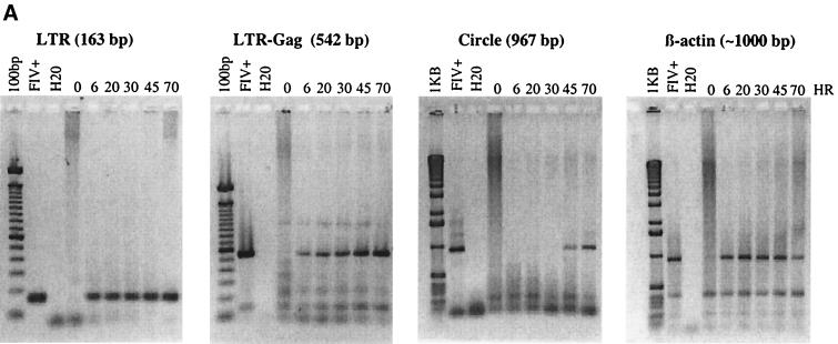
CrFK cell infection with FIV 34TF10 and detection of viral replication intermediates. (A) CrFK cells were infected with FIV 34TF10 in replicate wells and harvested from one well at 0, 6, 20, 30, 45, and 70 h PI. PCR was performed as described in the text, with FIV 2542-CRFK cellular DNA (FIV+) as the positive control and H2O plus reagents (H2O) as the negative control. The DNA marker is a 100-bp or 1-kb ladder, as indicated. PCR products were separated by agarose gel electrophoresis and visualized by ethidium bromide staining. (B) Schematic representation of primer positions for derivation of PCR products in FIV-infected cells derived from linear and circular viral DNA is shown. (C) One-LTR circle junction fragment homology (shaded segments) is indicated. As shown at the bottom of panel C, the fragments produced after FIV 34TF10 infection were colinear with that expected from the FIV 34TF10 genome.
CrFK cell infection with FIV 34TF10 and detection of viral replication intermediates. (A) CrFK cells were infected with FIV 34TF10 in replicate wells and harvested from one well at 0, 6, 20, 30, 45, and 70 h PI. PCR was performed as described in the text, with FIV 2542-CRFK cellular DNA (FIV+) as the positive control and H2O plus reagents (H2O) as the negative control. The DNA marker is a 100-bp or 1-kb ladder, as indicated. PCR products were separated by agarose gel electrophoresis and visualized by ethidium bromide staining. (B) Schematic representation of primer positions for derivation of PCR products in FIV-infected cells derived from linear and circular viral DNA is shown. (C) One-LTR circle junction fragment homology (shaded segments) is indicated. As shown at the bottom of panel C, the fragments produced after FIV 34TF10 infection were colinear with that expected from the FIV 34TF10 genome.
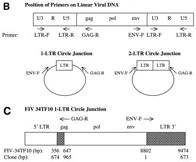
Lack of FIV DNA in viral stocks and impact of AZT on the production of viral DNA intermediates. (A) Viral inocula were examined for the presence of viral DNA by PCR. Viral stocks corresponded to FIV-A grown in CrFK cells (AC), FIV-B grown in CrFK cells (BC), FIV-B grown in PBMC (BP), FIV-C grown in PBMC (CP), and FIV 34TF10 (34) grown in CrFK cells. FIV+ corresponds to a positive control (FIV-infected CrFK DNA). (B) CrFK cells were infected with FIV 34TF10 in replicate wells, differing only by the addition of AZT-1MP. Cells were harvested at 0, 24, 72, and 144 h and immediately lysed and stored for subsequent DNA extraction. PCR was performed using 50 ng of DNA as a template. PCRs were separated by agarose gel electrophoresis and visualized by ethidium bromide staining (10 μl per sample). The marker (MW) for the LTR and LTR-gag fragments was a 100-bp ladder and for the circle and β-actin fragments was a 1-kB ladder. Controls were H2O plus reagents (H2O), CrFK DNA (CrFK), FIV 34TF10-infected CRFK DNA (FIV+), and p34TF10.

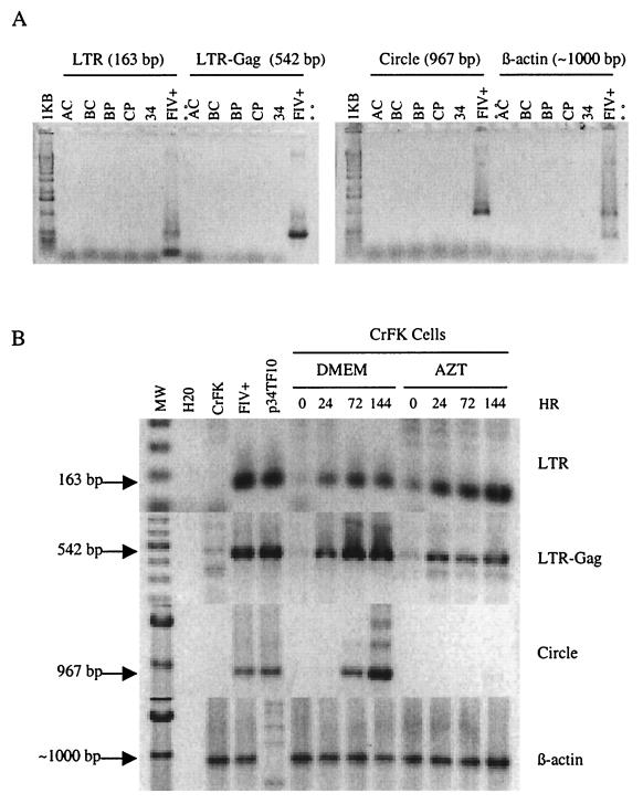
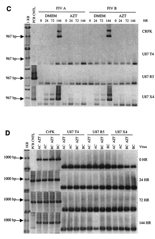
AZT can block circle formation in CrFK and U87-T4-CXCR4 cells infected with CrFK-derived FIV-A or FIV-B. FIV subtype A or FIV subtype B was used to infect CrFK, U87-T4, U87-T4-CCR5, and U87-T4-CXCR4 cell lines in replicate wells differing only by the addition of AZT. Cells were harvested, and viral sequences were PCR amplified and visualized as described in the legend to Fig. 2. The figure depicts the LTR PCR (A), the LTR-Gag PCR (B), the circle fragment PCR (C), and the β-actin PCR (D). The PCR for each fragment was performed concurrently for all four cell lines. Four controls were included for each amplification and were run in lane 2 (PCR CNTL) of the four gels in each panel, with the first gel of each panel containing the H2O control, the second gel containing the CrFK control, the third gel containing the 34TF10 CrFK control, and the fourth gel containing the 34TF10 plasmid. AC, FIV-A grown in CrFK cells; BC, FIV-B grown in CrFK cells.
AZT can block circle formation in CrFK and U87-T4-CXCR4 cells infected with CrFK-derived FIV-A or FIV-B. FIV subtype A or FIV subtype B was used to infect CrFK, U87-T4, U87-T4-CCR5, and U87-T4-CXCR4 cell lines in replicate wells differing only by the addition of AZT. Cells were harvested, and viral sequences were PCR amplified and visualized as described in the legend to Fig. 2. The figure depicts the LTR PCR (A), the LTR-Gag PCR (B), the circle fragment PCR (C), and the β-actin PCR (D). The PCR for each fragment was performed concurrently for all four cell lines. Four controls were included for each amplification and were run in lane 2 (PCR CNTL) of the four gels in each panel, with the first gel of each panel containing the H2O control, the second gel containing the CrFK control, the third gel containing the 34TF10 CrFK control, and the fourth gel containing the 34TF10 plasmid. AC, FIV-A grown in CrFK cells; BC, FIV-B grown in CrFK cells.
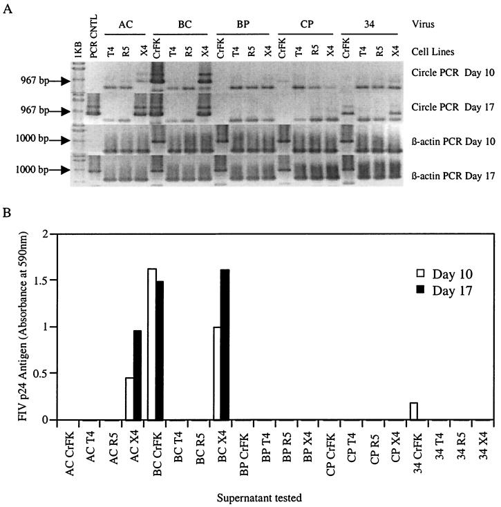
FIV antigen production corresponds to circle formation during FIV infection. Lysed cells and supernatants were examined at days 10 and 17 PI. (A) Circle and β-actin fragment PCR results from days 10 and 17 PI. The positive control was 34TF10 CrFK DNA and the negative control was an H2O reagent control. The PCR controls were run in lane 2 (PCR CNTL), with the H2O reagent control on the top gel for each fragment and the positive control on the bottom gel for each fragment. The DNA marker was the 1-kb ladder. (B) Results from the enzyme-linked immunosorbent assays for FIV antigen. T4, U87-T4 cells; R5, U87-T4-CCR5 cells; X4, U87-T4-CXCR4 cells. Virus designations are defined in the legend to Fig. 2.
Similar articles
-
Combined effect of zidovudine (ZDV), lamivudine (3TC) and abacavir (ABC) antiretroviral therapy in suppressing in vitro FIV replication.Antiviral Res. 2002 Jan;53(1):35-45. doi: 10.1016/s0166-3542(01)00190-5. Antiviral Res. 2002. PMID: 11684314
-
Binding of recombinant feline immunodeficiency virus surface glycoprotein to feline cells: role of CXCR4, cell-surface heparans, and an unidentified non-CXCR4 receptor.J Virol. 2001 May;75(10):4528-39. doi: 10.1128/JVI.75.10.4528-4539.2001. J Virol. 2001. PMID: 11312323 Free PMC article.
-
Bicyclams, selective antagonists of the human chemokine receptor CXCR4, potently inhibit feline immunodeficiency virus replication.J Virol. 1999 Aug;73(8):6346-52. doi: 10.1128/JVI.73.8.6346-6352.1999. J Virol. 1999. PMID: 10400726 Free PMC article.
-
The role of the chemokine receptor CXCR4 in infection with feline immunodeficiency virus.Mol Membr Biol. 1999 Jan-Mar;16(1):67-72. doi: 10.1080/096876899294779. Mol Membr Biol. 1999. PMID: 10332739 Review.
-
The virus-receptor interaction in the replication of feline immunodeficiency virus (FIV).Curr Opin Virol. 2013 Dec;3(6):670-5. doi: 10.1016/j.coviro.2013.08.003. Epub 2013 Aug 28. Curr Opin Virol. 2013. PMID: 23992667 Free PMC article. Review.
Cited by
-
Evolution of cell recognition by viruses: a source of biological novelty with medical implications.Adv Virus Res. 2003;62:19-111. doi: 10.1016/s0065-3527(03)62002-6. Adv Virus Res. 2003. PMID: 14719364 Free PMC article. Review.
-
FIV and neuroAIDS.J Neurovirol. 2002 Jun;8(3):155-7. doi: 10.1080/13550280290049714. J Neurovirol. 2002. PMID: 12053270 Review. No abstract available.
-
Interaction of short modified peptides deriving from glycoprotein gp36 of feline immunodeficiency virus with phospholipid membranes.Eur Biophys J. 2009 Sep;38(7):873-82. doi: 10.1007/s00249-009-0454-9. Epub 2009 May 5. Eur Biophys J. 2009. PMID: 19415263 Free PMC article.
-
Structural basis of antiviral activity of peptides from MPER of FIV gp36.PLoS One. 2018 Sep 21;13(9):e0204042. doi: 10.1371/journal.pone.0204042. eCollection 2018. PLoS One. 2018. PMID: 30240422 Free PMC article.
-
Lentiviral Gag assembly analyzed through the functional characterization of chimeric simian immunodeficiency viruses expressing different domains of the feline immunodeficiency virus capsid protein.PLoS One. 2014 Dec 2;9(12):e114299. doi: 10.1371/journal.pone.0114299. eCollection 2014. PLoS One. 2014. PMID: 25462889 Free PMC article.
References
-
- Cara A, Reitz M S., Jr New insight on the role of extrachromosomal retroviral DNA. Leukemia. 1997;11:1395–1399. - PubMed
-
- Dalgleish A G, Beverley P C, Clapham P R, Crawford D H, Greaves M F, Weiss R A. The CD4 (T4) antigen is an essential component of the receptor for the AIDS retrovirus. Nature. 1984;312:763–767. - PubMed
Publication types
MeSH terms
Substances
Grants and funding
LinkOut - more resources
Full Text Sources
Other Literature Sources
Research Materials
