Comparative Study
. 2001 Jun;75(11):5315-27.
doi: 10.1128/JVI.75.11.5315-5327.2001.

Analysis of a novel strain of murine gammaherpesvirus reveals a genomic locus important for acute pathogenesis

Affiliations
Comparative Study

Analysis of a novel strain of murine gammaherpesvirus reveals a genomic locus important for acute pathogenesis

A I Macrae et al. J Virol. 2001 Jun.

Abstract

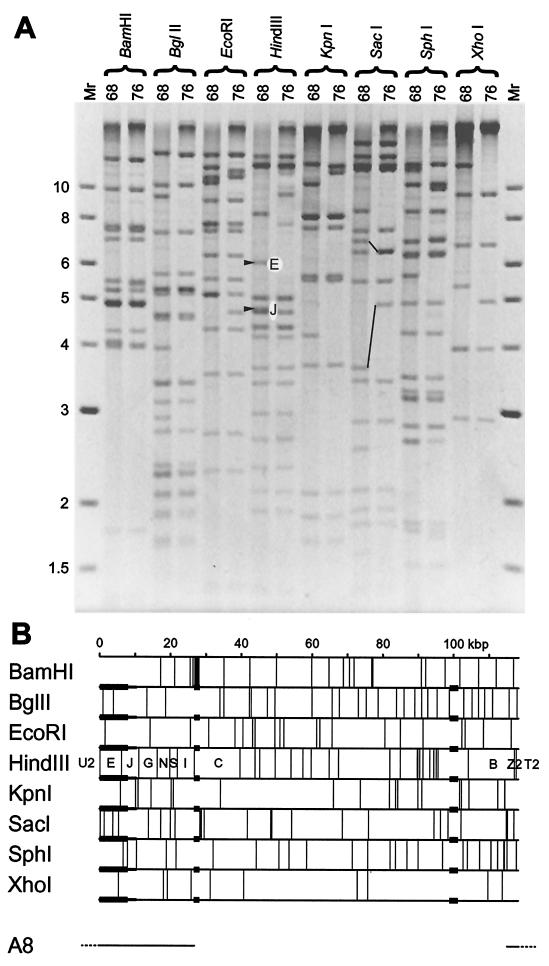
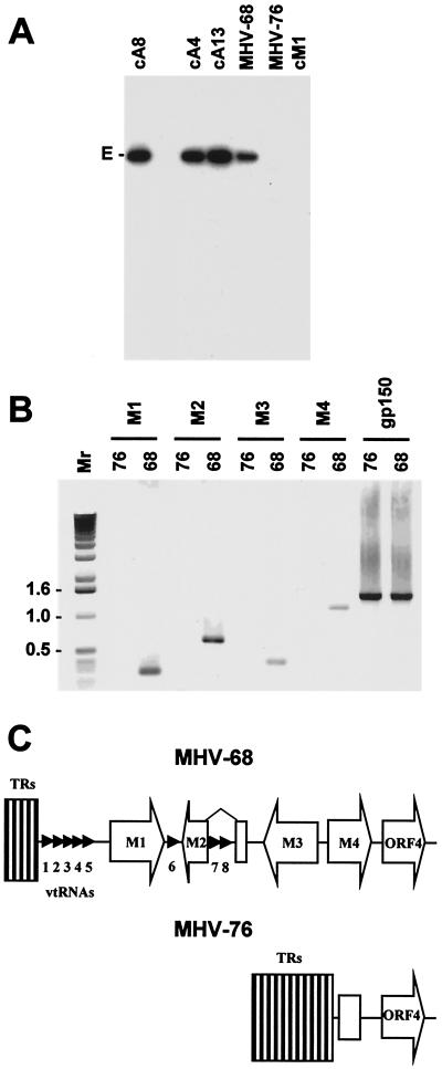
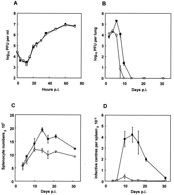
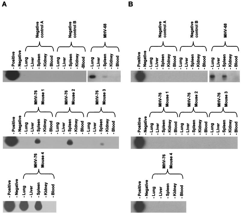
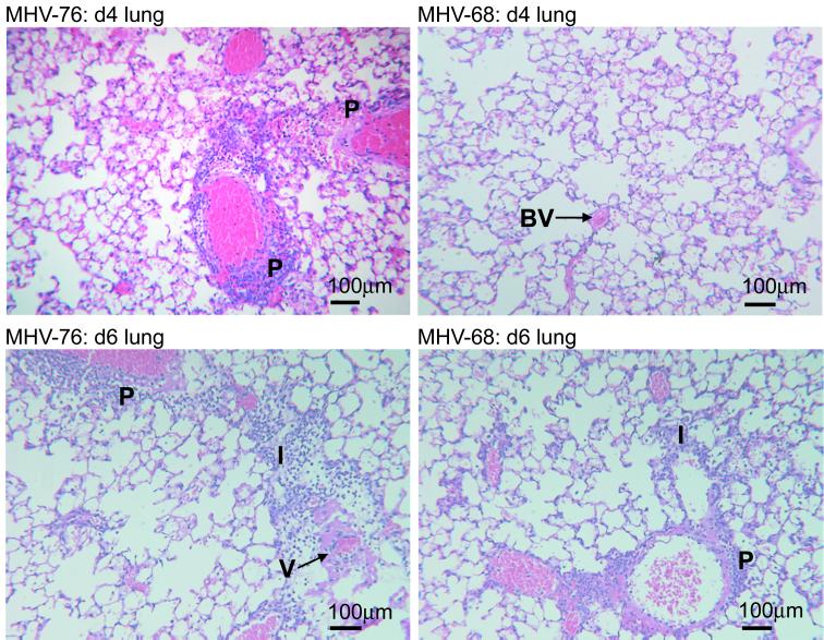
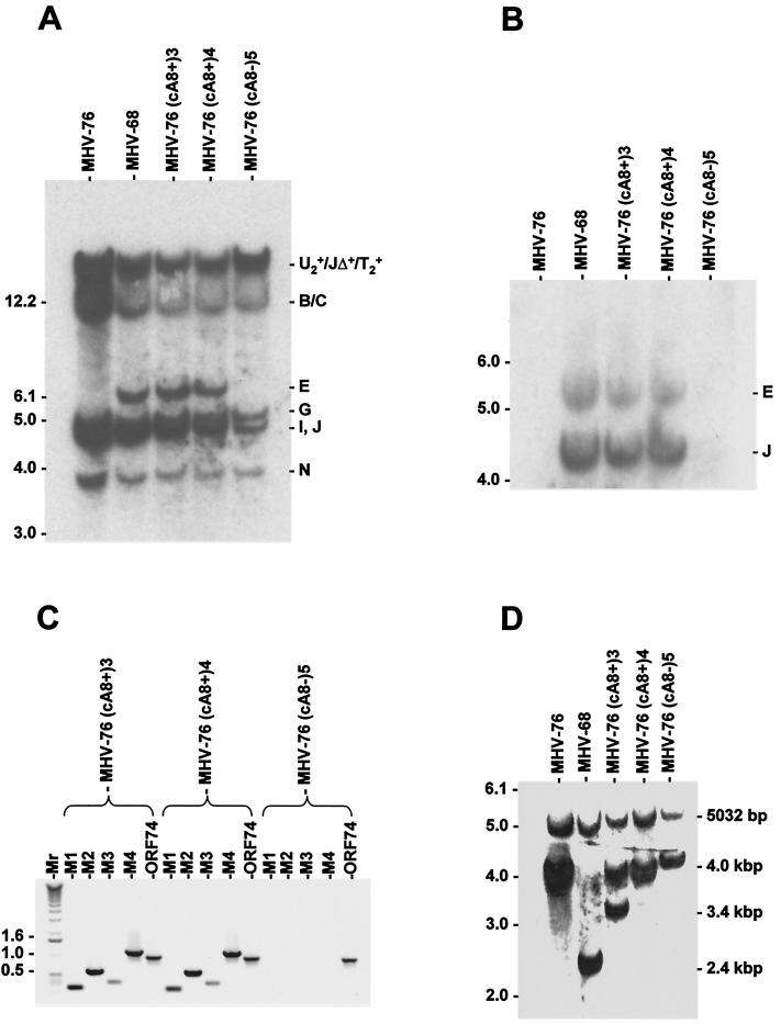
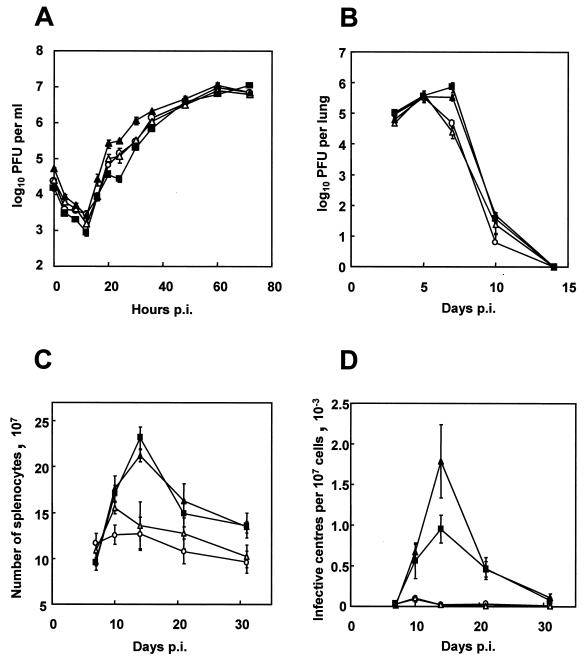
Infection of mice by murine gammaherpesvirus 68 (MHV-68) is an excellent small-animal model of gammaherpesvirus pathogenesis in a natural host. We have carried out comparative studies of another herpesvirus, murine herpesvirus 76 (MHV-76), which was isolated at the same time as MHV-68 but from a different murid host, the yellow-necked mouse (Apodemus flavicollis). Molecular analyses revealed that the MHV-76 genome is essentially identical to that of MHV-68, except for deletion of 9,538 bp at the left end of the unique region. MHV-76 is therefore a deletion mutant that lacks four genes unique to MHV-68 (M1, M2, M3, and M4) as well as the eight viral tRNA-like genes. Replication of MHV-76 in cell culture was identical to that of MHV-68. However, following infection of mice, MHV-76 was cleared more rapidly from the lungs. In line with this, there was an increased inflammatory response in lungs with MHV-76. Splenomegaly was also significantly reduced following MHV-76 infection, and much less latent MHV-76 was detected in the spleen. Nevertheless, MHV-76 maintained long-term latency in the lungs and spleen. We utilized a cosmid containing the left end of the MHV-68 genome to reinsert the deleted sequence into MHV-76 by recombination in infected cells, and we isolated a rescuant virus designated MHV-76(cA8+)4 which was ostensibly genetically identical to MHV-68. The growth properties of the rescuant in infected mice were identical to those of MHV-68. These results demonstrate that genetic elements at the left end of the unique region of the MHV-68 genome play vital roles in host evasion and are critical to the development of splenic pathology.


Figures

FIG. 1
FIG. 1
Structure of the MHV-76 genome. (A) Restriction endonuclease profiles of MHV-68 and MHV-76 virion DNAs. Samples were analyzed on a 0.8% agarose gel. Fragments E and J are indicated in the HindIII profile of MHV-68. These are absent from the HindIII profile of MHV-76. Fragments containing the 40-bp repeat (upper pair) and the 100-bp repeat (lower pair) are connected by lines in the SacI profiles of MHV-68 and MHV-76. Molecular size markers and their sizes in kilobase pairs are indicated to the left. (B) Restriction endonuclease maps of the unique region of the MHV-68 genome, highlighting the three regions that differ in MHV-76. Variable copy numbers of the terminal repeat (not shown) flank the unique region, resulting in heterogeneous populations of fragments from the genome termini differing by increments of 1.2 kbp. None of the restriction endonucleases for which maps are shown cleave the terminal repeat. Three regions in which MHV-76 and MHV-68 differ are denoted by thicker horizontal lines. Two correspond to variations in the copy number of reiterations in the 40-bp repeat (at kbp 27) and 100-bp repeat (at kbp 100). The third represents a deletion at the left end. Minimal and maximal extents of the deletion as deduced from panel A are indicated by thicker and thinner lines, respectively. The location of the insert in MHV-68 cosmid A8 is indicated at the bottom, with terminal repeat sequences dashed, and the corresponding fragment nomenclature is included in the HindIII map.
FIG. 2
FIG. 2
(A) Autoradiograph of a Southern blot of a 0.8% agarose gel showing the results of hybridizing radiolabeled HindIII E (isolated from a plasmid [11]) to the HindIII products of MHV-68 and MHV-76 DNAs and three MHV-68 cosmids (cA8, cA4, and cA13) and one MHV-76 cosmid (cM1) containing the left end of the genome. The HindIII E fragment (6,155 bp) is indicated. (B) PCR analysis of the MHV-68 and MHV-76 genomes. PCR amplification was performed on viral DNA templates using primers specific for M1, M2, M3, M4, and gp150 as indicated. Reaction products were analyzed on a 1% agarose gel. Molecular size markers and their sizes in kilobase pairs are indicated to the left. (C) Schematic diagram (not to scale) showing the structures of the left ends of the MHV-68 and MHV-76 genomes. The open arrows indicate protein-coding regions, and the small arrowheads indicate the vtRNA genes. The open rectangle in MHV-76 indicates residual M4 sequence. TRs, terminal repeats.
FIG. 3
FIG. 3
Biological characterization of MHV-76. (A) Single-step growth curves of MHV-68 (▪) and MHV-76 (○) on BHK-21 cells at an MOI of 5. Data are shown as mean log10 virus titer ± standard error and are representative of two separate experiments, each carried out in duplicate. (B) Viral replication in the lungs of BALB/c mice infected intranasally with 2 × 10⁵ PFU of MHV-68 (▪) or MHV-76 (○). The mean log10 virus titer ± standard error for four mice per group is shown for each time point. (C) Numbers of spleen cells during intranasal infection of BALB/c mice with 2 × 10⁵ PFU of MHV-68 (▪) or MHV-76 (○). The mean total number of splenocytes ± standard error for four mice per group is shown for each time point. (D) Latent virus in the spleens of BALB/c mice infected with 2 × 10⁵ PFU of MHV-68 (▪) or MHV-76 (○) as determined by infective-center assay. Infectious virus titers (less than 50 PFU per spleen) were subtracted from the infective-center results. The mean number of infective centers per spleen ± standard error for four mice per group is shown for each time point.
FIG. 4
FIG. 4
Detection of MHV-76 DNA in the tissues of mice at 5 months p.i. Four BALB/c mice were infected with 2 × 10⁵ PFU of MHV-76, and the lungs, livers, spleens, kidneys, and blood were harvested at 5 months p.i. Two uninfected mice were also harvested as negative controls, and one mouse infected 5 months previously with MHV-68 was harvested as a positive control. Viral DNA was extracted, and PCR amplification was performed on 1 μg of high-molecular-weight DNA. Samples containing MHV-68 viral DNA as a template (first lanes) or lacking DNA (second lanes) were used as positive and negative controls, respectively. (A) PCR amplification using primers specific for gp150. PCR products were analyzed by Southern blot hybridization with a 32P-labeled gp150 probe to confirm specificity. (B) PCR amplification using primers specific for M3 (a gene absent from MHV-76). PCR products were analyzed by Southern blot hybridization with a 32P-labeled M3 probe to confirm specificity.
FIG. 5
FIG. 5
Histopathological changes in the lungs of mice infected with MHV-76 or MHV-68. Representative sections of lung and spleen stained with hematoxylin and eosin are shown for mice infected for the times indicated. Salient pathological features are highlighted as follows: P, perivascular infiltration; BV, blood vessel; I, interstitial infiltration; V, vasculitis.
FIG. 6
FIG. 6
Molecular characterization of rescuant viruses. (A) Autoradiograph of a Southern blot of a 0.8% agarose gel showing the results of hybridizing radiolabeled MHV-68 cosmid A8 to the HindIII products of DNAs isolated from BHK-21 cells infected with MHV-76, MHV-68, MHV-76(cA8+)3, MHV-76(cA8+)4, and MHV-76(cA8-)5. The sizes of molecular size markers (in kilobase pairs) are shown on the left, and HindIII fragments are shown on the right (see Fig. 1B for a map). (B) Autoradiograph of a Southern blot of a 0.8% agarose gel showing the results of hybridizing radiolabeled M3 probe (nucleotides 6060 to 7277 in the MHV-68 genome) to the HindIII products of DNA isolated from BHK-21 cells infected with MHV-76, MHV-68, MHV-76(cA8+)3, MHV-76(cA8+)4, or MHV-76(cA8-)5. The sizes of molecular size markers (in kilobase pairs) are shown on the left, and HindIII fragments are shown on the right (see Fig. 1B for a map). (C) PCR analysis of viral DNA from MHV-76(cA8+)3, MHV-76(cA8+)4, or MHV-76(cA8-)5 using primers specific for the MHV-68 M1, M2, M3, and M4 genes and ORF 74. Reaction products were analyzed on a 1% agarose gel. Molecular size markers and their sizes are shown to the left, with sizes in kilobase pairs. (D) Autoradiograph of a Southern blot of a 0.8% agarose gel showing the results of hybridizing a radiolabeled BamHI M probe (which contains the 100-bp repeat) to the HincII products of DNA isolated from BHK-21 cells infected with MHV-76, MHV-68, MHV-76(cA8+)3, MHV-76(cA8+)4, or MHV-76(cA8-)5. The sizes of molecular size markers (in kilobase pairs) are shown on the left. Fragments containing the 100-bp repeat, with approximate sizes, are indicated on the right, along with the adjacent 5,032-bp fragment present in each genome.
FIG. 7
FIG. 7
Biological characterization of MHV-68, MHV-76, and the rescuant viruses. Data points (mean value ± standard error) are shown in each graph for MHV-68 (▪), MHV-76 (○), MHV-76(cA8+)4 (▴), and MHV-76(cA8-)5 (▵). (A) Single-step growth curves comparing growth of viruses on BHK-21 cells at an MOI of 5. The data are representative of two separate experiments, and each experiment was done in duplicate. (B) Viral replication in the lungs of BALB/c mice infected intranasally with 2 × 10⁵ PFU of virus. Data for four mice per group are shown at each time point. (C) Numbers of spleen cells during intranasal infection of BALB/c mice with 2 × 10⁵ PFU of virus. Data for four mice per group are shown at each time point. (D) Latent virus in the spleens of BALB/c mice infected with 2 × 10⁵ PFU of virus as determined by infective-center assay. Infectious virus titers (less than 50 PFU per spleen in every case) have been subtracted from the infective-center results. Data for four mice per group are shown at each time point.

Similar articles

Cited by

References

    1. Adler H, Messerle M, Wagner M, Koszinowski U H. Cloning and mutagenesis of the murine gammaherpesvirus 68 genome as an infectious bacterial artificial chromosome. J Virol. 2000;74:6964–6974. - PMC - PubMed
    1. Albrecht J C, Nicholas J, Biller D, Cameron K R, Biesinger B, Newman C, Wittmann S, Craxton M A, Coleman H, Fleckenstein B, et al. Primary structure of the herpesvirus saimiri genome. J Virol. 1992;66:5047–5058. - PMC - PubMed
    1. Blaskovic D, Stancekova M, Svobodova J, Mistrikova J. Isolation of five strains of herpesviruses from two species of free living small rodents. Acta Virol. 1980;24:468. - PubMed
    1. Bowden R J, Simas J P, Davis A J, Efstathiou S. Murine gammaherpesvirus 68 encodes tRNA-like sequences which are expressed during latency. J Gen Virol. 1997;78:1675–1687. - PubMed
    1. Cardin R D, Brooks J W, Sarawar S R, Doherty P C. Progressive loss of CD8+ T cell-mediated control of a gamma-herpesvirus in the absence of CD4+ T cells. J Exp Med. 1996;184:863–871. - PMC - PubMed

Publication types

Associated data