. 2001 Apr 30;153(3):585-98.
doi: 10.1083/jcb.153.3.585.

A new focal adhesion protein that interacts with integrin-linked kinase and regulates cell adhesion and spreading

Affiliations

A new focal adhesion protein that interacts with integrin-linked kinase and regulates cell adhesion and spreading

Y Tu et al. J Cell Biol. .

Abstract

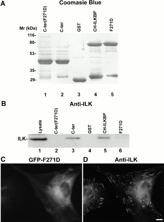
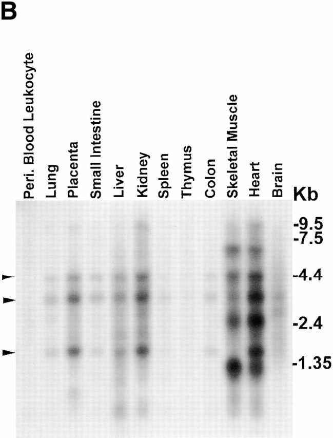
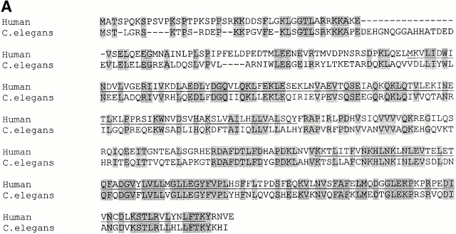
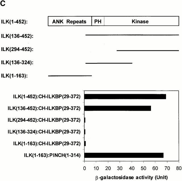
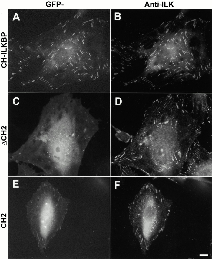
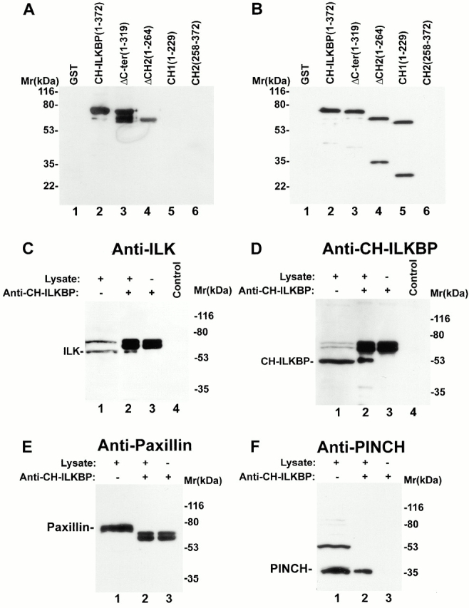
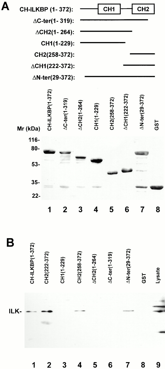
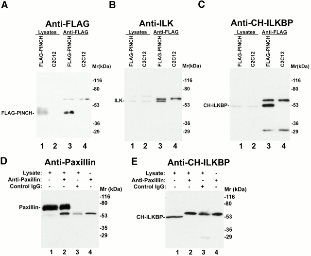
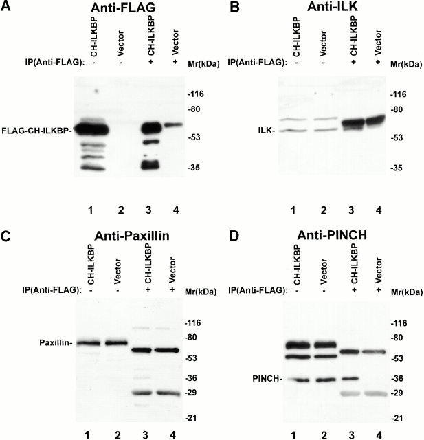
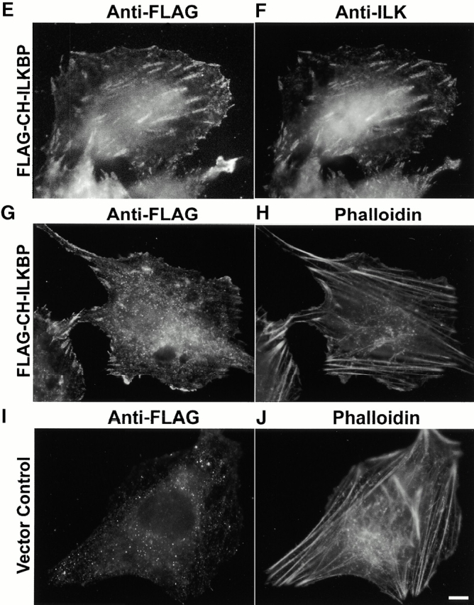
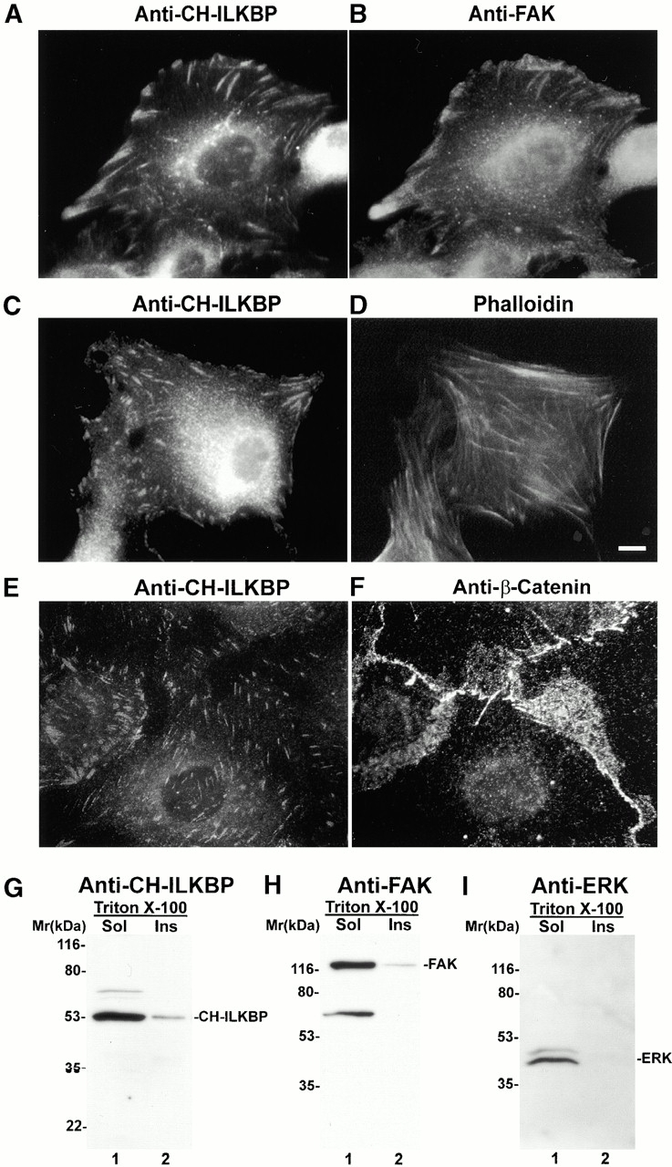
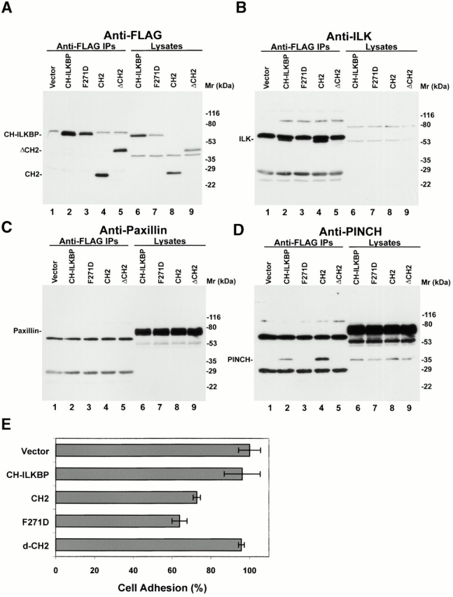
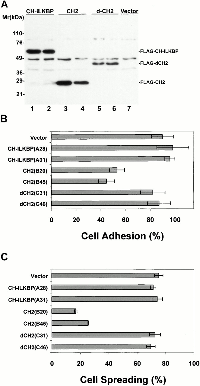
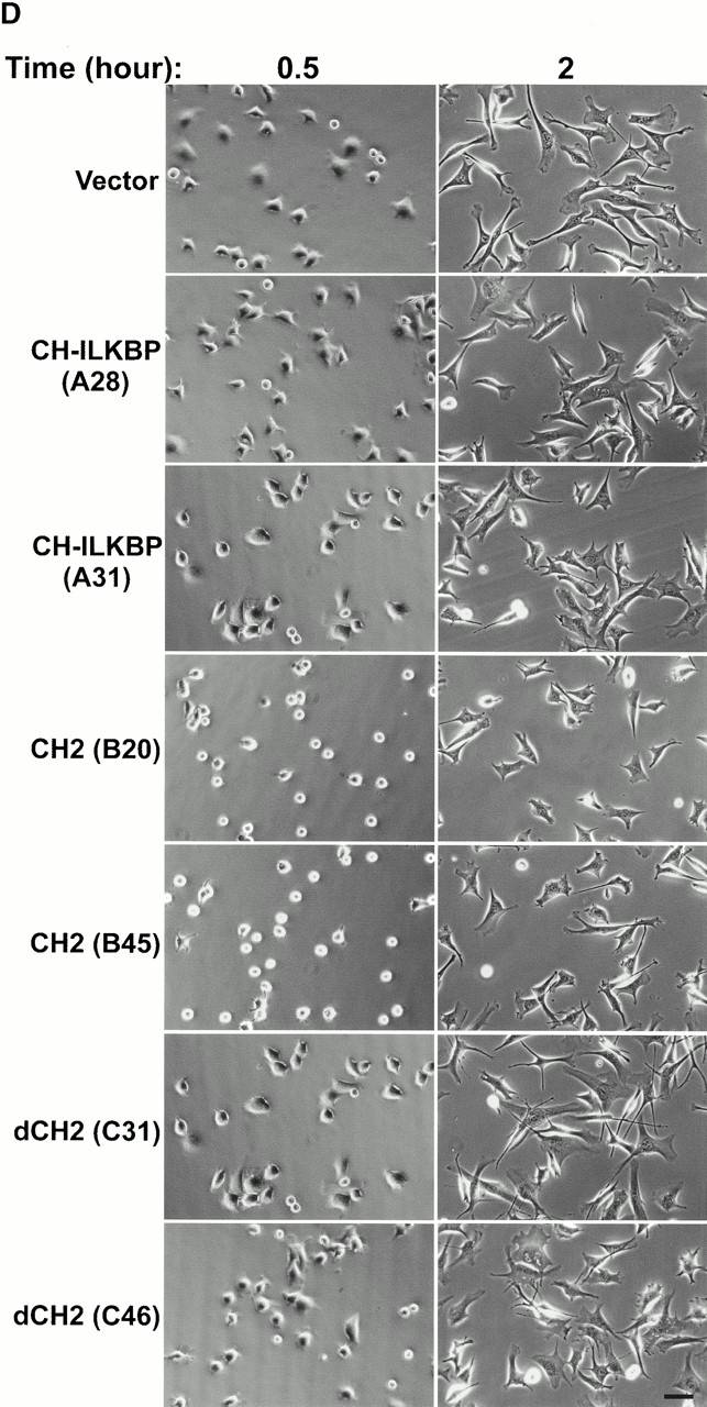
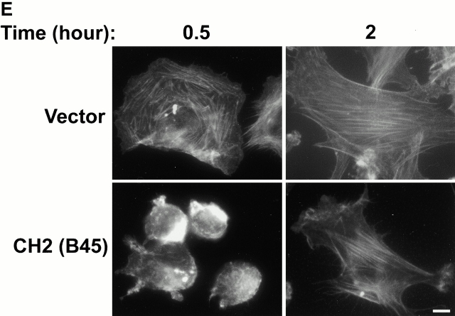
Integrin-linked kinase (ILK) is a multidomain focal adhesion (FA) protein that functions as an important regulator of integrin-mediated processes. We report here the identification and characterization of a new calponin homology (CH) domain-containing ILK-binding protein (CH-ILKBP). CH-ILKBP is widely expressed and highly conserved among different organisms from nematodes to human. CH-ILKBP interacts with ILK in vitro and in vivo, and the ILK COOH-terminal domain and the CH-ILKBP CH2 domain mediate the interaction. CH-ILKBP, ILK, and PINCH, a FA protein that binds the NH(2)-terminal domain of ILK, form a complex in cells. Using multiple approaches (epitope-tagged CH-ILKBP, monoclonal anti-CH-ILKBP antibodies, and green fluorescent protein-CH-ILKBP), we demonstrate that CH-ILKBP localizes to FAs and associates with the cytoskeleton. Deletion of the ILK-binding CH2 domain abolished the ability of CH-ILKBP to localize to FAs. Furthermore, the CH2 domain alone is sufficient for FA targeting, and a point mutation that inhibits the ILK-binding impaired the FA localization of CH-ILKBP. Thus, the CH2 domain, through its interaction with ILK, mediates the FA localization of CH-ILKBP. Finally, we show that overexpression of the ILK-binding CH2 fragment or the ILK-binding defective point mutant inhibited cell adhesion and spreading. These findings reveal a novel CH-ILKBP-ILK-PINCH complex and provide important evidence for a crucial role of this complex in the regulation of cell adhesion and cytoskeleton organization.


Figures

Figure 8
Figure 8
A point mutation that disrupts the ILK binding impairs the FA localization of CH-ILKBP. (A) GST–CH-ILKBP proteins. GST fusion proteins containing the mutant form (F271D) of CH2 domain (residues 258–372) (lane 1), the CH2 domain (residues 258–372) (lane 2), CH-ILKBP (lane 4), and the F271D point mutant (lane 5) and GST (lane 3) were separated on SDS-PAGE (10 μg/lane) and detected by Coomassie blue R-250 staining. (B) ILK binding. C2C12 cell lysates (260 μg) were incubated and precipitated with equal amount (10 μg) of GST or GST–CH-ILKBP proteins as indicated. ILK was detected by Western blotting with anti-ILK antibody 65.1. Lane 1 was loaded with C2C12 cell lysates (9 μg/lane). (C and D) FA localization. Rat mesangial cells expressing GFP fusion protein containing the full-length F271D point mutant were plated on fibronectin-coated coverslips and stained with anti-ILK antibody 65.1 and Rhodamine red™-conjugated anti–mouse IgG antibody. GFP-F271D (C) and ILK (D) were visualized under a fluorescence microscope equipped with GFP and rhodamine filters. Bar, 5 μm.
Figure 1
Figure 1
Identification of a new CH-containing ILK-binding protein. (A) Sequence alignment of human CH-ILKBP (sequence data available from GenBank/EMBL/DDBJ under accession number AF325830) with C. elegans T21D12.4 protein (sequence data available from GenBank/EMBL/DDBJ under accession no. AAC48090). Identical residues are indicated by gray background. The CH domains are underlined. (B) Northern blotting. Two micrograms of polyA+ RNA from human tissues were hybridized with a CH-ILKBP cDNA probe as described in Materials and Methods. Arrowheads indicate the positions of three transcripts (4.4, 3.5, and 1.7 kb). (C) ILK residues involved in the CH-ILKBP binding. The binding was determined by yeast two- hybrid binding assays as described in Materials and Methods. The numbers in parentheses indicate ILK, CH-ILKBP, and PINCH residues.
Figure 1
Figure 1
Identification of a new CH-containing ILK-binding protein. (A) Sequence alignment of human CH-ILKBP (sequence data available from GenBank/EMBL/DDBJ under accession number AF325830) with C. elegans T21D12.4 protein (sequence data available from GenBank/EMBL/DDBJ under accession no. AAC48090). Identical residues are indicated by gray background. The CH domains are underlined. (B) Northern blotting. Two micrograms of polyA+ RNA from human tissues were hybridized with a CH-ILKBP cDNA probe as described in Materials and Methods. Arrowheads indicate the positions of three transcripts (4.4, 3.5, and 1.7 kb). (C) ILK residues involved in the CH-ILKBP binding. The binding was determined by yeast two- hybrid binding assays as described in Materials and Methods. The numbers in parentheses indicate ILK, CH-ILKBP, and PINCH residues.
Figure 7
Figure 7
The ILK-binding CH2 domain mediates the localization of CH-ILKBP to FAs. Rat mesangial cells expressing GFP–CH-ILKBP (A and B), GFP-ΔCH2 (residues 1–229) (C and D), or GFP-CH2 (residues 222–372) (E and F) were plated on fibronectin-coated coverslips and stained with mouse anti-ILK antibody 65.1 and Rhodamine redTX-conjugated anti–mouse IgG antibody. GFP fusion proteins (A, C, and E) and ILK (B, D, and F) were visualized under a fluorescence microscope equipped with GFP and rhodamine filters. We have also expressed GFP–CH-ILKBP, GFP-ΔCH2, and GFP-CH2 in C2C12 cells and found that GFP–CH-ILKBP and GFP-CH2 but not GFP-ΔCH2 localized to FAs in these cells as well (not shown). Bar, 5 μm.
Figure 3
Figure 3
CH-ILKBP forms a complex with ILK and PINCH in mammalian cells. (A and B) GST and GST–CH-ILKBP proteins (0.1 μg protein/lane) were analyzed by Western blotting with anti–CH-ILKBP antibody 1D4 (A) and 3B5 (B). (C and D) Coimmunoprecipitation of ILK with CH-ILKBP. Anti–CH-ILKBP immunoprecipitates (lane 2) and control precipitates (lane 4) were prepared using C2C12 cell lysates as described in Materials and Methods. ILK and CH-ILKBP were detected by Western blotting with anti-ILK antibody 65.1 (C) and anti–CH-ILKBP antibody 3B5 (D), respectively. Lane 1 was loaded with C2C12 cell lysates (20 μg protein/lane). (E and F) Cell lysates (lane 1) and anti–CH-ILKBP immunoprecipitates (lane 2) were analyzed by Western blotting using mouse antipaxillin (E) or rabbit anti-PINCH (F) antibodies. Lane 3 was loaded with samples prepared like those of lane 2 except that the lysates were omitted.
Figure 2
Figure 2
The CH2 domain mediates the interaction with ILK. (A) GST–CH-ILKBP fusion proteins were separated on SDS-PAGE (10 μg/lane) and detected by Coomassie blue R-250 staining. The numbers in parentheses indicate CH-ILKBP residues. (B) ILK binding. C2C12 cell lysates (100 μg) were incubated with equal amount (10 μg) of GST or GST fusion proteins containing CH-ILKBP sequences as indicated in the figure. GST and the GST fusion proteins were precipitated with glutathione-Sepharose 4B beads. ILK was detected by Western blotting with anti-ILK antibody 65.1. (Lane 9) C2C12 cell lysates (10 μg/lane).
Figure 4
Figure 4
Coimmunoprecipitation of CH-ILKBP and ILK with FLAG-PINCH. (A and B) FLAG-PINCH was immunoprecipitated from lysates of C2C12 cells expressing FLAG-PINCH. FLAG-PINCH immunoprecipitates (lane 3) or control precipitates (lane 4) were analyzed by Western blotting with anti-FLAG antibody M2 (A), anti-ILK antibody 65.1 (B), or anti–CH-ILKBP antibody 3B5 (C). Lanes 1 and 2 were loaded with lysates (7.5 μg protein/lane) of the FLAG-PINCH–expressing C2C12 cells and the control C2C12 cells, respectively. (D and E) Antipaxillin immunoprecipitates (lane 2) or control IgG precipitates (lane 3) were blotted with antipaxillin antibody (D) or anti–CH-ILKBP antibody 3B5 (E). (Lane 1) 10 μg lysates; (lane 4) 0.4 μg antipaxillin IgG.
Figure 5
Figure 5
Complex formation and subcellular localization of FLAG–CH-ILKBP. (A–D) Coimmunoprecipitation of ILK and PINCH with FLAG–CH-ILKBP. The FLAG–CH-ILKBP immunoprecipitates (lane 3) and control precipitates (lane 4) were prepared as described in Materials and Methods and analyzed by Western blotting with anti-FLAG antibody M2 (A), anti-ILK antibody 65.1 (B), and antipaxillin antibody (clone 349) (C), respectively. Lanes 1 and 2 were loaded with cell lysates (20 μg protein/lane). In D, the membrane used in C was reprobed (without striping) with anti-PINCH antibodies. (E–J) FA localization of FLAG–CH-ILKBP. Rat mesangial cells were transfected with the FLAG–CH-ILKBP vector (E–H) or a FLAG vector as a control (I and J). The FLAG–CH-ILKBP, FAK, and actin stress fibers were detected by staining the cells with mouse anti-FLAG antibody M2 (E, G, and I), rabbit anti-FAK antibody (F), or rhodamine-labeled phalloidin (H and J). We also analyzed the subcellular localization of FLAG–CH-ILKBP in C2C12 cells and obtained similar results (not shown). Bar, 5 μm.
Figure 5
Figure 5
Complex formation and subcellular localization of FLAG–CH-ILKBP. (A–D) Coimmunoprecipitation of ILK and PINCH with FLAG–CH-ILKBP. The FLAG–CH-ILKBP immunoprecipitates (lane 3) and control precipitates (lane 4) were prepared as described in Materials and Methods and analyzed by Western blotting with anti-FLAG antibody M2 (A), anti-ILK antibody 65.1 (B), and antipaxillin antibody (clone 349) (C), respectively. Lanes 1 and 2 were loaded with cell lysates (20 μg protein/lane). In D, the membrane used in C was reprobed (without striping) with anti-PINCH antibodies. (E–J) FA localization of FLAG–CH-ILKBP. Rat mesangial cells were transfected with the FLAG–CH-ILKBP vector (E–H) or a FLAG vector as a control (I and J). The FLAG–CH-ILKBP, FAK, and actin stress fibers were detected by staining the cells with mouse anti-FLAG antibody M2 (E, G, and I), rabbit anti-FAK antibody (F), or rhodamine-labeled phalloidin (H and J). We also analyzed the subcellular localization of FLAG–CH-ILKBP in C2C12 cells and obtained similar results (not shown). Bar, 5 μm.
Figure 6
Figure 6
FA localization and cytoskeleton association of CH-ILKBP. (A–F) Subcellular localization of CH-ILKBP. Primary rat mesangial cells were stained with mouse anti–CH-ILKBP antibody 1D4 and rabbit anti-FAK antibodies (A and B) or anti–CH-ILKBP antibody 1D4 and phalloidin (C and D). We have also stained IEC18 rat intestinal epithelial cells, CHO cells, and mouse C2C12 cells with anti–CH-ILKBP antibodies and found that CH-ILKBP localizes to FA in these cells as well. Immunofluorescence images of IEC18 cells stained with mouse anti–CH-ILKBP antibody 1D4 (E) and rabbit anti–β-catenin antibody (F). (G–I) Association of CH-ILKBP with Triton X-100–insoluble cytoskeleton fractions. CH-ILKBP, FAK, and p44/42 ERK in the Triton X-100–soluble and –insoluble fractions were detected by Western blotting with anti–CH-ILKBP antibody 3B5 (G), anti-FAK antibody (H), and anti-p44/42 ERK antibody (New England Biolabs, Inc.) (I), respectively. Bar, 5 μm.
Figure 9
Figure 9
Effects of CH-ILKBP on CHO cell adhesion. (A–D) FLAG-tagged CH-ILKBP (lane 2), F271D (lane 3), CH2 (residues 222–372) (lane 4), and ΔCH2 (residues 1–229) (lane 5) were immunoprecipitated from CHO cells transfected with the corresponding FLAG expression vectors. (Lane 1) Anti-FLAG immunoprecipitates obtained with the vector control cells. The immunoprecipitates were analyzed by Western blotting with anti-FLAG (A), anti-ILK (B), or antipaxillin (C) antibodies. In D, the membrane used in C was reprobed (without striping) with anti-PINCH antibodies. Positions of FLAG–CH-ILKBP (or F271D), FLAG-ΔCH2, FLAG–CH2, ILK, paxillin, and PINCH were indicated. (Lanes 6–9) Lysates (15 μg/lane) of the transfectants as indicated. (E) Cell adhesion. Cells (as indicated in the figure) were allowed to adhere to collage IV–coated 96-well plates for 90 min, and the adhered cells were quantified as described in Materials and Methods. Under the condition used, ∼70% of the vector control cells adhered. The adhesion efficiency of CHO cells expressing different forms of CH-ILKBP was compared with that of the vector control cells and presented as percentages of that of the control cells. Data represent the mean ± SD of two separate experiments.
Figure 10
Figure 10
Effects of CH-ILKBP on C2C12 cell adhesion and spreading. (A) Lysates (15 μg/lane) of independent C2C12 clones expressing the FLAG-tagged proteins were probed with anti–FLAG-antibody M2. (Lane 1) FLAG–CH-ILKBP clone A28; (lane 2) FLAG–CH-ILKBP clone A31; (lane 3) FLAG-CH2 (residues 222–372) clone B20; (lane 4) FLAG-CH2 clone B45; (lane 5) FLAG-ΔCH2 (residues 1–229) clone C31; (lane 6) FLAG-ΔCH2 clone C46; (lane 7) lysates (15 μg) from the vector control cells. (B) Cell adhesion. Cells (as indicated in the figure) were allowed to adhere to collage I–coated 12-well plates for 30 min and were then washed. Cell adhesion is presented as the number of adhered cells (after wash) divided by the number of total cells (before wash). Data represent the mean ± SD of four randomly selected fields. (C and D) Cell spreading. C2C12 cells expressing different forms of CH-ILKBP were seeded in collage I–coated 12-well plates. The cell morphology was recorded 0.5 and 2 h after seeding (D). The percentage of cells adopting spread morphology 0.5 h after seeding was quantified by analyzing >300 cells from three randomly selected fields (C). Data represent mean ± SD. (E) C2C12 cells overexpressing FLAG-CH2 (clone B45) and the vector control cells were plated on collage I–coated slides for 0.5 or 2 h and stained with rhodamine-labeled phalloidin. Identical results were obtained with clone B20. Bars: (D) 100 μm; (E) 5 μm.
Figure 10
Figure 10
Effects of CH-ILKBP on C2C12 cell adhesion and spreading. (A) Lysates (15 μg/lane) of independent C2C12 clones expressing the FLAG-tagged proteins were probed with anti–FLAG-antibody M2. (Lane 1) FLAG–CH-ILKBP clone A28; (lane 2) FLAG–CH-ILKBP clone A31; (lane 3) FLAG-CH2 (residues 222–372) clone B20; (lane 4) FLAG-CH2 clone B45; (lane 5) FLAG-ΔCH2 (residues 1–229) clone C31; (lane 6) FLAG-ΔCH2 clone C46; (lane 7) lysates (15 μg) from the vector control cells. (B) Cell adhesion. Cells (as indicated in the figure) were allowed to adhere to collage I–coated 12-well plates for 30 min and were then washed. Cell adhesion is presented as the number of adhered cells (after wash) divided by the number of total cells (before wash). Data represent the mean ± SD of four randomly selected fields. (C and D) Cell spreading. C2C12 cells expressing different forms of CH-ILKBP were seeded in collage I–coated 12-well plates. The cell morphology was recorded 0.5 and 2 h after seeding (D). The percentage of cells adopting spread morphology 0.5 h after seeding was quantified by analyzing >300 cells from three randomly selected fields (C). Data represent mean ± SD. (E) C2C12 cells overexpressing FLAG-CH2 (clone B45) and the vector control cells were plated on collage I–coated slides for 0.5 or 2 h and stained with rhodamine-labeled phalloidin. Identical results were obtained with clone B20. Bars: (D) 100 μm; (E) 5 μm.

Similar articles

Cited by

References

    1. Burridge K., Chrzanowska-Wodnicka M. Focal adhesions, contractility, and signaling. Annu. Rev. Cell Dev. Biol. 1996;12:463–518. - PubMed
    1. Calderwood D.A., Shattil S.J., Ginsberg M.H. Integrins and actin filamentsreciprocal regulation of cell adhesion and signaling. J. Biol. Chem. 2000;275:22607–22610. - PubMed
    1. Clark E.A., Brugge J.S. Integrins and signal transduction pathwaysthe road taken. Science. 1995;268:233–239. - PubMed
    1. Dedhar S., Hannigan G.E. Integrin cytoplasmic interactions and bidirectional transmembrane signalling. Curr. Opin. Cell Biol. 1996;8:657–669. - PubMed
    1. Dedhar S., Williams B., Hannigan G. Integrin linked kinase (ILK)a regulator of integrin and growth-factor signaling. Trends Cell Biol. 1999;9:319–323. - PubMed

Publication types

MeSH terms