. 2001 May;21(10):3564-75.
doi: 10.1128/MCB.21.10.3564-3575.2001.

Localization of the Rsp5p ubiquitin-protein ligase at multiple sites within the endocytic pathway

Affiliations

Localization of the Rsp5p ubiquitin-protein ligase at multiple sites within the endocytic pathway

G Wang et al. Mol Cell Biol. 2001 May.

Abstract

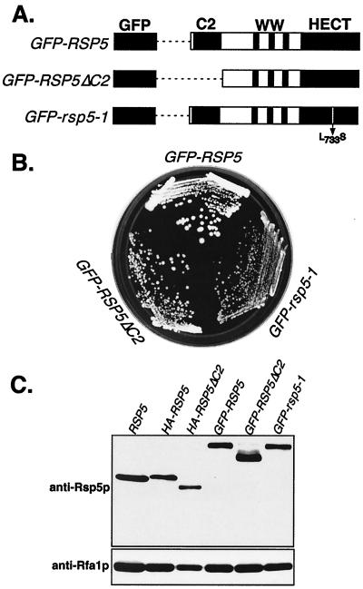
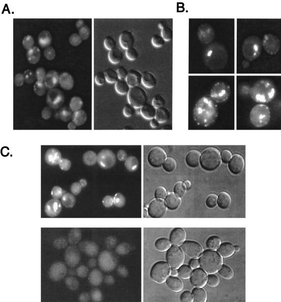
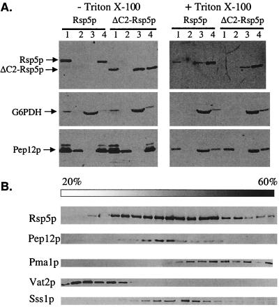
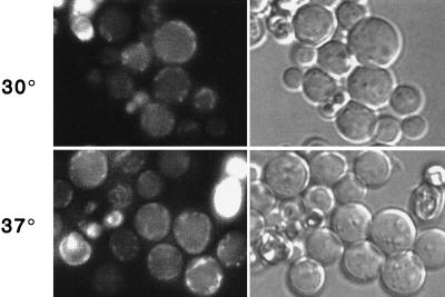
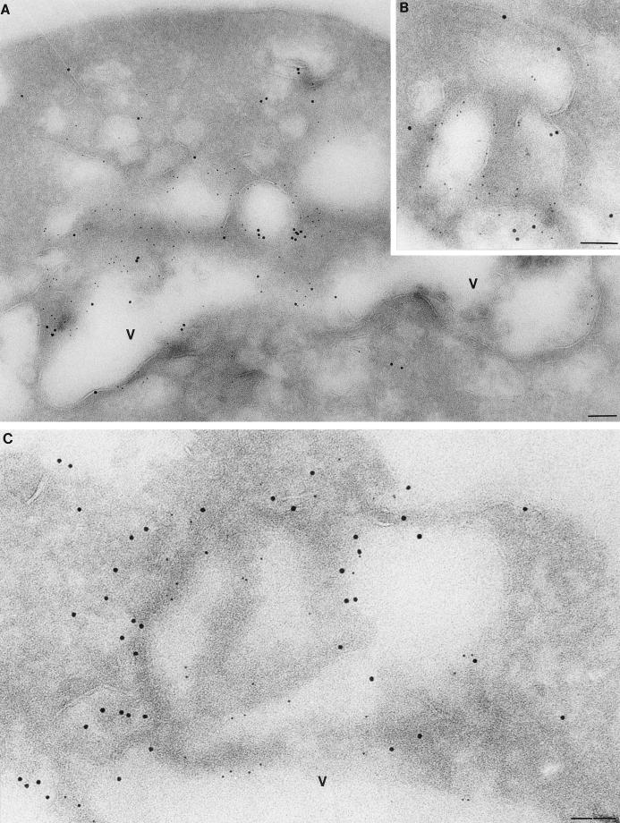
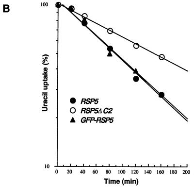
The Saccharomyces cerevisiae RSP5 gene encodes an essential HECT E3 ubiquitin-protein ligase. Rsp5p contains an N-terminal C2 domain, three WW domains in the central portion of the molecule, and a C-terminal catalytic HECT domain. A diverse group of substrates of Rsp5p and vertebrate C2 WW-domain-containing HECT E3s have been identified, including both nuclear and membrane-associated proteins. We determined the intracellular localization of Rsp5p and the determinants necessary for localization, in order to better understand how Rsp5p activities are coordinated. Using both green fluorescent protein fusions to Rsp5p and immunogold electron microscopy, we found that Rsp5p was distributed in a punctate pattern at the plasma membrane, corresponding to membrane invaginations that are likely sites of endosome formation, as well as at perivacuolar sites. The latter appeared to correspond to endocytic intermediates, as these structures were not seen in a sla2/end4-1 mutant, and double-immunogold labeling demonstrated colocalization of Rsp5p with the endosomal markers Pep12p and Vps32p. The C2 domain was an important determinant of localization; however, mutations that disrupted HECT domain function also caused mislocalization of Rsp5p, indicating that enzymatic activity is linked to localization. Deletion of the C2 domain partially stabilized Fur4p, a protein previously shown to undergo Rsp5p- and ubiquitin-mediated endocytosis; however, Fur4p was still ubiquitinated at the plasma membrane when the C2 domain was deleted from the protein. Together, these results indicate that Rsp5p is located at multiple sites within the endocytic pathway and suggest that Rsp5p may function at multiple steps in the ubiquitin-mediated endocytosis pathway.


Figures

FIG. 1
FIG. 1
GFP-Rsp5p fusion proteins are functional in yeast. (A) Schematic representation of GFP fused to wild-type Rsp5p, Rsp5ΔC2p, and the rsp5-1p temperature-sensitive mutant allele. (B) Growth of haploid rsp5Δ cells expressing the indicated plasmid-borne GFP fusion proteins. Cells expressing GFP alone were inviable. (C) Western analysis to confirm the expression of full-length GFP fusion proteins, by using anti-Rsp5p antibody. The first lane shows Rsp5p expression in the undisrupted W303 strain; all other lanes are in the rsp5Δ background expressing the indicated plasmid-borne HA- or GFP-tagged Rsp5p proteins. The lower panel shows levels of a control protein, Rfa1p.
FIG. 2
FIG. 2
Localization of GFP-Rsp5p and GFP-Rsp5p-ΔC2. GW072 cells expressing GFP-Rsp5p were examined by fluorescence (A and B) and DIC (A, right panel) microscopy. (C) Top panels show GW072 cells (GFP-Rsp5p) and bottom panels show GW073 cells (GFP- Rsp5p-ΔC2) as seen with fluorescence (left panels) or DIC (right panels) microscopy.
FIG. 3
FIG. 3
Fractionation of Rsp5p and Rsp5p-ΔC2. (A) GW047 and GW050 cells expressing HA-Rsp5p and HA-Rsp5ΔC2p were spheroplasted and lysed, and lysates were incubated without (left panel) or with (right panel) 1% Triton X-100 prior to sequential differential centrifugation. Immunoblotting of fractions was performed with antibodies against Rsp5p, G6PDH, and Pep12p. G6PDH and Pep12p served as controls for soluble and vacuolar proteins, respectively. Lanes 1 to 4 represent the 300 × g pellet (lane 1), the 13,000 × g pellet (lane 2), the 100,000 × g supernatant (lane 3), and the 100,000 × g pellet (lane 4). Each lane represents protein from an equivalent number of cells. (B) Cell lysates were prepared from exponentially growing wild-type RSP5 cells (W303) in yeast nitrogen base medium with glucose as a carbon source. Lysate was fractionated on a 20-to-60% sucrose density gradient. Aliquots of the various fractions were analyzed by immunoblotting for Rsp5p, Pma1p (plasma membrane marker), Pep12p (late endosome marker), Vat2p (vacuolar marker), and Sss1p (endoplasmic reticulum marker).
FIG. 4
FIG. 4
Localization of temperature-sensitive GFP–rsp5-1p. Fluorescence and phase-contrast microscopy of cells at 30 and 37°C (top and bottom, respectively), expressing GFP-Rsp5p (GW072; left) or GFP–Rsp5-1p (GW082; right).
FIG. 5
FIG. 5
Localization of GFP-Rsp5p in the sla2/end4-1 mutant. Plasmid pRS414gal-GFP-RSP5 was introduced into sla2/end4-1 cells, and cells were grown in dextrose-containing medium. Cells were switched into galactose-containing medium for 4 h at 30 and 37°C, respectively, and then examined by fluorescence and phase-contrast microscopy.
FIG. 6
FIG. 6
Localization of Rsp5p by immunogold EM. GW047 cells harboring the plasmid expressing HA-Rsp5p were prepared for electron microscopy and probed with anti-HA antibody. Labeling was in the cytoplasm and adjacent to the vacuole (A and B) and at sites of invaginations at the plasma membrane (C and D). V, vacuole; pm, plasma membrane. Bar = 90 nm.
FIG. 7
FIG. 7
Double-label immunogold EM. GW072 (GFP-HA-Rsp5p) cells were prepared for EM and probed with anti-GFP antibody to detect Rsp5p, using a 5-nm-diameter Au bead-conjugated secondary antibody (small gold particles), and either anti-Pep12p (A and B) or anti-Vps32p (C) with a 10-nm-diameter Au bead-conjugated secondary antibody. V, vacuole. Scale bar = 85 nm.
FIG. 8
FIG. 8
Normal turnover of Fur4p, but not ubiquitination, is dependent on C2 domain. Strains GW047 (RSP5), GW050 (RSP5-ΔC2), and GW072 (GFP-RSP5) were transformed with plasmid p38gF (GAL10-FUR4) and grown as described in Materials and Methods. (A) Cycloheximide (CHX; 100 μg/ml) was added to log-phase cultures, and extracts were prepared at the indicated times after CHX addition. Fur4p protein levels were determined by immunoblotting. A control protein remained stable during the experiment. (B) Uracil uptake was measured at the times indicated after addition of CHX. Results are expressed as a percentage of the activity at time zero. ●, GW047 (RSP5), ○, GW050 (RSP5-ΔC2), ▴, GW072 (GFP-RSP5). (C) Deletion of C2 domain does not impair Fur4p ubiquitination. GWO47 (RSP5) and GW050 (RSP5ΔC2) cells transformed with YEp96fF (2μm URA3 FUR4 CUP1-UB) were grown in yeast nitrogen base medium plus glucose as carbon source, and ubiquitin overexpression was induced for 1 h in the presence of CuSO4 (0.1 mM). Cells were collected during the exponential growth phase (A600, 0.8) and used to prepare membrane-enriched fractions (P13). Aliquots of the P13 pellets were analyzed by Western immunoblotting for uracil permease. Longer exposure of the blot revealed in both types of cells a small amount of additional Ub-Fur4p species of higher molecular weight.
FIG. 8
FIG. 8
Normal turnover of Fur4p, but not ubiquitination, is dependent on C2 domain. Strains GW047 (RSP5), GW050 (RSP5-ΔC2), and GW072 (GFP-RSP5) were transformed with plasmid p38gF (GAL10-FUR4) and grown as described in Materials and Methods. (A) Cycloheximide (CHX; 100 μg/ml) was added to log-phase cultures, and extracts were prepared at the indicated times after CHX addition. Fur4p protein levels were determined by immunoblotting. A control protein remained stable during the experiment. (B) Uracil uptake was measured at the times indicated after addition of CHX. Results are expressed as a percentage of the activity at time zero. ●, GW047 (RSP5), ○, GW050 (RSP5-ΔC2), ▴, GW072 (GFP-RSP5). (C) Deletion of C2 domain does not impair Fur4p ubiquitination. GWO47 (RSP5) and GW050 (RSP5ΔC2) cells transformed with YEp96fF (2μm URA3 FUR4 CUP1-UB) were grown in yeast nitrogen base medium plus glucose as carbon source, and ubiquitin overexpression was induced for 1 h in the presence of CuSO4 (0.1 mM). Cells were collected during the exponential growth phase (A600, 0.8) and used to prepare membrane-enriched fractions (P13). Aliquots of the P13 pellets were analyzed by Western immunoblotting for uracil permease. Longer exposure of the blot revealed in both types of cells a small amount of additional Ub-Fur4p species of higher molecular weight.

Similar articles

Cited by

References

    1. Americk A Y, Nowak J, Swaminathan S, Hochstrasser M. The Doa4 deubiquitinating enzyme is functionally linked to the vacuolar protein-sorting and endocytic pathways. Mol Biol Cell. 2000;11:3365–3380. - PMC - PubMed
    1. Babst M, Wendland B, Estepa E J, Emr S D. The Vps4p AAA ATPase regulates membrane association of a Vps protein complex required for normal endosomal function. EMBO J. 1998;17:2982–2993. - PMC - PubMed
    1. Beaudenon S L, Huacani M R, Wang G, McDonnell D P, Huibregtse J M. Rsp5 ubiquitin-protein ligase mediates DNA damage-induced degradation of the large subunit of RNA polymerase II in Saccharomyces cerevisiae. Mol Cell Biol. 1999;19:6972–6979. - PMC - PubMed
    1. Becherer K A, Rieder S E, Emr S D, Jones E W. Novel syntaxin homologue, Pep12p, required for the sorting of lumenal hydrolases to the lysosome-like vacuole in yeast. Mol Biol Cell. 1996;7:579–594. - PMC - PubMed
    1. Beck T, Schmidt A, Hall M N. Starvation induces vacuolar targeting and degradation of the tryptophan permease in yeast. J Cell Biol. 1999;146:1227–1238. - PMC - PubMed

Publication types

MeSH terms

Substances

LinkOut - more resources