

Tumor necrosis factor-related apoptosis-inducing ligand in T cell development: sensitivity of human thymocytes
- PMID: 11309507
- PMCID: PMC33180
- DOI: 10.1073/pnas.091100398
Tumor necrosis factor-related apoptosis-inducing ligand in T cell development: sensitivity of human thymocytes
Abstract
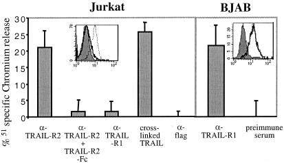
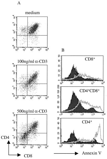
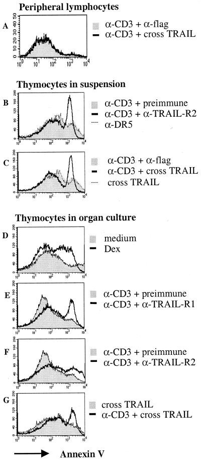
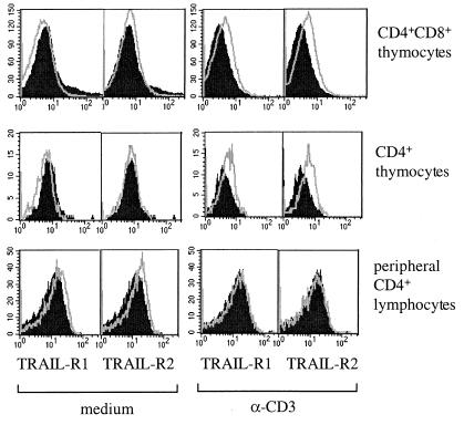
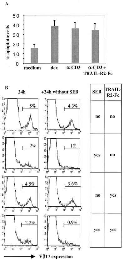
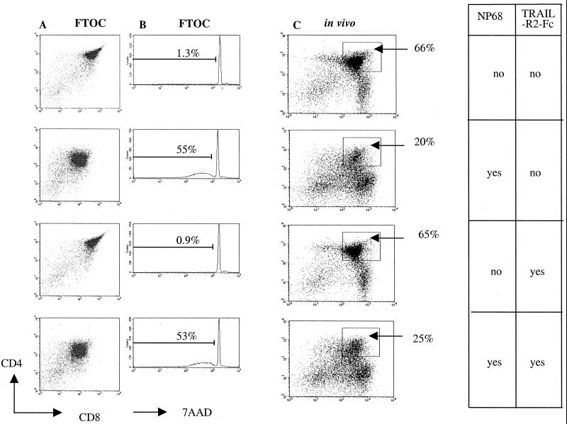
TRAIL (tumor necrosis factor-related apoptosis-inducing ligand) is a recently identified member of the tumor necrosis factor cytokine superfamily. TRAIL has been shown to induce apoptosis in various tumor cell lines, whereas most primary cells seem to be resistant. These observations have raised considerable interest in the use of TRAIL in tumor therapy. Yet little is known about the physiological function of TRAIL. This is particularly the case in the immune system, where TRAIL has been suggested by some to be involved in target cell killing and lymphocyte death. We have developed a panel of mAbs and soluble proteins to address the role of TRAIL in lymphocyte development. These studies demonstrate activation-induced sensitization of thymocytes to TRAIL-mediated apoptosis and expression of the apoptosis-inducing TRAIL receptors. However, with the use of several model systems, our subsequent experiments rule out the possibility that TRAIL plays a major role in antigen-induced deletion of thymocytes. In contrast to thymocytes, there is no up-regulation of TRAIL receptors in peripheral T cells on activation, which remain resistant to TRAIL. Thus, susceptibility to TRAIL-induced apoptosis is controlled differently by central and peripheral T cells.
Figures
Similar articles
-
Activated human NK and CD8+ T cells express both TNF-related apoptosis-inducing ligand (TRAIL) and TRAIL receptors but are resistant to TRAIL-mediated cytotoxicity.Blood. 2004 Oct 15;104(8):2418-24. doi: 10.1182/blood-2004-04-1294. Epub 2004 Jun 17. Blood. 2004. PMID: 15205263
-
Expression of TRAIL receptors in human autoreactive and foreign antigen-specific T cells.Cell Death Differ. 2000 Jul;7(7):637-44. doi: 10.1038/sj.cdd.4400692. Cell Death Differ. 2000. PMID: 10889508
-
Enhanced proliferation and increased IFN-gamma production in T cells by signal transduced through TNF-related apoptosis-inducing ligand.J Immunol. 2001 Aug 1;167(3):1347-52. doi: 10.4049/jimmunol.167.3.1347. J Immunol. 2001. PMID: 11466352
-
TNF-related apoptosis inducing ligand (TRAIL) and its receptors in tumor surveillance and cancer therapy.Apoptosis. 2002 Oct;7(5):449-59. doi: 10.1023/a:1020039225764. Apoptosis. 2002. PMID: 12207178 Review.
-
TRAIL-induced signalling and apoptosis.Toxicol Lett. 2003 Apr 4;139(2-3):89-97. doi: 10.1016/s0378-4274(02)00422-8. Toxicol Lett. 2003. PMID: 12628743 Review.
Cited by
-
Repositioning the Role of Tumor Necrosis Factor-Related Apoptosis-Inducing Ligand (TRAIL) on the TRAIL to the Development of Diabetes Mellitus: An Update of Experimental and Clinical Evidence.Int J Mol Sci. 2022 Mar 17;23(6):3225. doi: 10.3390/ijms23063225. Int J Mol Sci. 2022. PMID: 35328646 Free PMC article. Review.
-
Attenuated Bordetella pertussis protects against highly pathogenic influenza A viruses by dampening the cytokine storm.J Virol. 2010 Jul;84(14):7105-13. doi: 10.1128/JVI.02542-09. Epub 2010 May 5. J Virol. 2010. PMID: 20444902 Free PMC article.
-
TRAIL protects the immature lung from hyperoxic injury.Cell Death Dis. 2022 Jul 15;13(7):614. doi: 10.1038/s41419-022-05072-5. Cell Death Dis. 2022. PMID: 35840556 Free PMC article.
-
Genes and genomics of autoimmune inflammation: from Rel to TRAIL.Immunol Res. 2003;27(2-3):169-78. doi: 10.1385/IR:27:2-3:169. Immunol Res. 2003. PMID: 12857966 Review.
-
TRAIL-R1 and TRAIL-R2 Mediate TRAIL-Dependent Apoptosis in Activated Primary Human B Lymphocytes.Front Immunol. 2019 Apr 30;10:951. doi: 10.3389/fimmu.2019.00951. eCollection 2019. Front Immunol. 2019. PMID: 31114586 Free PMC article.
References
-
- Wiley S R, Schooley K, Smolak P J, Din W S, Huang C P, Nicholl J K, Sutherland G R, Smith T D, Rauch C, Smith C A, et al. Immunity. 1995;3:673–682. - PubMed
-
- Ashkenazi A, Dixit V M. Curr Opin Cell Biol. 1999;11:255–260. - PubMed
-
- Pan G, O'Rourke K, Chinnaiyan A M, Gentz R, Ebner R, Ni J, Dixit V M. Science. 1997;276:111–113. - PubMed
-
- Pan G, Ni J, Wei Y F, Yu G L, Gentz R, Dixit V M. Science. 1997;277:815–818. - PubMed
Publication types
MeSH terms
Substances
LinkOut - more resources
Full Text Sources
Other Literature Sources
Research Materials
