. 2001 Apr 15;29(8):1703-14.
doi: 10.1093/nar/29.8.1703.

Treble clef finger--a functionally diverse zinc-binding structural motif

Affiliations

Treble clef finger--a functionally diverse zinc-binding structural motif

N V Grishin. Nucleic Acids Res. .

Abstract

Detection of similarity is particularly difficult for small proteins and thus connections between many of them remain unnoticed. Structure and sequence analysis of several metal-binding proteins reveals unexpected similarities in structural domains classified as different protein folds in SCOP and suggests unification of seven folds that belong to two protein classes. The common motif, termed treble clef finger in this study, forms the protein structural core and is 25-45 residues long. The treble clef motif is assembled around the central zinc ion and consists of a zinc knuckle, loop, beta-hairpin and an alpha-helix. The knuckle and the first turn of the helix each incorporate two zinc ligands. Treble clef domains constitute the core of many structures such as ribosomal proteins L24E and S14, RING fingers, protein kinase cysteine-rich domains, nuclear receptor-like fingers, LIM domains, phosphatidylinositol-3-phosphate-binding domains and His-Me finger endonucleases. The treble clef finger is a uniquely versatile motif adaptable for various functions. This small domain with a 25 residue structural core can accommodate eight different metal-binding sites and can have many types of functions from binding of nucleic acids, proteins and small molecules, to catalysis of phosphodiester bond hydrolysis. Treble clef motifs are frequently incorporated in larger structures or occur in doublets. Present analysis suggests that the treble clef motif defines a distinct structural fold found in proteins with diverse functional properties and forms one of the major zinc finger groups.


Figures

Figure 1
Figure 1
Structural comparisons of representative treble clef fingers. (A) Structural diagrams of ribosomal protein S14 (1fjf, chain N, residues 21–53), ribosomal protein L24E (1ffk, chain R, residues 3–45), RING finger of RAG1 (dimerization domain) (1rmd, residues 23–60), ARF-GAP domain of Pyk2-associated protein β (1dcq, chain A, residues 262–297), Cys2 activator-binding domain of protein kinase Cδ (1ptq, residues 241–280), FYVE domain of Vps27p protein, first treble clef finger (1vfy, chain A, residues 172–207), FYVE domain of Vps27p protein, second treble clef finger (1vfy, chain A, residues 190–235), retinoid X receptor α DNA-binding domain (2nll, chain B, residues 300–336), recombination endonuclease VII (1en7, chain A, residues 19–79), MH1 domain of Smad (1mhd, chain A, residues 93–132) showing the treble clef finger domains from each protein. In each protein, N- and C-termini are labeled with N and C. β-Strands and α-helices are labeled in lower and upper case letters, respectively. The color of the letter corresponds to the color of the element. The short β-strands in the zinc knuckle region are shown in purple (a and b) with the zinc knuckle turn colored red. Side-chains of zinc ligands and corresponding residues in 1mhd are shown in ball-and-stick representation. Zinc ions are shown as orange balls. The ribbon diagrams were rendered by Bobscript (23), a modified version of Molscript (24). (B) Stereo diagram of superimposed Cα-traces of the 10 structures from (A) shown in the same orientation. The Cα-traces of proteins, side chains of zinc ligands and Zn2+ are shown. Superpositions were made using Insight II package (MSI). The regions used in r.m.s.d. minimization are outlined in thicker lines. Color coding of structures corresponds to the dot color scheme (in front of each PDB entry) in (C). (C) Structure-based sequence alignment of treble clef motif regions of the 10 proteins illustrated in panel (A). For each sequence, the PDB entry name and chain ID, starting and ending residue numbers are given. Zinc ligands are boxed in black. Color shading and labels of secondary structure elements correspond to those in (A). Long insertions are not displayed: the number of omitted residues is specified in brackets.
Figure 2
Figure 2
Structure-based sequence alignment of treble clef fingers. For each sequence, gene identification (gi) number of the NCBI/GenBank protein sequence database, PDB entry name, chain ID (if any), fragment number (if more than one fragment is shown), and starting and ending residue numbers are given. The numbers correspond to the numbering in the PDB file. Sequences are divided into families with the family numbers shown on the left. Families are separated from each other by a larger spacing between the sequences. The families are: I, ribosomal protein L24E; II, ribosomal protein S14; III, RING finger; IV, Pyk2-associated protein β-ARF-GAP domain; V, protein kinase cysteine-rich domain; VI, phosphatidylinositol-3-phosphate-binding domain; VII, GATA-1, LIM and DNA repair factor XPA zinc-binding domains; VIII, nuclear receptor DNA-binding domain; IX, His-Me finger endonucleases/MH1 domain of Smad. Zinc ligands in the signature of the treble clef motif are boxed in black, non-zinc-binding residues in corresponding positions are shown in blue, Zn2+ ligands in other sites are boxed in dark gray, Mg2+ ligands are boxed in olive, active site histidine in endonucleases is shown in green, uncharged residues (all amino acids except D,E,K,R) in mostly hydrophobic sites are highlighted in yellow, non-hydrophobic residues (all amino acids except W,F,Y,M,L,I,V) at mostly hydrophilic sites are highlighted in light gray, small residues (G,P,A,S,C,T,V) at positions occupied by mostly small residues are shown in red letters. Secondary structure consensus is shown below the alignment. β-Strands are displayed as arrows, α-helix is shown as a cylinder. Colors and labels are according to the scheme from Figure 1. Arcs connect hydrogen-bonded residues in the β-hairpins. The sites used in r.m.s.d. minimization are marked with asterisks above the alignment. Red asterisks are for the crucial six sites used in the alignment construction.
Figure 3
Figure 3
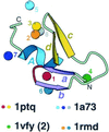
Metal-binding sites in treble clef fingers. All 42 treble clef finger structures (Fig. 2) are superimposed and metal ions bound at different sites in the structural motif (having zinc ligands from different sites in sequence alignment) are displayed. Metal ions are shown as balls and are numbered from 1 to 8. Protein ribbon corresponds to Cys2 activator-binding domain of protein kinase Cδ (1ptq, residues 241–280). Color coding and labeling of secondary structural elements correspond to Figure 1. PDB codes of representative structures that cover all distinct zinc-binding sites are shown below and colored dots indicate the sites that are present in the structures.
Figure 4
Figure 4
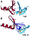
Arrangement of treble clef pairs. (A) A pair of tandem treble clef motifs in the LIM domain of cysteine-rich protein CRIP (1qli, residues 117–145). (B) Two zinc-binding motifs of the FYVE domain of vps27p protein (1vfy, chain A, residues 173–235). One treble clef finger is colored in blue, the other is colored in red. Secondary structural elements are named according to Figure 1. Indices 1 and 2 refer to the first and second treble clef finger, respectively. The segment that belongs to both treble clef fingers is colored in purple. Side chains of zinc ligands are shown in ball-and-stick representation. Zn2+ is shown as an orange ball.
Figure 5
Figure 5
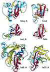
Treble clef fingers inside larger structures. Structural diagrams of ARF-GAP domain of Pyk2-associated protein β (1dcq, chain A, residues 248–365), RING finger of RAG1 (dimerization domain) (1rmd, residues 1–87), retinoid X receptor α DNA-binding domain (2nll, chain B, residues 300–369), Cys2 activator-binding domain of protein kinase Cδ (1ptq, residues 243–280), S.marcescens endonuclease (1ql0, chain A, residues 6–245) and intron-encoded homing endonuclease I-PpoI (1a73, chain A, residues 21–139). The treble clef finger motif is outlined in red. β-Strands and α-helices that are not part of the motif are shown in yellow and blue, respectively. Side-chains of zinc ligands, residues in sites corresponding to ligands of zinc #1, and active site residues in 1a73 are shown in ball-and-stick representation. Active site residues are colored in green. Zn2+ and Mg2+ are displayed as orange and green balls, respectively. In each protein, N- and C-termini are labeled with N and C. β-Strands and α-helices are labeled in lower and upper case letters, respectively. The color of the letter corresponds to the color of the element. Zinc ions are labeled with numbers corresponding to those in Figure 3.
Figure 6
Figure 6
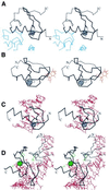
Functional properties of treble clef fingers. Stereo diagrams of (A) RING finger domain of signal transduction protein Cbl (black) in complex with ubiquitin-conjugating enzyme Ubch7 (blue) (1fbv, chain A, residues 376–431 in black, segments of the chain C in blue); (B) Cys2 activator-binding domain of protein kinase Cδ (black) in complex with phorbol ester (orange) (1ptr, residues 231–280); (C) retinoid X receptor α DNA-binding domain (black) in complex with DNA (red) (2nll, chain B residues 300–336 in black, chains C and D in red); (D) intron-encoded homing endonuclease I-PpoI (black) in complex with DNA (red) (1a73, chain A, residues 49–125 in black, chains C and D in red). Cα traces of treble-clef-containing proteins are displayed in black with N- and C-termini labeled. The treble clef motif is shown in thicker lines. Zinc ions are represented by a gray ball. Side chains of zinc ligands or residues in corresponding sites are shown in black. Side chains of active site residues and an active site Mg2+ ion are shown in green. Cα traces of the polypeptide chains interacting with the treble clef domain are dark blue, small molecules are in orange, DNA chains are in red.

Similar articles

Cited by

References

    1. Alberts I.L., Nadassy,K. and Wodak,S.J. (1998) Analysis of zinc binding sites in protein crystal structures. Protein Sci., 7, 1700–1716. - PMC - PubMed
    1. Mackay J.P. and Crossley,M. (1998) Zinc fingers are sticking together. Trends Biochem. Sci., 23, 1–4. - PubMed
    1. Murzin A.G., Brenner,S.E., Hubbard,T. and Chothia,C. (1995) SCOP: a structural classification of proteins database for the investigation of sequences and structures. J. Mol. Biol., 247, 536–540. - PubMed
    1. Bateman A., Birney,E., Durbin,R., Eddy,S.R., Finn,R.D. and Sonnhammer,E.L. (1999) Pfam 3.1: 1313 multiple alignments and profile HMMs match the majority of proteins. Nucleic Acids Res., 27, 260–262. - PMC - PubMed
    1. Teplyakov A., Polyakov,K., Obmolova,G., Strokopytov,B., Kuranova,I., Osterman,A., Grishin,N., Smulevitch,S., Zagnitko,O., Galperina,O. et al. (1992) Crystal structure of carboxypeptidase T from Thermoactinomyces vulgaris. Eur. J. Biochem., 208, 281–288. - PubMed

MeSH terms