. 2001 Apr;75(8):3859-72.
doi: 10.1128/JVI.75.8.3859-3872.2001.

Evaluation of interactions of human cytomegalovirus immediate-early IE2 regulatory protein with small ubiquitin-like modifiers and their conjugation enzyme Ubc9

Affiliations

Evaluation of interactions of human cytomegalovirus immediate-early IE2 regulatory protein with small ubiquitin-like modifiers and their conjugation enzyme Ubc9

J H Ahn et al. J Virol. 2001 Apr.

Abstract

The human cytomegalovirus (HCMV) major immediate-early protein IE2 is a nuclear phosphoprotein that is believed to be a key regulator in both lytic and latent infections. Using yeast two-hybrid screening, small ubiquitin-like modifiers (SUMO-1, SUMO-2, and SUMO-3) and a SUMO-conjugating enzyme (Ubc9) were isolated as IE2-interacting proteins. In vitro binding assays with glutathione S-transferase (GST) fusion proteins provided evidence for direct protein-protein interaction. Mapping data showed that the C-terminal end of SUMO-1 is critical for interaction with IE2 in both yeast and in vitro binding assays. IE2 was efficiently modified by SUMO-1 or SUMO-2 in cotransfected cells and in cells infected with a recombinant adenovirus expressing HCMV IE2, although the level of modification was much lower in HCMV-infected cells. Two lysine residues at positions 175 and 180 were mapped as major alternative SUMO-1 conjugation sites in both cotransfected cells and an in vitro sumoylation assay and could be conjugated by SUMO-1 simultaneously. Although mutations of these lysine residues did not interfere with the POD (or ND10) targeting of IE2, overexpression of SUMO-1 enhanced IE2-mediated transactivation in a promoter-dependent manner in reporter assays. Interestingly, many other cellular proteins identified as IE2 interaction partners in yeast two-hybrid assays also interact with SUMO-1, suggesting that either directly bound or covalently conjugated SUMO moieties may act as a bridge for interactions between IE2 and other SUMO-1-modified or SUMO-1-interacting proteins. When we investigated the intracellular localization of SUMO-1 in HCMV-infected cells, the pattern changed from nuclear punctate to predominantly nuclear diffuse in an IE1-dependent manner at very early times after infection, but with some SUMO-1 protein now associated with IE2 punctate domains. However, at late times after infection, SUMO-1 was predominantly detected within viral DNA replication compartments containing IE2. Taken together, these results show that HCMV infection causes the redistribution of SUMO-1 and that IE2 both physically binds to and is covalently modified by SUMO moieties, suggesting possible modulation of both the function of SUMO-1 and protein-protein interactions of IE2 during HCMV infection.


Figures

FIG. 1
FIG. 1
Interaction of IE2 with SUMO-1, SUMO-2, SUMO-3, and Ubc9 in yeast two-hybrid assays. Plasmids encoding GAL4-DB fusions and GAL4-A fusions were introduced together into Y190 cells. Transformants were selected on plates lacking Trp and Leu, and the β-galactosidase activities of the transformants were measured as described in Materials and Methods. (A) Interaction of GAL4-BD/IE2(87–542) with GAL4-A fusion proteins. The cDNAs used to make GAL4-A fusions were SUMO-1(1–101), SUMO-1(ΔGG)(1–95), SUMO-2(1–103), SUMO-3(1–95), Ubc9(1–158), and IE2(290–542). (B) Interaction of GAL4-DB/IE2(87–542, K175/180R) with GAL4-A fusion proteins. Solid arrowheads indicate the position after the double-glycine motif, which is cleaved by protease to activate the SUMO moiety for conjugation to the substrate proteins (see also Fig. 2).
FIG. 2
FIG. 2
Comparison of amino acid sequences for SUMO-1, SUMO-2, and SUMO-3 isolated as IE2-interacting proteins from a human B-lymphocyte cDNA library. The amino acid sequences of SUMO-1, SUMO-2 (hsSmt3A), and SUMO-3 (hsSmt3B) isolated in this study were aligned. Residues that are identical in either two or three of the proteins are boxed. The arrow after the double-glycine motif indicates the position of proteolytic cleavage for conjugation. The numbers in parentheses are the numbers of amino acids presumably conjugated to the substrates.
FIG. 3
FIG. 3
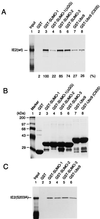
Direct interactions of IE2 with SUMO-1, SUMO-2, SUMO-3, and Ubc9. (A) In vitro binding assay of wild-type IE2 with GST fusion proteins. The same amounts of GST or GST fusion proteins immobilized to glutathione-Sepharose beads were incubated with [35S]methionine-labeled full-length IE2 proteins. One-fifth of the labeled IE2 proteins used in each binding reaction was loaded as an input control (lane 1). After in vitro binding and five washing steps, purified proteins were fractionated by electrophoresis on SDS–8% polyacrylamide gels and visualized by autoradiography. (B) Relative sizes and purity of the GST or GST fusion proteins used. The same gel as in panel A was stained with Coomassie blue before autoradiography. (C) In vitro binding assay of IE2(S203A) with GST fusion proteins. The assay conditions were exactly the same as in panel A.
FIG. 4
FIG. 4
Covalent modification of IE2 by SUMO-1 or SUMO-2. (A) Detection of SUMO-1-conjugated IE2 by Western immunoblotting in transfected 293T cells. Total cell extracts prepared from 293T cells transfected with 1 μg of empty plasmid vector DNA (lane 1) or with 1 μg of pJHA124 (encoding IE2) DNA alone (lane 2) or cotransfected with 1 μg of pJHA124 plus 2 μg of pJHA312 encoding Flag–SUMO-1 (lane 3) were fractionated on SDS–8% polyacrylamide gels, and immunoblot analysis was carried out with mouse anti-IE2 12E2 MAb. (B) Samples of the same IE2 and IE2/Flag–SUMO-1–transfected 293T cell extracts were subjected to analysis with mouse anti-IE2 CH810 MAb. Lanes 3 and 4 contained three times more loaded extract than lanes 1 and 2 and were exposed longer. (C) Samples of IE2 and IE2 plus Flag–SUMO-1–transfected 293T cell extracts were immunoblotted with mouse anti-IE2 mouse MAb CH810 (lanes 1 and 2), washed, and reprobed with mouse MAb specific for Flag epitope (lanes 3 and 4). (D) Transfected 293T cell lysates (same sample set as in panel A) were immunoprecipitated with MAb CH810, and the immunoprecipitates were fractionated by size and reacted with anti-SUMO-1 MAb 21C7. (E) U373-MG cells were transfected with empty vector (lane 1) or 1 μg of pJHA124 (encoding IE2) alone (lane 2) or cotransfected with 1 μg of pJHA124 plus 2 μg of pJHA312 (Flag–SUMO-1) (lane 3), pJHA342 (Flag–SUMO-2) (lane 4), or pJHA344 [Flag–SUMO-1(ΔGG)] (lane 5). Total cell extracts were prepared and fractionated as described above, and immunoblot analysis was carried out with anti-IE2-specific MAb 12E2. (F) HF cells were mock infected or infected with HCMV(Towne) at an MOI of 5.0 for 24, 48, or 72 h. Total cell extracts were prepared and fractionated as described above, and immunoblot analysis was performed with MAb CH810 recognizing both IE1 and IE2. (G) HF cells were infected with Ad-IE1 (lane 1) or Ad-IE2 (lane 2) at an MOI of 20 for 24 h, and immunoblot analysis of the total cell extracts with anti-IE1/IE2 CH810 MAb was carried out as in panel F. The SUMO-conjugated 90-kDa form of IE1 and 105-kDa forms of IE2 are designated IE1-S and IE2-S, and the IE2 form modified by the putative endogenous SUMO moiety is indicated by ⧫ in panels B, D and E.
FIG. 5
FIG. 5
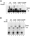
Mapping of the lysine residues in IE2 that are conjugated by SUMO-1. (A) Total cell extracts were prepared from 293T cells transfected with 1 μg of vector alone (lane 1), or 1 μg of pJHA124 encoding wild-type IE2 (lane 2), pYX105 encoding IE2(K175R) (lane 4), pYX106 encoding IE2(K180R) (lane 6), or pYX104 encoding IE2(K175/180R) (lane 8) or the same four plasmids plus 2 μg of pJHA312 encoding Flag–SUMO-1 (lanes 3, 5, 7, and 9). Total cell extracts were prepared and subjected to electrophoresis on SDS–8% polyacrylamide gels, and immunoblot analysis was carried out with MAb CH810. (B) In vitro SUMO-1 conjugation assays. [35S]methionine-labeled wild-type and mutant IE2 proteins were incubated at 37°C in the presence or absence of SUMO-1 modification reaction mixtures (Rxn Mix) as described in Materials and Methods. The reaction products were visualized by autoradiography. IE2-S, Flag–SUMO-1 conjugated IE2; ⧫, IE2 forms conjugated by an endogenous SUMO moiety.
FIG. 6
FIG. 6
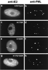
Intracellular localization patterns of mutant IE2 proteins unable to be modified by SUMO-1. Vero cells were transfected with pJHA124 encoding wild-type IE2 (a and b), pYX105 encoding IE2(K175R) (c and d), pYX106 encoding IE2(K180R) (e and f), or pYX104 encoding IE2(K175/180R) (g and h). At 48 h after transfection, the cells were fixed by the paraformaldehyde procedure and double-label IFA was carried out with MAb 12E2 for IE2 (a, c, e, and g) or PAb PML(C) for endogenous PML (b, d, f, and h). FITC-labeled anti-mouse IgG and rhodamine-coupled anti-rabbit IgG were used for visualization.
FIG. 7
FIG. 7
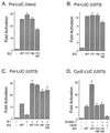
Effect of sumoylation on transactivation by IE2. (A and B) Comparison of transactivation by wild-type and sumoylation mutant forms of IE2 on the HCMV Pol promoter. Vero (A) and U373-MG (B) cells were cotransfected with 1 μg of plasmid DNA containing a reporter gene driven by the HCMV Pol promoter (Pol-LUC) and 2 μg of pJHA124 (encoding intact IE2) (WT), pYX105(K175R), pYX106(K180R), or pYX104(K175/180R). At 48 h after transfection, total-cell extracts were prepared and assayed for luciferase activity. (C) Comparison of cooperative transactivation with IE1 by wild-type and sumoylation mutant forms of IE2 on the HCMV Pol promoter. U373-MG cells were cotransfected with 0.5 μg of reporter plasmid (Pol-LUC) and with 1 μg of either pJHA303 (encoding IE1) or pJHA124 (IE2) alone or with pJHA303 plus wild-type or mutant IE2 plasmid. (D) Comparison of transactivation of the cyclin E promoter by wild-type and sumoylation mutant IE2. U373-MG cells were cotransfected with 0.5 μg of plasmid DNA containing a reporter gene driven by the cellular cyclin E promoter (CycE-LUC) and with various combinations of plasmid pJHA312 encoding Flag–SUMO-1, pWJ5 encoding Flag-Ubc9, and plasmids for wild-type or mutant IE2. Luciferase activities are indicated as fold activation over the basal level of each reporter gene and shown as an average of duplicated experiments.
FIG. 8
FIG. 8
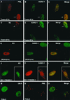
Displacement of SUMO-1 in HCMV-infected cells and colocalization of IE2 with SUMO-1. (A to I) HF cells were infected with HCMV(Towne) at a low MOI (<1.0 PFU/cell) or with HCMV(CR208) at an MOI of 5.0. At 6 h after infection, the cells were fixed in methanol and double-label IFA was carried out with anti-peptide rabbit PAb PML(C) against PML and mouse MAb 21C7 against SUMO-1 (A to C) or with anti-peptide rabbit PAb P3 against IE2 and MAb 21C7 (D to I). (J to L) Vero cells were cotransfected with pMP18 plasmid DNA encoding wild-type intact IE2 and pJHA312 encoding Flag–SUMO-1 (F/SUMO-1) and then stained for double-labeled IFA for IE2 and Flag–SUMO-1 with mouse MAb and anti-Flag rabbit PAb. (M to O) Vero cells were transfected with plasmid pWJ5 encoding Flag-Ubc9 (F/Ubc9) and double-labeled for Flag-Ubc9 and the endogenous PML with anti-Flag mouse MAb and PAb PML(C). For color IFA visualization, FITC-labeled anti-mouse IgG (green) and rhodamine-coupled anti-rabbit IgG (red) were used.
FIG. 9
FIG. 9
Localization of SUMO-1 within viral DNA replication compartments in HCMV-infected cells. HF cells were infected with HCMV(Towne) at an MOI of 3.0. At 60 h after infection, the cells were fixed by the paraformaldehyde procedure. (A to D) Single-label IFA for SUMO-1 was carried out either with mouse MAb 21C7 (A) or with rabbit PAb FL-101 (C). (E to H) Double-label IFA for both SUMO-1 and IE2 was performed with both rabbit PAb FL-101 and mouse MAb 12E2 (for IE2). SUMO-1, IE2, and the merge images from a single microscopic field are shown in panels E, F, and G, respectively. For color IFA visualization, FITC-labeled anti-mouse IgG (green) and rhodamine-coupled anti-rabbit IgG (red) were used. DAPI staining for the same fields of panels A, C, and E to G are shown in panels B, D, and H, respectively. HCMV-infected cells showing viral DNA replication compartments are indicated by arrows. The rabbit PAb gives nonspecific cytoplasmic staining in addition to specific nuclear staining in late-stage HCMV-infected cells.
FIG. 10
FIG. 10
Possible role for SUMO-1 as a bridge for the interaction of IE2 with cellular proteins. SUMO-1 can be either covalently attached to lysine residues (K) or physically bound to the putative SUMO-1-binding consensus sequence (IVISDSEEE, designated SXS) within IE2. Both cellular proteins containing SUMO-1-binding consensus sequences (X) or those conjugated by SUMO-1 (Y) appear capable of interacting with IE2 via the SUMO-1 bridging molecules.

Similar articles

Cited by

References

    1. Ahn J H, Brignole III E J, Hayward G S. Disruption of PML subnuclear domains by the acidic IE1 protein of human cytomegalovirus is mediated through interaction with PML and may modulate a RING finger-dependent cryptic transactivator function of PML. Mol Cell Biol. 1998;18:4899–4913. - PMC - PubMed
    1. Ahn J H, Chiou C J, Hayward G S. Evaluation and mapping of the DNA binding and oligomerization domains of the IE2 regulatory protein of human cytomegalovirus using yeast one and two hybrid interaction assays. Gene. 1998;210:25–36. - PubMed
    1. Ahn J H, Hayward G S. Disruption of PML-associated nuclear bodies by IE1 correlates with efficient early stages of viral gene expression and DNA replication in human cytomegalovirus infection. Virology. 2000;274:39–55. - PubMed
    1. Ahn J H, Hayward G S. The major immediate-early proteins IE1 and IE2 of human cytomegalovirus colocalize with and disrupt PML-associated nuclear bodies at very early times in infected permissive cells. J Virol. 1997;71:4599–4613. - PMC - PubMed
    1. Ahn J H, Jang W J, Hayward G S. The human cytomegalovirus IE2 and UL112–113 proteins accumulate in viral DNA replication compartments that initiate from the periphery of promyelocytic leukemia protein-associated nuclear bodies (PODs or ND10) J Virol. 1999;73:10458–10471. - PMC - PubMed

Publication types

MeSH terms

Substances