Pentoxifylline treatment of mice with chronic pulmonary tuberculosis accelerates the development of destructive pathology
- PMID: 11260331
- PMCID: PMC1783172
- DOI: 10.1046/j.1365-2567.2001.01161.x
Pentoxifylline treatment of mice with chronic pulmonary tuberculosis accelerates the development of destructive pathology
Abstract
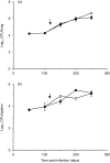
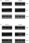
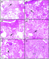
It is well established in animal models that production of the cytokine tumour necrosis factor-alpha (TNF-alpha) is essential to the proper expression of acquired specific resistance following infection with Mycobacterium tuberculosis. This gives rise to an apparent state of chronic disease which over the next 100-200 days is characterized by slowly worsening pathological changes in the lung. To determine whether continued TNF-alpha production was harmful during this phase mice were treated with a TNF-alpha inhibitor, pentoxifylline. It was observed that although this therapy did not alter the numbers of bacteria recovered from the lungs of the infected mice, tissue damage within the lung was accelerated. These data thus demonstrate that production of TNF-alpha, already known to be important during the early expression of resistance to tuberculosis, remains important and beneficial during the chronic stage of the disease.
Figures


Similar articles
-
In vivo IL-10 production reactivates chronic pulmonary tuberculosis in C57BL/6 mice.J Immunol. 2002 Dec 1;169(11):6343-51. doi: 10.4049/jimmunol.169.11.6343. J Immunol. 2002. PMID: 12444141
-
Cytokine profile during latent and slowly progressive primary tuberculosis: a possible role for interleukin-15 in mediating clinical disease.Clin Exp Immunol. 2006 Jan;143(1):180-92. doi: 10.1111/j.1365-2249.2005.02976.x. Clin Exp Immunol. 2006. PMID: 16367949 Free PMC article.
-
Analysis of the local kinetics and localization of interleukin-1 alpha, tumour necrosis factor-alpha and transforming growth factor-beta, during the course of experimental pulmonary tuberculosis.Immunology. 1997 Apr;90(4):607-17. doi: 10.1046/j.1365-2567.1997.00193.x. Immunology. 1997. PMID: 9176116 Free PMC article.
-
Effects of tumor necrosis factor alpha on host immune response in chronic persistent tuberculosis: possible role for limiting pathology.Infect Immun. 2001 Mar;69(3):1847-55. doi: 10.1128/IAI.69.3.1847-1855.2001. Infect Immun. 2001. PMID: 11179363 Free PMC article.
-
Differential influence of nutrient-starved Mycobacterium tuberculosis on adaptive immunity results in progressive tuberculosis disease and pathology.Infect Immun. 2015 Dec;83(12):4731-9. doi: 10.1128/IAI.01055-15. Epub 2015 Sep 28. Infect Immun. 2015. PMID: 26416911 Free PMC article.
Cited by
-
Reduced immunopathology and mortality despite tissue persistence in a Mycobacterium tuberculosis mutant lacking alternative sigma factor, SigH.Proc Natl Acad Sci U S A. 2002 Jun 11;99(12):8330-5. doi: 10.1073/pnas.102055799. Proc Natl Acad Sci U S A. 2002. PMID: 12060776 Free PMC article.
-
Correlates of Vaccine-Induced Protection against Mycobacterium tuberculosis Revealed in Comparative Analyses of Lymphocyte Populations.Clin Vaccine Immunol. 2015 Oct;22(10):1096-108. doi: 10.1128/CVI.00301-15. Epub 2015 Aug 12. Clin Vaccine Immunol. 2015. PMID: 26269537 Free PMC article.
-
Advancing host-directed therapy for tuberculosis.Nat Rev Immunol. 2015 Apr;15(4):255-63. doi: 10.1038/nri3813. Epub 2015 Mar 13. Nat Rev Immunol. 2015. PMID: 25765201 Review.
-
Host-directed therapies for bacterial and viral infections.Nat Rev Drug Discov. 2018 Jan;17(1):35-56. doi: 10.1038/nrd.2017.162. Epub 2017 Sep 22. Nat Rev Drug Discov. 2018. PMID: 28935918 Free PMC article. Review.
References
-
- Orme IM, Cooper AM. Cytokine/chemokine cascades in immunity to tuberculosis. Immunol Today. 1999;20:307–12. - PubMed
-
- Kindler V, Sappino A-P, Grau GE, Piguet P-F, Vassalli P. The inducing role of tumor necrosis factor in the development of bactericidal granulomas during BCG infection. Cell. 1989;56:731–40. - PubMed
-
- Bean AG, Roach DR, Briscoe H, France MP, Korner H, Sedgwick JD, Britton WJ. Structural deficiencies in granuloma formation in TNF gene-targeted mice underlie the heightened susceptibility to aerosol Mycobacterium tuberculosis infection, which is not compensated for by lymphotoxin. J Immunol. 1999;162:3504–3511. - PubMed
-
- Kaneko H, Yamada H, Mizuno S, Udagawa T, Kazumi Y, Sekikawa K, Sugawara I. Role of tumor necrosis factor-alpha in Mycobacterium-induced granuloma formation in tumor necrosis factor-alpha-deficient mice. Lab Invest. 1999;79:379–86. - PubMed
Publication types
MeSH terms
Substances
Grants and funding
LinkOut - more resources
Full Text Sources

