. 2001 Apr;21(7):2393-403.
doi: 10.1128/MCB.21.7.2393-2403.2001.

Protein tyrosine phosphatase CD148-mediated inhibition of T-cell receptor signal transduction is associated with reduced LAT and phospholipase Cgamma1 phosphorylation

Affiliations

Protein tyrosine phosphatase CD148-mediated inhibition of T-cell receptor signal transduction is associated with reduced LAT and phospholipase Cgamma1 phosphorylation

J E Baker et al. Mol Cell Biol. 2001 Apr.

Abstract

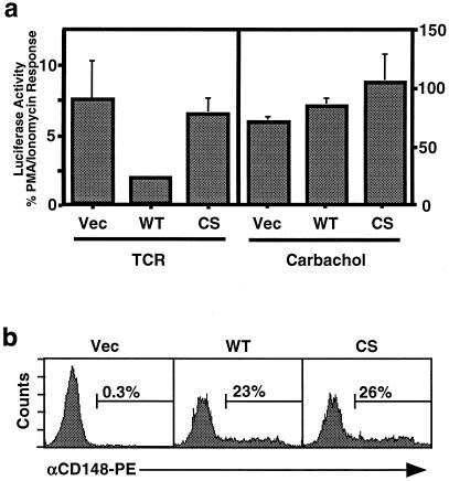
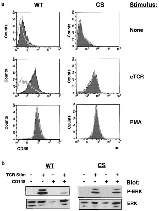
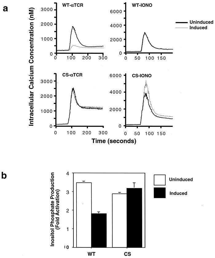
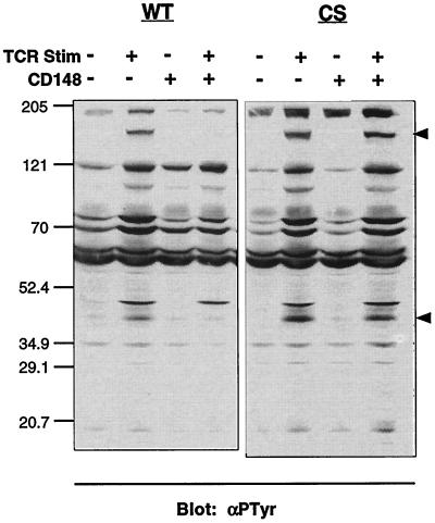
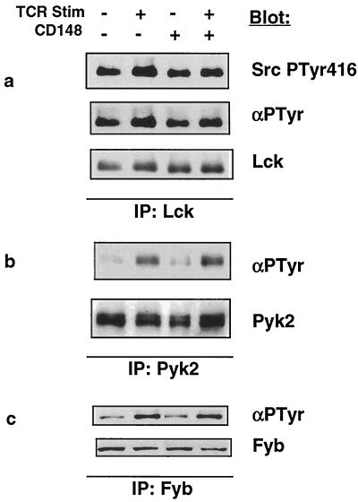
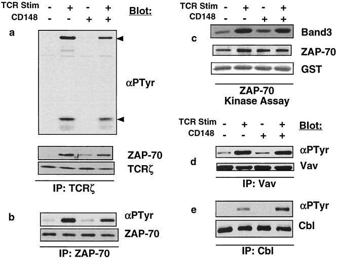
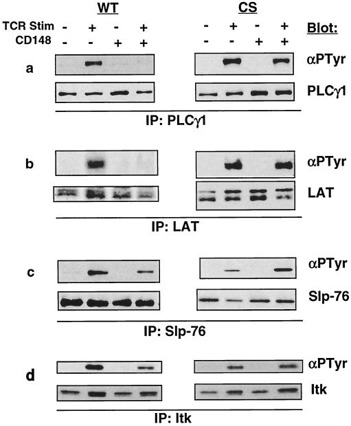
In this study, we investigate the role of the receptor-like protein tyrosine phosphatase CD148 in T-cell activation. Overexpression of CD148 in the Jurkat T-cell line inhibited activation of the transcription factor nuclear factor of activated T cells following T-cell receptor (TCR) stimulation but not following stimulation through a heterologously expressed G protein-coupled receptor, the human muscarinic receptor subtype 1. Using a tetracycline-inducible expression system, we show that the TCR-mediated activation of both the Ras and calcium pathways was inhibited by expression of CD148 at levels that approximate those found in activated primary T cells. These effects were dependent on the phosphatase activity of CD148. Analysis of TCR-induced protein tyrosine phosphorylation demonstrated that most phosphoproteins were unaffected by CD148 expression. However, phospholipase Cgamma1 (PLCgamma1) and LAT were strikingly hypophosphorylated in CD148-expressing cells following TCR stimulation, whereas the phosphorylation levels of Slp-76 and Itk were modestly reduced. Based on these results, we propose that CD148 negatively regulates TCR signaling by interfering with the phosphorylation and function of PLCgamma1 and LAT.


Figures

FIG. 1
FIG. 1
Effect of CD148 on NFAT activation in J-HM1–2.2 cells. (a) J-HM1–2.2 cells were cotransfected with 20 μg of NFAT-luciferase reporter construct together with 20 μg of empty pEF-BOS expression vector (Vec) or the expression plasmid pEF-BOS/CD148 or pEF-BOS/CD148CS containing cDNA encoding wild-type CD148 (WT) or a catalytically inactive mutant (CS), respectively. The following day, equivalent numbers of cells were stimulated in triplicate for 6 h with anti-TCR MAb, carbachol, or PMA plus ionomycin. The results are reported as the TCR response or the carbachol response as a percentage of the PMA-plus-ionomycin response. Data are representative of at least three independent experiments. (b) CD148 expression levels in the transfectants were examined by staining the transfectants with a PE-conjugated antibody specific for CD148, followed by flow cytometry. The numbers above the bars refer to the percentage of live cells expressing CD148.
FIG. 2
FIG. 2
Analysis of inducible CD148 expression in stably transfected Jurkat cell lines. (a) Stably transfected cell lines containing the rtTA and either wild-type CD148 (WT) or catalytically inactive CD148 (CS) driven by a tetracycline-responsive promoter were left untreated or were treated with 1 μg of doxycycline/ml. After 48 h, the cells were stained with a PE-conjugated antibody specific for CD148 and were analyzed by flow cytometry. The shaded histogram represents the untreated cells, and the empty histogram represents the doxycycline-treated cells. L19 and L12 represent two individual WT lines. (b) Human peripheral blood leukocytes stimulated for 2 days with phytohemagglutinin were stained with an FITC-conjugated anti-CD3 antibody and either a PE-conjugated antibody to CD148 (empty histogram) or a PE-conjugated isotype matched control antibody (shaded histogram) and were subsequently analyzed by flow cytometry. The histograms represent CD3-positive cells.
FIG. 3
FIG. 3
Expression of CD148 inhibits TCR-mediated CD69 upregulation and ERK phosphorylation. (a) The wild-type CD148 (WT) and catalytically inactive CD148 (CS) stable lines were either untreated or were induced for 48 h with doxycycline. Subsequently, the cells were stimulated with anti-TCR MAb (1:1,000), 25 ng of PMA/ml or were left unstimulated. Fourteen hours later, the cells were stained with an FITC-conjugated antibody to CD69 followed by flow cytometry. The shaded histogram represents the uninduced cells, while the empty histogram represents the doxycycline-induced cells. The stimulus is noted to the right of the histograms. (b) The stable lines were induced with doxycycline as described for panel a. Subsequently, the cells were left unstimulated or were stimulated for 2 min with anti-TCR MAb, and postnuclear lysate was analyzed by Western blotting with an antiserum against phospho-ERK (P-ERK). The blot was then stripped and reprobed with an antiserum against total cellular ERK. The blotting antisera are noted to the right of the blots. Stim, stimulation. + and − represent presence and absence of TCR stimulation or of CD148.
FIG. 4
FIG. 4
CD148 inhibits calcium mobilization and inositol phosphate production following TCR stimulation. The stable lines were induced with doxycyline as described for Fig. 3a. (a) The cells were loaded with the calcium indicator dye Indo-1, stimulated at the 60-s time point with either anti-TCR MAb (αTCR) or 1 μM ionomycin (IONO), and the concentration of intracellular calcium was calculated based on the fluorescence at 400- and 500-nm wavelengths. The dotted line represents the doxycycline-induced cells, while the solid line represents the uninduced cells. WT, wild type; CS, catalytically inactive mutant. (b) Equivalent numbers of cells were loaded with [3H]myo-inositol and were left unstimulated or were stimulated with anti-TCR MAb for 10 min. Soluble inositol phosphates were extracted, and their levels were measured by scintillation counting. The graphs indicate the fold increase in the total amount of inositol phosphates following stimulation. The empty bars represent the uninduced cells, while the solid bars represent the induced cells. The experiment was performed in triplicate twice with the WT cells and once with the CS cells.
FIG. 5
FIG. 5
Effect of CD148 on inducible tyrosine phosphorylation. The stable cell lines were induced with doxycycline as described for Fig. 3a, and equivalent numbers of cells were left unstimulated or were stimulated with anti-TCR MAb for 3 min. Postnuclear lysates were separated by SDS-PAGE and analyzed by Western blotting with the antiphosphotyrosine antibody 4G10 (αPTyr). The molecular weight markers (in thousands) are noted to the left of the blot, and the upper and lower arrows to the right of the blot correspond to the molecular weights of PLCγ1 and LAT, respectively. WT; wild type; CS; catalytically inactive mutant; Stim, stimulation; −, absence of TCR or CD148; +, presence of TCR or CD148.
FIG. 6
FIG. 6
Analysis of Lck, Pyk2, and SLAP-130/Fyb tyrosine phosphorylation. The wild-type (WT) CD148 stable cell line was induced with doxycycline as described for Fig. 3a. Equivalent numbers of cells were left unstimulated or were stimulated for 3 min with anti-TCR MAb, and postnuclear lysates were prepared. Immunoprecipitations (IPs) were performed with antibodies against Lck (a), Pyk2 (b), or SLAP-130/Fyb (c); the immunoprecipitates were separated by SDS-PAGE and were analyzed by Western blotting using antiphosphotyrosine antibodies 4G10 (Lck and SLAP-130/Fyb) and RC20 (Pyk2). The blots were subsequently stripped and reprobed with the antibodies used in the immunoprecipitation to control for protein level. The blotting antibodies are noted to the right of the blots. Stim, stimulation; −, absence of TCR or CD148; +, presence of TCR or CD148.
FIG. 7
FIG. 7
Analysis of TCRζ, ZAP-70, Vav, and Cbl phosphorylation and of ZAP-70 kinase activity. The wild-type (WT) CD148 stable cell line was induced with doxycycline as described for Fig. 3a. Equivalent numbers of cells were left unstimulated or were stimulated for 3 min with anti-TCR MAb, and postnuclear lysates were prepared. Immunoprecipitations (IPs) were performed with antibodies against TCRζ (a), ZAP-70 (b), Vav (d), or Cbl (e); the immunoprecipitates were separated by SDS-PAGE and were analyzed by Western blotting using the antiphosphotyrosine antibody 4G10 (αPTyr). The blots were subsequently stripped and were reprobed with the antibodies used in the immunoprecipitation to control for protein level. The TCRζ immunoprecipitates were also blotted with ZAP-70 to demonstrate that nearly equivalent amounts of ZAP-70 coimmunoprecipitate with TCRζ. The upper and lower arrows to the right of the blot in panel a correspond to ZAP-70 and TCRζ, respectively. The blotting antibodies are noted to the right of the blots. (c) To assess ZAP-70 kinase activity, immunoprecipitations were performed with antibodies against ZAP-70, followed by an in vitro kinase assay with GST-band 3 as a substrate. The kinase assay was separated by SDS-PAGE and transferred to an Immobilon-P membrane. The in vitro phosphorylated band 3 was detected by autoradiography (top panel). The membrane was then probed with antibodies to ZAP-70 (middle panel) and to GST (bottom panel) to control for protein levels of the kinase and the substrate. Similar results were obtained using GST-LAT as a substrate (data not shown). Stim, stimulation; −, absence of TCR or CD148; +, presence of TCR or CD148.
FIG. 8
FIG. 8
Analysis of PLCγ1, LAT, Slp-76, and Itk phosphorylation. The stable cell lines were induced with doxycycline as described for Fig. 3a. Equivalent numbers of cells were left unstimulated or were stimulated for 3 min with anti-TCR MAb, and postnuclear lysates were made. Immunoprecipitations (IPs) were performed with antibodies against PLCγ1 (a), LAT (b), Slp-76 (c), or Itk (d); the immunoprecipitates were separated by SDS-PAGE and were analyzed by Western blotting using the antiphosphotyrosine antibody 4G10 (αPTyr). The blots were subsequently stripped and reprobed with the antibodies used in the immunoprecipitation to control for protein level. The blotting antibodies are noted to the right of the blots. WT, wild type CD148; CS, catalytically inactive mutant; Stim, stimulation; −, absence of TCR or CD148; +, presence of TCR or CD148.
FIG. 9
FIG. 9
CD148 can dephosphorylate LAT and PLCγ1 in vitro. Myc-tagged LAT-reconstituted JCaM2 cells (a) and Jurkat cells (b) were stimulated with pervanadate for 10 min, and postnuclear lysates were prepared. Immunoprecipitations (IPs) were performed with antibodies against Myc (a) or PLCγ1 (b). The immunoprecipitates were divided into three, and an in vitro phosphatase assay was performed by adding either wild-type GST-CD148 (WT), a catalytically inactive GST-CD148 (DA), or phosphatase buffer (−). The proteins were then resolved by SDS-PAGE and analyzed by Western blotting with the antiphosphotyrosine antibody 4G10 (αPTyr) or antibodies against LAT and PLCγ1 to control for protein levels. The blotting antibodies are noted to the right of the blots.

Similar articles

Cited by

References

    1. Asada H, Ishii N, Sasaki Y, Endo K, Kasai H, Tanaka N, Takeshita T, Tsuchiya S, Konno T, Sugamura K. Grf40, a novel Grb2 family member, is involved in T cell signaling through interaction with SLP-76 and LAT. J Exp Med. 1999;189:1383–1390. - PMC - PubMed
    1. Ashwell J D, D'Oro U. CD45 and Src-family kinases: and now for something completely different. Immunol Today. 1999;20:412–416. - PubMed
    1. Carpenter G, Ji Q. Phospholipase C-gamma as a signal-transducing element. Exp Cell Res. 1999;253:15–24. - PubMed
    1. Clements J L, Yang B, Ross-Barta S E, Eliason S L, Hrstka R F, Williamson R A, Koretzky G A. Requirement for the leukocyte-specific adapter protein SLP-76 for normal T cell development. Science. 1998;281:416–419. - PubMed
    1. D'Ambrosio D, Cantrell D A, Frati L, Santoni A, Testi R. Involvement of p21ras activation in T cell CD69 expression. Eur J Immunol. 1994;24:616–620. - PubMed

Publication types

MeSH terms