. 2001 Mar;12(3):539-49.
doi: 10.1091/mbc.12.3.539.

The nuclear export receptor Xpo1p forms distinct complexes with NES transport substrates and the yeast Ran binding protein 1 (Yrb1p)

Affiliations
Free PMC article

The nuclear export receptor Xpo1p forms distinct complexes with NES transport substrates and the yeast Ran binding protein 1 (Yrb1p)

P Maurer et al. Mol Biol Cell. 2001 Mar.
Free PMC article

Abstract

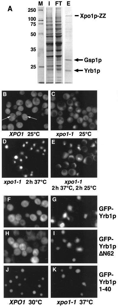
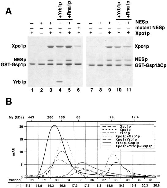
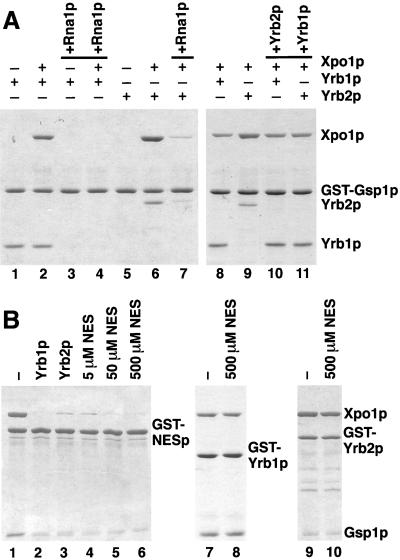
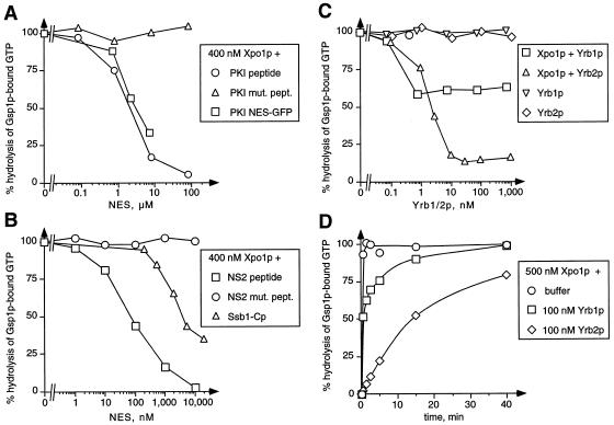
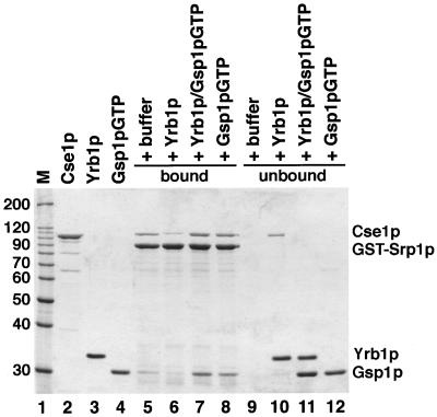
Xpo1p (Crm1p) is the nuclear export receptor for proteins containing a leucine-rich nuclear export signal (NES). Xpo1p, the NES-containing protein, and GTP-bound Ran form a complex in the nucleus that translocates across the nuclear pore. We have identified Yrb1p as the major Xpo1p-binding protein in Saccharomyces cerevisiae extracts in the presence of GTP-bound Gsp1p (yeast Ran). Yrb1p is cytoplasmic at steady-state but shuttles continuously between the cytoplasm and the nucleus. Nuclear import of Yrb1p is mediated by two separate nuclear targeting signals. Export from the nucleus requires Xpo1p, but Yrb1p does not contain a leucine-rich NES. Instead, the interaction of Yrb1p with Xpo1p is mediated by Gsp1p-GTP. This novel type of export complex requires the acidic C-terminus of Gsp1p, which is dispensable for the binding to importin beta-like transport receptors. A similar complex with Xpo1p and Gsp1p-GTP can be formed by Yrb2p, a relative of Yrb1p predominantly located in the nucleus. Yrb1p also functions as a disassembly factor for NES/Xpo1p/Gsp1p-GTP complexes by displacing the NES protein from Xpo1p/Gsp1p. This Yrb1p/Xpo1p/Gsp1p complex is then completely dissociated after GTP hydrolysis catalyzed by the cytoplasmic GTPase activating protein Rna1p.


Figures

Figure 1
Figure 1
(A) Yrb1p is a major Xpo1p-binding protein. Xpo1p-ZZ was purified from yeast extracts in the presence of recombinant Gsp1pQ71L-GTP as described in Materials and Methods, and proteins bound to immobilized Xpo1p were eluted with 500 mM KCl. The input (I), flow-through (FT), and eluate (E) were analyzed by SDS PAGE and Coomassie blue staining. The two major proteins in the eluate were identified by MALDI and Western blotting and correspond to Gsp1p and Yrb1p. Molecular weight markers (M) are in kDa. (B-E) Yrb1p shuttles between the cytoplasm and the nucleus. Wild-type cells (B) or xpo1–1 cells (C-E) expressing GFP-YRB1 were grown in liquid medium at 25°C. The cultures were kept at 25°C (C), shifted to 37°C for 2 h (D), or shifted to 37°C for 2 h and then shifted back to 25°C for 2 h (E). To inhibit protein synthesis, cycloheximide (final concentration 0.1 mg/ml) was added to the cultures before the temperature shift. The cells were viewed by fluorescence microscopy to visualize GFP-Yrb1p. The perinuclear staining in wild-type cells is indicated by arrows. (F-K) Yrb1p has two nuclear targeting sequences. Wild-type cells (F, H, and J) or xpo1–1 mutants (G, I, and K) were transformed with plasmids encoding GFP-Yrb1p, GFP-Yrb1ΔN62p, or GFP-Yrb1p 1–40, as indicated. The cultures were incubated at 30°C (XPO1 cells) or 25°C (xpo1–1 cells) in raffinose-containing medium. Expression of the GFP fusions was induced by 2% galactose and repressed by addition of 2% glucose after 1 h. Cells were kept at 30°C (XPO1) or shifted to 37°C for 2 h (xpo1–1) and viewed by fluorescence microscopy.
Figure 2
Figure 2
Association of importin β-like proteins with Gsp1p-GTP and Yrb1p. (A) GST-Gsp1p-GTP (5 μg per reaction) was immobilized on glutathione Sepharose and incubated for 30 min at 4°C with 3 μg of Yrb1p or 12 μg of Xpo1p, Cse1p, Kap95p, Pse1p, Yrb4p, or Kap104p, as indicated. The reactions containing the export receptors Xpo1p and Cse1p received additionally 4 μg of PKI-NES-2GFP (NESp) or 5 μg of importin α (Srp1p). Molecular weight markers (M) are in kDa. (B and C) GST-Yrb1p (4 μg per reaction) was immobilized on glutathione Sepharose and incubated for 30 min at 4°C with 12 μg of each importin β-like protein and with 10 μg of Gsp1pQ71L loaded with either GTP (B) or GDP (C). The load of Gsp1p (50% of the input) is shown in the left lane. Asterisks indicate weak binding of Kap104p and Kap95p to Yrb1p in the presence of Gsp1p-GDP. The bound material of all reactions was washed three times and analyzed by SDS PAGE and Coomassie blue staining.
Figure 3
Figure 3
(A) Yrb1p but not an NES protein requires the acidic C-terminus of Gsp1p for complex formation. GST-Gsp1p-GTP (lanes 1–6) and GST-Gsp1ΔCp-GTP (lanes 7–11)(4 μg per reaction) were immobilized on glutathione Sepharose and incubated for 30 min at 4°C with 8 μg of PKI-NES-2GFP (NESp), 8 μg of P12-NES-2GFP (mutant NESp), and/or 12 μg of Xpo1p, as indicated. Bound material was washed three times and either eluted with SDS sample buffer (lanes 1–3 and 6–9) or incubated further for 15 min at 4°C with 4 μg of Yrb1p (lanes 4 and 10) or 1 μg of Rna1p (lanes 5 and 11) and then washed again three times. All samples were analyzed by SDS PAGE and Coomassie blue staining. (B) Xpo1p, Yrb1p, and Gsp1p-GTP form a stable 1:1:1 complex. Xpo1p (125 kDa), Yrb1p (23 kDa), and 6His-Gsp1pQ71L-GTP (26 kDa) were mixed as indicated and incubated for 30 min at 4°C. The reactions were gelfiltrated with a Superose 6 column (Pharmacia) in PBSKMT buffer (see Materials and Methods) at a flow rate of 0.4 ml/min. Absorption units at 280 nm were recorded using the Äkta Explorer 100 software (Pharmacia) and plotted against the fraction numbers and the volume after sample injection. Molecular weight markers (Sigma): apoferritin (443 kDa), β-amylase (200 kDa), alcohol dehydrogenase (150 kDa), serum albumin (66 kDa), carbonic anhydrase (29 kDa), cytochrome c (12.4 kDa).
Figure 4
Figure 4
The association of Yrb1p and Yrb2p with Xpo1p/Gsp1p-GTP is sensitive to Rna1p but resistant to excess amounts of NES peptides. (A) GST-Gsp1p-GTP (4 μg per reaction) was immobilized on glutathione Sepharose and incubated for 30 min at 4°C with 5 μg of Yrb1p, 5 μg of Yrb2p, and/or 12 μg of Xpo1p, as indicated. Bound material was washed three times and either eluted with SDS sample buffer (lanes 1, 2, 5, 6, 8, and 9) or incubated further for 15 min at 4°C with 1 μg of Rna1p (lanes 3, 4, and 7), 10 μg of Yrb2p (lane 10), or 10 μg of Yrb1p (lane 11), and then washed again three times. All samples were analyzed by SDS PAGE and Coomassie blue staining. (B) GST fusions (6 μg per reaction) to PKI-NES-2GFP (GST-NESp)(lanes 1–6), Yrb1p (lanes 7 and 8), or Yrb2p (lanes 9 and 10) were immobilized on glutathione Sepharose and incubated for 30 min at 4°C with 10 μg of Gsp1p-GTP and 12 μg of Xpo1p. After three washes, the reactions were further incubated for 15 min at 4°C with buffer alone (lanes 1, 7, and 9), with 6 μg of Yrb1p (lane 2), with 6 μg of Yrb2p (lane 3), or with the indicated amounts of peptides corresponding to the PKI-NES (lanes 4–6, 8, and 10). After three washes, bound material was analyzed by SDS PAGE and Coomassie blue staining.
Figure 5
Figure 5
(A-C) Cooperative binding of NES, Yrb1p, and Yrb2p to Xpo1p/Gsp1p-GTP. Gsp1p[γ-32P]GTP (50 pM) was incubated with 400 nM Xpo1p and the indicated concentrations of NES peptides or proteins (A and B) or Yrb1p or Yrb2p (C). Reactions containing Gsp1p[γ-32P]GTP and Yrb1p or Yrb2p alone served as controls. After 30 min, the GTPase reactions were started by addition of 40 nM Rna1p. Hydrolysis of GTP by Gsp1p was determined as released [32P]phosphate after 30 s by the charcoal method. (D) Kinetics of Rna1p-induced complex disassembly. Gsp1p[γ-32P]GTP (50 pM) was incubated for 30 min with 500 nM Xpo1p alone, with Xpo1p and 100 nM Yrb1p, or with Xpo1p and 100 nM Yrb2p. After Rna1p addition, the reactions were incubated further for the indicated time periods. GTP hydrolysis was then determined by the charcoal method.
Figure 6
Figure 6
The Srp1p/Cse1p/Gsp1p-GTP complex is sensitive to Yrb1p but resistant to Yrb1p complexed to Gsp1p-GTP. GST-Srp1p (8 μg per reaction) was immobilized on glutathione Sepharose and incubated with 12 μg of Cse1p and 4 μg of Gsp1pQ71L-GTP for 30 min at 4°C. After three washes, bound material was incubated with buffer alone (lane 5), with 5 μg of Yrb1p (lane 6), with a preincubated mixture of Yrb1p and Gsp1pQ71L-GTP (lane 7), or with 10 μg of Gsp1pQ71L-GTP (lane 8). After further incubation for 30 min at 4°C, bound (lanes 5–8) and unbound (lanes 9–12) material was separated by SDS PAGE and analyzed by Coomassie blue staining. The molecular weight markers (M) and protein loads are shown in lanes 1–4.

Similar articles

Cited by

References

    1. Aitchison JD, Blobel G, Rout MP. Kap104p: a karyopherin involved in the nuclear transport of messenger RNA binding proteins. Science. 1996;274:624–627. - PubMed
    1. Askjaer P, Bachi A, Wilm M, Bischoff FR, Weeks DL, Ogniewski V, Ohno M, Niehrs C, Kjems J, Mattaj IW, Fornerod M. RanGTP-regulated interactions of CRM1 with nucleoporins and a shuttling DEAD-box helicase. Mol Cell Biol. 1999;19:6276–6285. - PMC - PubMed
    1. Bischoff FR, Krebber H, Smirnova E, Dong W, Ponstingl H. Co-activation of RanGTPase and inhibition of GTP dissociation by Ran-GTP binding protein RanBP1. EMBO J. 1995a;14:705–715. - PMC - PubMed
    1. Bischoff FR, Krebber H, Kempf T, Hermes I, Ponstingl H. Human RanGTPase activating protein RanGap1. is a homologue of yeast Rna1p involved in mRNA processing and transport. Proc Natl Acad Sci USA. 1995b;92:1749–1753. - PMC - PubMed
    1. Bischoff FR, Görlich D. RanBP1 is crucial for the release of RanGTP from importin β-related nuclear transport factors. FEBS Lett. 1997;419:249–254. - PubMed

Publication types

MeSH terms