. 2001 Apr;75(7):3250-8.
doi: 10.1128/JVI.75.7.3250-3258.2001.

Kaposi's sarcoma-associated herpesvirus latency-associated nuclear antigen 1 mediates episome persistence through cis-acting terminal repeat (TR) sequence and specifically binds TR DNA

Affiliations

Kaposi's sarcoma-associated herpesvirus latency-associated nuclear antigen 1 mediates episome persistence through cis-acting terminal repeat (TR) sequence and specifically binds TR DNA

M E Ballestas et al. J Virol. 2001 Apr.

Abstract

Kaposi's sarcoma (KS)-associated herpesvirus (KSHV) (also known as human herpesvirus 8) latently infects KS tumors, primary effusion lymphomas (PELs), and PEL cell lines. In latently infected cells, KSHV DNA is maintained as circularized, extrachromosomal episomes. To persist in proliferating cells, KSHV episomes must replicate and efficiently segregate to progeny nuclei. In uninfected B-lymphoblastoid cells, KSHV latency-associated nuclear antigen (LANA1) is necessary and sufficient for persistence of artificial episomes containing specific KSHV DNA. In previous work, the cis-acting sequence required for episome persistence contained KSHV terminal-repeat (TR) DNA and unique KSHV sequence. We now show that cis-acting KSHV TR DNA is necessary and sufficient for LANA1-mediated episome persistence. Furthermore, LANA1 binds TR DNA in mobility shift assays and a 20-nucleotide LANA1 binding sequence has been identified. Since LANA1 colocalizes with KSHV episomes along metaphase chromosomes, these results are consistent with a model in which LANA1 may bridge TR DNA to chromosomes during mitosis to efficiently segregate KSHV episomes to progeny nuclei.


Figures

FIG. 1
FIG. 1
Schematic diagram of KSHV DNAs assayed for episome persistence. Approximate KSHV genome coordinates (in kilobases) are shown for Z6 (43). Vertical lines separate the ∼0.8-kb TR units. The diagrams are not drawn to scale. Episome persistence in LANA1-expressing cells is indicated by +, and lack of episome persistence is indicated by −. Z6, Z6-13, Z6-7, and Z6-11 were assayed in earlier experiments for episome persistence (4). The asterisk indicates that initial G418-resistant outgrowth of BJAB/F-LANA1 cells transfected with Z6-1TR was slower than after transfection of BJAB/F-LANA1 cells with other DNAs that scored positive for episome persistence.
FIG. 2
FIG. 2
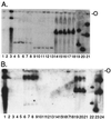
LANA1 acts on cis-acting unit-length KSHV TR DNA to mediate episome persistence. (A) G418-resistant BJAB/F-LANA1 cells (2 × 10⁶) transfected with Z6-BE, Z6-3TRA, or Z6-2TR were lysed in situ in wells of Gardella gels, electrophoresis was performed, DNA was transferred to a nylon membrane, and KSHV TR DNA was detected. Lanes: 1, BC-1; 2, BJAB; 3, BCBL-1; 4 to 18, G418-resistant BJAB/F-LANA1 cells transfected with Z6-3TRA (lanes 4 to 8), Z6-2TR (lanes 9 to 13) or Z6-BE (lanes 14 to 18); 19, Z6-3TRA plasmid; 20, Z6-2TR plasmid; 21, Z6-BE plasmid. The upper bands in lanes 19 to 21 are from nicked plasmid DNA. The lower bands in lanes 1 and 3 are from linear and degraded virus DNA. The data shown are representative of two experiments. (B) G418-resistant BJAB or BJAB/F-LANA1 cells transfected with Z6-BE, Z6-3TRA, or Z6-A were analyzed in Gardella gels. Lanes: 1 BCBL-1; 2, BJAB; 3 to 5, Z6-3TRA-transfected BJAB cells; 6 to 9, Z6-3TRA-transfected BJAB/F-LANA1 cells; 10 to 12, Z6-A-transfected BJAB cells; 13 to 15, Z6-A-transfected BJAB/F-LANA1 cells; 16 to 18, Z6-BE-transfected BJAB cells; 19 to 21, Z6-BE-transfected BJAB/F-LANA1 cells; 22, Z6-3TRA plasmid; 23, Z6-A plasmid; 24, Z6-BE plasmid. The upper bands in lanes 22 to 24 are from nicked plasmid DNA. The lower band and smear in lane 1 is from linear and degraded DNA. The data shown are representative of two experiments. O, well origins.
FIG. 3
FIG. 3
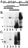
KSHV TR DNA restriction fragments are bound by immunoprecipitated F-LANA1. Radiolabeled KSHV TR restriction fragments were incubated with F-LANA1 or control immunoprecipitates from BJAB/F-LANA1 cells. Bound restriction fragments were eluted and resolved on 5% nondenaturing polyacrylamide gels. (A) Restriction map of the KSHV TR. Numbers indicate the the length of restriction fragments in nucleotides. (The NotI restriction site is at TR nt 383 [43].) Only the nonmethylated AvaII site is shown. (B) F-LANA1 (lanes 1 and 3) or control (lane 2 and 4) immunoprecipitates were incubated with the 266-bp NotI-AscI probe (lanes 1 and 2) or the 535-bp NotI-AscI probe (lanes 3 and 4); input 266-bp NotI-AscI probe (lane 5) and input 535-bp NotI-AscI probe (lane 6) are also shown. The arrow indicates the 535-bp NotI-AscI probe, and the arrowhead indicates the 266-bp NotI-AscI probe. All lanes are from the same gel and have the same exposure time. (C) F-LANA1 (lanes 1 and 3) or control (lane 2 and 4) immunoprecipitates were incubated with the 297-bp NotI-AvaII probe (lanes 1 and 2) or the 370-bp Sau3A-AscI probe (lanes 3 and 4); the input 297-bp NotI-AvaII probe (lane 5) and the input 370-bp Sau3A-AscI probe (lane 6) are also shown. All lanes are from the same gel and have the same exposure time. The arrow indicates the 370-bp Sau3A-AscI probe, and the arrowhead indicates the 297-bp NotI-AvaII probe. The data shown are representative of three experiments.
FIG. 4
FIG. 4
KSHV TR oligonucleotides. (A) The positions of TR-8, TR-7, TR-2, and TR-3 within the 132-bp Sau3A-AvaII fragment of the KSHV TR are shown schematically, TR-8 extends 7 nt 5′ to the Sau3A site. The sequence between TR-7 and TR-8 and the sequence 3′ to TR-3 was not synthesized. The sequences of TR-8, TR-7, TR-2 and TR-3 are shown. Sequences common to TR-7 and TR-2 are underlined, and the overlapping sequence between TR-2 and TR-3 is shown in bold type. (B) The positions of TR-11, TR-12, and TR-13 within TR-2 are shown schematically. Identical sequence is aligned.
FIG. 5
FIG. 5
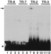
In vitro-translated F-LANA1 gel shifts TR-2. TR-8 (lanes 1 to 3), TR-7 (lanes 4 to 6), TR-2 (lanes 7 to 9) and TR-3 (lanes 10 to 12) 32P-labeled oligonucleotides were each incubated with in vitro-translated F-LANA1 (lanes 1, 2, 4, 5, 7, 8, 10, and 11) or RBP-Jκ (lanes 3, 6, 9, and 12), and EMSA was performed. A 50-fold excess of unlabeled TR-8 (lane 2), TR-7 (lane 5), TR-2 (lane 8), or TR-3 (lane 11) oligonucleotide was included in the incubation. The arrow indicates the specific gel shift in lane 7, and the asterisk indicates free probe. Data shown are representative of two experiments.
FIG. 6
FIG. 6
Delineation of F-LANA1 binding within TR-2. (A) Competition for TR-2 binding to F-LANA1 with oligonucleotides comprising TR-2. In vitro-translated F-LANA1 was incubated with TR-2 prior to EMSA in all lanes except lane 3, where RBP-Jκ was used. A 50-fold molar excess of unlabeled TR-2 was incubated with F-LANA1 for 5 min before addition of TR-2 probe (lane 2). A 50-fold (lanes 4, 6, and 8) or 100-fold (lanes 5, 7, and 9) molar excess of unlabeled TR-11, TR-12, or TR-13 was incubated with F-LANA 1 before the addition of TR-2 probe. The arrow indicates specific gel shifts, and the asterisk indicates free probe. (B) F-LANA1 specifically gel shifts TR-13. In vitro-translated F-LANA1 (lanes 1, 2, 4, 5, 7, and 8) or RBP-Jκ (lanes 3, 6, and 9) was incubated with radiolabeled TR-11 (lanes 1 to 3), TR-12 (lanes 4 to 6), or TR-13 (lanes 7 to 9) (indicated at the top). A 50-fold excess of unlabeled oligonucleotide was included in the incubation (lanes 2, 5, and 8). Asterisk indicates unbound probe. NSB, nonspecific band. (C) Excess nonradiolabeled TR-13 specifically competes the TR-13 gel shift. In vitro-translated F-LANA1 was incubated with radiolabeled TR-13. A 10-, 50-, or 100-fold excess of unlabeled TR-13 was included in the incubation in lanes 2, 3, and 4 respectively. A 50- or 100-fold excess of unlabeled TR-12 was included in the incubations in lanes 5 and 6, respectively. Free probe was run off the gel. U, upper F-LANA1 TR-13 gel-shifted complex; L, lower F-LANA1 TR-13 gel-shifted complex. Unlabeled competitor oligonucleotides are indicated at the bottom. Data shown are representative of two experiments.
FIG. 7
FIG. 7
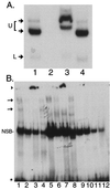
Supershift analyses of F-LANA1 and PEL cell nuclear extracts with TR-13. (A) Anti-FLAG antibody supershifts F-LANA1 TR-13 complexes. In vitro-translated F-LANA1 was incubated with TR-13 probe for all lanes. A 50-fold excess of unlabeled TR-13 oligonucleotide (lane 2), monoclonal anti-FLAG antibody (lane 3), or isotype-matched control antibody (lane 4) was added 15 min prior to the addition of 50,000 cpm of TR-13. Arrows indicate specific gel shifts. U, upper F-LANA1 shifted complex seen in Fig. 6B and C, which is resolved into two complexes here after a longer gel run; L, lower F-LANA1 gel-shifted complex. Data shown are representative of three experiments. Free probe was run off the gel. (B) LANA1 from PEL cells gel shifts TR-13. EMSA was performed using TR-13 probe and nuclear extracts from BCBL-1 (lanes 1 to 4), BC-1 (lanes 5 to 8) or (uninfected) BJAB cells (lanes 9 to 12). A 50-fold molar excess of unlabeled TR-13 (lanes 2, 6, and 10), anti-LANA1 monoclonal antibody (lanes 3, 7, and 11), or control antibody (lanes 4, 8, and 12) were included in incubations prior to the addition of 50,000 cpm of TR-13 probe. The arrows indicate specific gel shifts, and the arrowhead indicates supershifted probe near the gel origin (lanes 3 and 7). NSB, nonspecific band. Free probe is indicated by an asterisk. Data shown are representative of three experiments.

Similar articles

Cited by

References

    1. Aiyar A, Tyree C, Sugden B. The plasmid replicon of EBV consists of multiple cis-acting elements that facilitate DNA synthesis by the cell and a viral maintenance element. EMBO J. 1998;17:6394–6403. - PMC - PubMed
    1. Ambinder R F, Mullen M A, Chang Y N, Hayward G S, Hayward S D. Functional domains of Epstein-Barr virus nuclear antigen EBNA-1. J Virol. 1991;65:1466–1478. - PMC - PubMed
    1. Ausubel F M. Current protocols in molecular biology. New York, N.Y: John Wiley & Sons, Inc.; 1998.
    1. Ballestas M E, Chatis P A, Kaye K M. Efficient persistence of extrachromosomal KSHV DNA mediated by latency-associated nuclear antigen. Science. 1999;284:641–644. - PubMed
    1. Bastien N, McBride A A. Interaction of the papillomavirus E2 protein with mitotic chromosomes. Virology. 2000;270:124–134. - PubMed

Publication types

LinkOut - more resources