. 2001 Mar 1;29(5):1027-33.
doi: 10.1093/nar/29.5.1027.

Sp1 phosphorylation regulates inducible expression of platelet-derived growth factor B-chain gene via atypical protein kinase C-zeta

Affiliations

Sp1 phosphorylation regulates inducible expression of platelet-derived growth factor B-chain gene via atypical protein kinase C-zeta

L A Rafty et al. Nucleic Acids Res. .

Abstract

Platelet-derived growth factor (PDGF) is a broadly expressed mitogenic and chemotactic factor with diverse roles in a number of physiologic and pathologic settings. The zinc finger transcription factors Sp1, Sp3 and Egr-1 bind to overlapping elements in the proximal PDGF B-chain promoter and activate transcription of this gene. The anthracycline nogalamycin has previously been reported to inhibit the capacity of Egr-1 to bind DNA in vitro. Here we used electrophoretic mobility shift assays to show that nogalamycin added to cells in culture did not alter the interaction of Egr-1 with the PDGF-B promoter. Instead, it enhanced the capacity of Sp1 to bind DNA. Nogalamycin increased PDGF-B mRNA expression at the level of transcription, which was abrogated by mutation of the Sp1 binding site in the PDGF-B promoter or overexpression of mutant Sp1. Rather than increasing total levels of Sp1, nogalamycin altered the phosphorylation state of the transcription factor. Overexpression of dominant-negative PKC-zeta blocked nogalamycin-inducible Sp1 phosphorylation and PDGF-B promoter-dependent expression. Nogalamycin stimulated the phosphorylation of PKC-zeta (on residue Thr(410)). These findings demonstrate for the first time that PKC-zeta and Sp1 phosphorylation mediate the inducible expression of this growth factor.


Figures

Figure 1
Figure 1
PDGF-B mRNA expression is induced in WKY12-22 cells exposed to nogalamycin. WKY12-22 cells were exposed to 10 µM nogalamycin for various times, and total RNA was prepared using TRIzol reagent according to the manufacturer’s instructions. The RNA was reverse-transcribed and the cDNA was used as the template for PCR. (A) Time-course of nogalamycin-inducible PDGF-B expression. (B) Actinomycin D pretreatment (10 µg/ml for 2 h) blocks PDGF-B expression inducible by nogalamycin. Cycle-based PDGF-B PCR (bottom panel) was performed on 8 h samples without actinomycin D preincubation. The data are representative of two independent determinations.
Figure 1
Figure 1
PDGF-B mRNA expression is induced in WKY12-22 cells exposed to nogalamycin. WKY12-22 cells were exposed to 10 µM nogalamycin for various times, and total RNA was prepared using TRIzol reagent according to the manufacturer’s instructions. The RNA was reverse-transcribed and the cDNA was used as the template for PCR. (A) Time-course of nogalamycin-inducible PDGF-B expression. (B) Actinomycin D pretreatment (10 µg/ml for 2 h) blocks PDGF-B expression inducible by nogalamycin. Cycle-based PDGF-B PCR (bottom panel) was performed on 8 h samples without actinomycin D preincubation. The data are representative of two independent determinations.
Figure 2
Figure 2
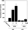
Nogalamycin increases PDGF-B promoter-dependent gene transcription. WKY12-22 cells were transiently transfected with 8 µg of PDGF-B promoter–CAT construct d26 or d75 and CAT activity in the cell lysates was determined after 48 h. CAT activity was normalised to the concentration of protein in the lysates. Nogalamycin was added (2 and 10 µM final concentration) to the transfectants 24 h prior to harvesting. The data are representative of two independent determinations.
Figure 3
Figure 3
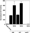
The zinc finger binding site in the proximal PDGF-B promoter mediates nogalamycin-inducible PDGF-B promoter-dependent gene expression. WKY12-22 cells were transiently transfected with construct d26, d77 or md77. After 24 h, the transfectants were incubated with 10 µM of nogalamycin for a further 24 h prior to harvest. The cells were lysed and CAT activity was normalised to the concentration of protein. The data are representative of two independent determinations.
Figure 4
Figure 4
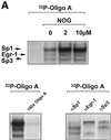
Nogalamycin increases Sp1 binding to a GC-rich oligonucleotide probe. 32P-labelled oligonucleotide bearing consensus elements for Sp1, Egr-1 and Sp3 (32P-Oligo A, 5′-GGG GGG GGC GGG GGC GGG GGC GGG GGA GG-3′, two overlapping consensus Egr-1 binding sites italicised). (A) EMSA was performed as described in Materials and Methods. Supershift analysis shows a distinct pattern of nucleoprotein complexes (Sp1, Egr-1, Sp3), whereas competition analysis demonstrates the specificity of these complexes. (B) Densitometric quantitation of band intensity from EMSA. (C) Western blot analysis for AP2 using the same extracts as those used for the EMSA. The data are representative of two independent determinations.
Figure 4
Figure 4
Nogalamycin increases Sp1 binding to a GC-rich oligonucleotide probe. 32P-labelled oligonucleotide bearing consensus elements for Sp1, Egr-1 and Sp3 (32P-Oligo A, 5′-GGG GGG GGC GGG GGC GGG GGC GGG GGA GG-3′, two overlapping consensus Egr-1 binding sites italicised). (A) EMSA was performed as described in Materials and Methods. Supershift analysis shows a distinct pattern of nucleoprotein complexes (Sp1, Egr-1, Sp3), whereas competition analysis demonstrates the specificity of these complexes. (B) Densitometric quantitation of band intensity from EMSA. (C) Western blot analysis for AP2 using the same extracts as those used for the EMSA. The data are representative of two independent determinations.
Figure 5
Figure 5
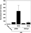
PDGF-B transcription inducible by nogalamycin is Sp1-dependent. WKY12-22 cells were cotransfected with 8 µg of d26 and 5 µg of either pEBG or pEBG-DN Sp1. After 24 h, 10 µM nogalamycin was added to transfectants. CAT activity in the lysates was determined 48 h after transfection and normalised to the concentration of protein. The data are representative of two independent determinations.
Figure 6
Figure 6
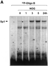
Nogalamycin increases Sp1 DNA binding to the proximal PDGF-B promoter. (A) EMSA was performed using nuclear extracts of WKY12-22 cells exposed to 10 µM nogalamycin for the indicated times and 32P-labelled Oligo B. Binding reactions and non-denaturing gel electrophoresis were performed as described in Materials and Methods. (B) Nogalamycin increases Sp1 DNA binding in a dose-dependent manner. Nuclear extracts from WKY12-22 cells exposed to various concentrations of nogalamycin for 5 h were incubated with 32P-labelled Oligo B (5′-GCT GTC TCC ACC CAC CTC TCG CAC TCT-3′). Binding reactions and non-denaturing gel electrophoresis were performed as described in Materials and Methods. Nucleoprotein complexes were visualised by autoradiography. The data are representative of two independent determinations.
Figure 6
Figure 6
Nogalamycin increases Sp1 DNA binding to the proximal PDGF-B promoter. (A) EMSA was performed using nuclear extracts of WKY12-22 cells exposed to 10 µM nogalamycin for the indicated times and 32P-labelled Oligo B. Binding reactions and non-denaturing gel electrophoresis were performed as described in Materials and Methods. (B) Nogalamycin increases Sp1 DNA binding in a dose-dependent manner. Nuclear extracts from WKY12-22 cells exposed to various concentrations of nogalamycin for 5 h were incubated with 32P-labelled Oligo B (5′-GCT GTC TCC ACC CAC CTC TCG CAC TCT-3′). Binding reactions and non-denaturing gel electrophoresis were performed as described in Materials and Methods. Nucleoprotein complexes were visualised by autoradiography. The data are representative of two independent determinations.
Figure 7
Figure 7
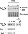
Nogalamycin phosphorylates endogenous Sp1 but not Egr-1 or Sp3. Nuclear extracts from WKY12-22 cells exposed to 10 µM nogalamycin for various times were resolved by electrophoresis prior to transfer to PVDF membranes and exposure to polyclonal antibodies to Sp1, Sp3 or Egr-1. Where indicated, the 8 h sample was incubated with CIP prior to western blot analysis for Sp1. The data are representative of two independent determinations.
Figure 8
Figure 8
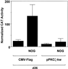
Nogalamycin activation of the PDGF-B promoter is PKC-ζ-dependent. WKY12-22 cells were cotransfected with 8 µg of d26 and 5 µg of either CMV-Flag or pPKC-ζ-kw. Nogalamycin (10 µM) was added to the transfectants 24 h post-transfection prior to harvest 24 h later. CAT activity was normalised to the concentration of protein in the extracts. The data are representative of two independent determinations.
Figure 9
Figure 9
DN PKC-ζ ablates Sp1 phosphorylation in WKY12-22 cells. Cells were transfected with 30 µg of CMV-Flag or pPKC-ζ-kw 24 h prior to the addition of nogalamycin (2 µM). Nuclear extracts were prepared 24 h later. Nuclear extracts were resolved by electrophoresis prior to transfer to PVDF membranes and exposure to polyclonal Sp1 antibodies. The data are representative of two independent determinations.
Figure 10
Figure 10
Nogalamycin stimulates PKC-ζ (Thr410) phosphorylation in WKY12-22 cells. Cells were exposed to 10 µM of nogalamycin for various times prior to resolution of the cytoplasmic extracts by electrophoresis and western blot analysis using antibodies to the Thr410 phosphorylated form of PKC-ζ. The data are representative of two independent determinations.

Similar articles

Cited by

References

    1. Antoniades H.N., Scher,C.D. and Stiles,C.D. (1979) Purification of platelet-derived growth factor. J. Biol. Chem., 76, 1809–1813. - PMC - PubMed
    1. Deuel T.F., Huang,J.S., Proffit,R.T., Baenzinger,J.U., Chang,D. and Kennedy,B.B. (1981) Human platelet-derived growth factor: purification and resolution into two active protein fractions. J. Biol. Chem., 256, 8896–8899. - PubMed
    1. Soma Y., Takehara,K. and Ishibashi,Y. (1994) Alteration of the chemotactic response of human skin fibroblasts to PDGF by growth factors. Exp. Cell Res., 212, 274–277. - PubMed
    1. Rydziel S., Ladd,C., McCarthy,T.L., Centrella,M. and Canalis,E. (1992) Determination and expression of platelet-derived growth factor-AA in bone cell cultures. Endocrinology, 130, 1916–1922. - PubMed
    1. Thommen R., Humar,R., Misevic,G., Pepper,M.S., Hahn,A.W., John,M. and Battegay,E.J. (1997) PDGF-BB increases endothelial migration on cord movements during angiogenesis in vitro. J. Cell. Biochem., 64, 403–413. - PubMed

Publication types

MeSH terms