. 2001 Jan;12(1):85-100.
doi: 10.1091/mbc.12.1.85.

Structure and function of a vimentin-associated matrix adhesion in endothelial cells

Affiliations
Free PMC article

Structure and function of a vimentin-associated matrix adhesion in endothelial cells

M Gonzales et al. Mol Biol Cell. 2001 Jan.
Free PMC article

Abstract

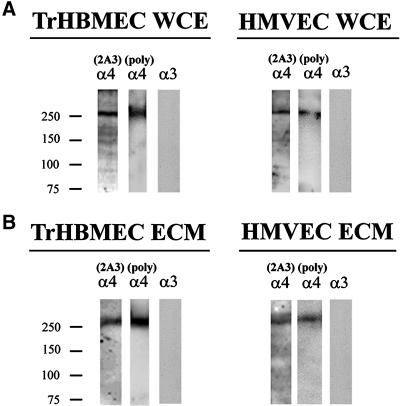
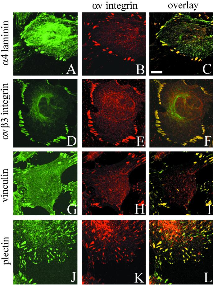
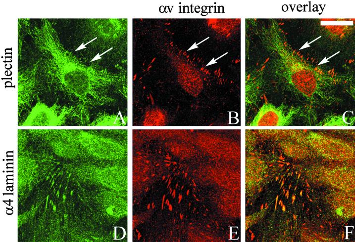
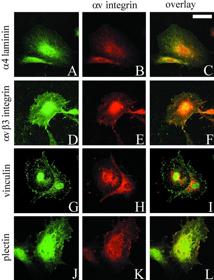
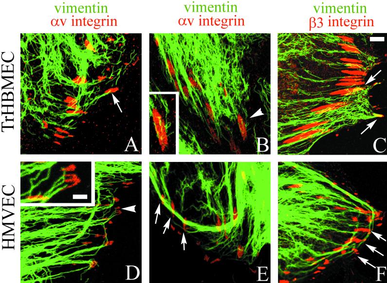
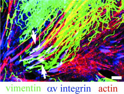
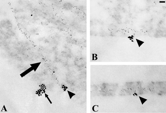
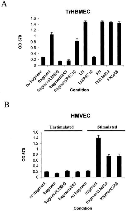
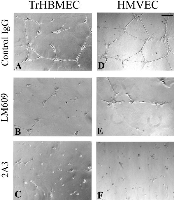
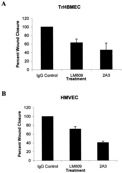
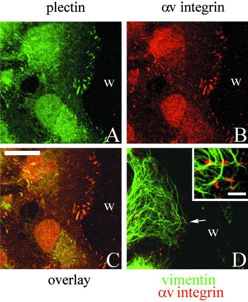
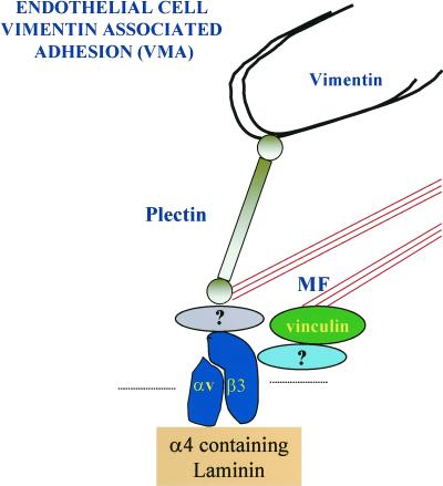
The alpha4 laminin subunit is a component of endothelial cell basement membranes. An antibody (2A3) against the alpha4 laminin G domain stains focal contact-like structures in transformed and primary microvascular endothelial cells (TrHBMECs and HMVECs, respectively), provided the latter cells are activated with growth factors. The 2A3 antibody staining colocalizes with that generated by alphav and beta3 integrin antibodies and, consistent with this localization, TrHBMECs and HMVECs adhere to the alpha4 laminin subunit G domain in an alphavbeta3-integrin-dependent manner. The alphavbeta3 integrin/2A3 antibody positively stained focal contacts are recognized by vinculin antibodies as well as by antibodies against plectin. Unusually, vimentin intermediate filaments, in addition to microfilament bundles, interact with many of the alphavbeta3 integrin-positive focal contacts. We have investigated the function of alpha4-laminin and alphavbeta3-integrin, which are at the core of these focal contacts, in cultured endothelial cells. Antibodies against these proteins inhibit branching morphogenesis of TrHBMECs and HMVECs in vitro, as well as their ability to repopulate in vitro wounds. Thus, we have characterized an endothelial cell matrix adhesion, which shows complex cytoskeletal interactions and whose assembly is regulated by growth factors. Our data indicate that this adhesion structure may play a role in angiogenesis.


Figures

Figure 1
Figure 1
An antibody termed 2A3, against the G-domain of the α4 laminin subunit, was characterized by Western immunoblotting using SDS-PAGE preparations of whole cell extracts (WCE) (A) and matrix (ECM) (B) of TrHBMECs and HMVECs. In the Western blots of the whole cell extract and the matrix preparations, 2A3 antibody primarily reacts with a protein of an approximate molecular mass of 250 kDa. A polyclonal serum against the α4 laminin subunit recognizes a similar sized polypeptide in these preparations. A monoclonal antibody (RG13) against the α3 laminin subunit fails to recognize any polypeptides in either the extract or matrix of either cell type.
Figure 2
Figure 2
TrHBMECs were processed for double-label immunofluorescence using a combination of antibodies against α4 laminin (2A3) and αv integrin subunit (A and B), the αvβ3 integrin complex and the αv-subunit (D and E), vinculin and the αv integrin subunit (G and H), and plectin and the αv integrin subunit (J and K). Cells were viewed by confocal microscopy. The focal plane is close to the substratum-attached surface of the cells. C, F, I, and L are overlays of the fluorescence images. Bar in C, 10 μm.
Figure 3
Figure 3
TrHBMECs were maintained on glass coverslips until the cells reached confluence. Approximately 24 h later the cells were processed for double-label immunofluorescence microscopy using an antibody against plectin (A) or 2A3 antibody against the α4 laminin subunit (D) in combination with an antiserum against αv-integrin (B and E). Cells were viewed by confocal microscopy. The focal plane is close to the substratum-attached surface of the cells. C and F show the overlays of the fluorescence images. Arrows in A–C indicate areas of focal contacts stained by αv-antibodies in B that are not recognized by plectin antibodies in A. Rather, the plectin antibodies in A generate a predominantly filamentous staining pattern. Bar in C, 20 μm.
Figure
Figure
s 4 and 5. HMVECs maintained in medium lacking growth factors (Figure 4) or medium supplemented with bFGF (Figure 5) were processed for double labeling using a combination of antibodies against α4-laminin (2A3) and αv integrin subunit (A and B), the αvβ3 integrin complex and the αv integrin subunit (D and E), vinculin and the αv integrin subunit (G and H), and plectin and the αv integrin subunit (J and K). Cells were viewed by confocal microscopy. The focal plane is close to the substratum-attached surface of the cells. C, F, I, and L are overlays of the fluorescence images. Bar in Figure 4C, 20 μm. Bar in Figure 5C, 10 μm.
Figure
Figure
s 4 and 5. HMVECs maintained in medium lacking growth factors (Figure 4) or medium supplemented with bFGF (Figure 5) were processed for double labeling using a combination of antibodies against α4-laminin (2A3) and αv integrin subunit (A and B), the αvβ3 integrin complex and the αv integrin subunit (D and E), vinculin and the αv integrin subunit (G and H), and plectin and the αv integrin subunit (J and K). Cells were viewed by confocal microscopy. The focal plane is close to the substratum-attached surface of the cells. C, F, I, and L are overlays of the fluorescence images. Bar in Figure 4C, 20 μm. Bar in Figure 5C, 10 μm.
Figure 6
Figure 6
TrHBMECs (A–C) and HMVECs (D–F) were processed for double-label immunofluorescence using a monoclonal antibody against vimentin (green) in combination either with an antiserum against the αv integrin subunit (red) (A, B, D, and E) or an antiserum against β3 integrin (red) (C and F). Overlays of the staining patterns are shown. In TrHBMECs some vimentin filament bundles can be seen to terminate at αv-integrin–containing focal contacts (arrow in A). The focal contact indicated by the arrowhead in B is shown at higher magnification in the inset. Vimentin filaments appear to be wrapped around the focal contact. In HMVECs vimentin bundles are also seen in association with αv-integrin–containing focal contacts (arrowhead and arrows in D and E). The inset in D shows a higher power view of one focal contact (arrowhead) in the HMVEC cell shown in D. Note that three individual vimentin filament bundles appear to terminate at the site of the focal contact. Vimentin filaments and bundles also associate with focal contacts stained by β3 integrin antibodies in both TrHBMECs and HMVECs (arrows in C and F). Bar in C, 5 μm; bar in D (insets), 1 μm.
Figure 7
Figure 7
TrHBMECs were processed for triple-label fluorescence microscopy using antibody probes against vimentin (green) and αv-integrin (blue), together with rhodamine-conjugated phalloidin (red). The overlay of the three labels is shown. White color indicates an overlap in the staining patterns (arrows). Bar, 5 μm.
Figure 8
Figure 8
TrHBMECs were processed for double-label immunogold localization using a mixture of mouse monoclonal antibodies against vimentin and a rabbit antiserum against β3-integrin. The binding of these probes was visualized with 6-nm gold-conjugated goat anti-mouse IgG and 18-nm gold-conjugated goat anti-rabbit IgG. Sections of the cells were prepared either parallel (A) or perpendicular (B and C) to their substratum-attached surface. In A, linear arrays of 6-nm gold particles arise from a cluster of 18-nm gold particles. A second cluster of 18-nm gold particles shows no association with the smaller sized gold particles in A. In B and C, strings of 6-nm particles interact with 18-nm gold clusters along the basal surface of the cells. Bar, 60 nm.
Figure 9
Figure 9
The TrHBMECs and HMVECs attach to a laminin α4-fragment in an αvβ3-integrin–dependent manner. (A) TrHBMECs were plated in a 96-well plate in uncoated wells, wells coated with the α4 laminin subunit fragment, laminin-1 (LN), or fibronectin (FN) in the presence or absence of antibody LM609 against αvβ3, antibody 2A3 against the α4 laminin subunit, P4C10 against the β1-integrin or control IgG. TrHBMECs attach efficiently to the fragment, laminin-1, and to fibronectin within 90 min after plating. Antibodies 2A3 and LM609 inhibit cell attachment to the α4 laminin fragment, whereas the same antibodies do not inhibit cell attachment to fibronectin. P4C10 has a minor inhibitory effect on attachment of TrHBMECs to the α4 chain fragment, whereas the same antibody significantly inhibits attachment to laminin-1. (B) HMVECs were plated onto uncoated wells or wells coated with the recombinant fragment in the presence or absence of growth factors. The attachment of cells was assayed after 120 min. Attachment of HMVECs to the α4 laminin fragment is stimulated by growth factors but can be inhibited by antibody 2A3 and LM609.
Figure 10
Figure 10
TrHBMECs (A–C) and HMVECs (D–F) were plated onto Matrigel, and after 18 h were photographed. In A and D the cells were maintained in the presence of control IgG, and both cell types show assembly into tube-like structures. This morphogenesis is inhibited by LM609 antibodies against αvβ3 (B and E) and antibody 2A3 against the α4 laminin chain (C and F). Bar in D, 100 μm.
Figure 11
Figure 11
Confluent monolayers of TrHBMECs (A–C) and HMVECs (D–F) were wounded by scraping with a pipette-man tip. The cultures were allowed to recover for 18 h in the presence of control IgG (A and D), antibody LM609 against αvβ3-integrin (B and E), and antibody 2A3 against the α4 laminin subunit (C and F). The cells in A and D have completely covered the wound site, whereas in B, C, E, and F the wound sites (w) are incompletely closed (as indicated by the line). Bar in D, 100 μm.
Figure 12
Figure 12
The extent of wound closure in the experiments in Figure 11 was quantitated. Wounds were photographed 0 and 18 h after wounding. For each trial 40 measurements across the wound at distances at least 100 μm apart were made 18 h after wounding. These measurements were used to determine the average wound closure (average width of the wound at 0 h minus average width of the wound at 18 h). This is presented as a percentage of the average width of the wound at 0 h.
Figure 13
Figure 13
A confluent monolayer of TrHBMECs was wounded by scraping with a pipette-man tip. The culture was allowed to recover for 4 h, at which time cells had begun to migrate into the wound area. The preparation was then processed for double-label immunofluorescence microscopy using antibodies against plectin (A) together with an antiserum against αv-integrin (B). Cells were viewed by confocal microscopy. The focal plane is close to the substratum-attached surface of the cells. C is the overlay of the two fluorescence images. Preparations were also made of wounded cultures for double-label immunofluorescence using the antiserum against the αv-integrin (red) combined with a monoclonal vimentin antibody (green). The overlay of the two images is shown in D. Inset in D shows a higher power view of several αv-integrin positively stained focal contacts with which vimentin interacts in a cell that has migrated into the wound site (w). Bar in C, 20 μm; bar in D, inset, 3 μm.
Figure 14
Figure 14
This diagram shows the molecular components of the VMA we have begun to characterize. The αvβ3-integrin associates with a laminin subunit at the core of the structure. We speculate that vimentin-type intermediate filaments interact with the VMA via an association with plectin (Wiche et al., 1982; Steinböck et al., 2000). Because plectin possesses an actin-binding motif, plectin, together with focal contact proteins such as vinculin, may be involved in mediating microfilament (MF) bundle-cell surface association (Elliot et al., 1997). The protein that links vinculin to the αvβ3-integrin may be α-actinin (Simon and Burridge, 1994), whereas the identity of the protein that links plectin to αvβ3 is yet to be determined.

Similar articles

Cited by

References

    1. Aumailley M, Smyth N. The role of laminins in basement membrane function. J Anat. 1998;193:1–21. - PMC - PubMed
    1. Babic AM, Chen C-C, Lau LF. Fisp12/mouse connective tissue growth factor mediates endothelial cell adhesion and migration through integrin αvβ3, promotes endothelial cell survival, and induces angiogenesis in vivo. Mol Cell Biol. 1999;19:2958–2966. - PMC - PubMed
    1. Baker SE, Hopkinson SB, Fitchmun M, Andreason GL, Frasier F, Plopper G, Quaranta V, Jones JCR. Laminin-5 and hemidesmosomes: role of the alpha 3 chain subunit in hemidesmosome stability and assembly. J Cell Sci. 1996;109:2509–2520. - PubMed
    1. Bershadsky AD, Tint IS, Svitkina TM. Association of intermediate filaments with vinculin-containing adhesion plaques of fibroblasts. Cell Motil Cytoskeleton. 1987;8:274–283. - PubMed
    1. Brooks PC, Clark RAF, Cheresh DA. Requirement of vascular integrin αvβ3 for angiogenesis. Science. 1994a;264:569–571. - PubMed

Publication types

MeSH terms

LinkOut - more resources