. 2001 Feb;107(3):295-304.
doi: 10.1172/JCI11706.

Indian hedgehog couples chondrogenesis to osteogenesis in endochondral bone development

Affiliations

Indian hedgehog couples chondrogenesis to osteogenesis in endochondral bone development

U I Chung et al. J Clin Invest. 2001 Feb.

Abstract

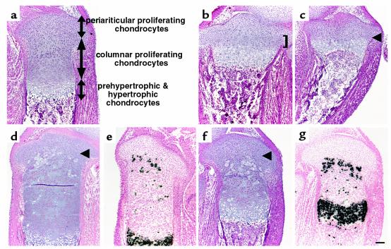
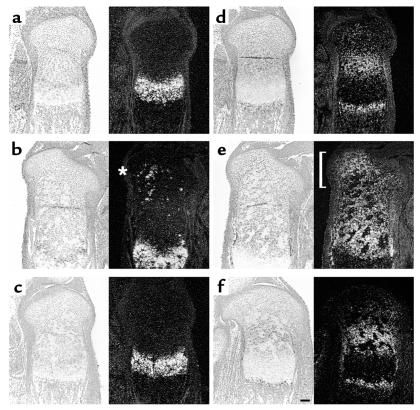
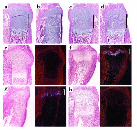
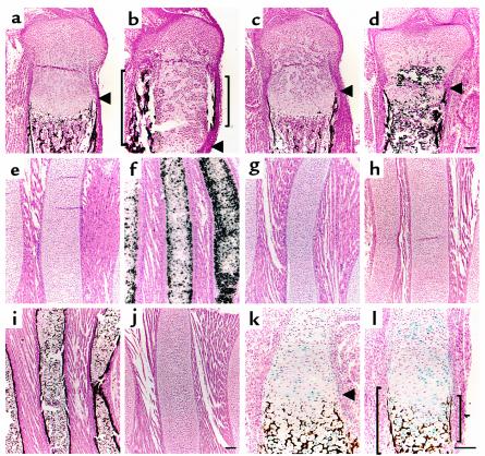
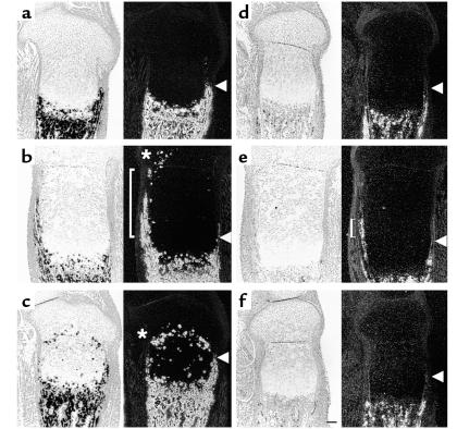
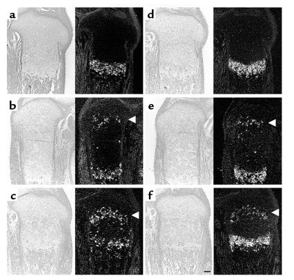
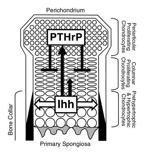
Vertebrate skeletogenesis requires a well-coordinated transition from chondrogenesis to osteogenesis. Hypertrophic chondrocytes in the growth plate play a pivotal role in this transition. Parathyroid hormone-related peptide (PTHrP), synthesized in the periarticular growth plate, regulates the site at which hypertrophy occurs. By comparing PTH/PTHrP receptor(-/-)/wild-type (PPR(-/-)/wild-type) chimeric mice with IHH(-/-);PPR(-/-)/wild-type chimeric and IHH(-/-)/wild-type chimeric mice, we provide in vivo evidence that Indian hedgehog (IHH), synthesized by prehypertrophic and hypertrophic chondrocytes, regulates the site of hypertrophic differentiation by signaling to the periarticular growth plate and also determines the site of bone collar formation in the adjacent perichondrium. By providing crucial local signals from prehypertrophic and hypertrophic chondrocytes to both chondrocytes and preosteoblasts, IHH couples chondrogenesis to osteogenesis in endochondral bone development.


Figures

Figure 1
Figure 1
Ectopic differentiation of growth plate chondrocytes deficient in PTHrP signaling. (ac) Sections of the tibiae from d17.5 wild-type (a), PTHrP–/– (b), and PPR–/– (c) embryos were stained with H&E. The growth plates of PTHrP–/– mice and PPR–/– mice have a shorter layer of columnar proliferating chondrocytes. In PTHrP–/– mice, they represent less than 30% of wild-type (b, bracket), and in PPR–/– mice, they are almost completely absent (c, arrowhead). (dg) H&E staining and in situ hybridization with a mouse type X collagen antisense probe of the sections of the tibiae from d17.5 PPR–/–/wild-type chimera (d and e, respectively) and Ihh–/–;PPR–/–/wild-type chimera (f and g, respectively) embryos. In the absence of PTHrP signaling, mutant cells ectopically hypertrophy when they move from the layer of periarticular proliferating chondrocytes into the layer of columnar proliferating chondrocytes (arrowheads). Staining for β-galactosidase activity of both chimeric growth plates shows that all the mutant chondrocytes undergo ectopic hypertrophy, whereas wild-type cells do not (data not shown). Horizontal bar = 100 μm.
Figure 2
Figure 2
Ihh signaling by ectopically hypertrophied PPR–/– and Ihh–/–;PPR–/– chondrocytes. (ac) In situ hybridization of the sections of the tibiae from d17.5 embryos with a mouse Ihh antisense probe. In the wild-type growth plate (a), Ihh mRNA is expressed in prehypertrophic and hypertrophic chondrocytes. In the chimeric growth plates, ectopically differentiated PPR–/– chondrocytes also express Ihh mRNA (b, asterisk), whereas ectopically differentiated Ihh–/–;PPR–/– cells do not (c). (df) In situ hybridization of the sections of the tibiae from d17.5 embryos with a mouse Ptc1 antisense probe. Ptc1 is a transcriptional target of Ihh signaling. In the wild-type growth plate (d), Ptc1 mRNA is expressed most strongly in columnar proliferating chondrocytes adjacent to prehypertrophic chondrocytes with the expression decreasing toward the end of bone. Ptc1 mRNA is also expressed in the perichondrium and the primary spongiosa. In the PPR–/–/wild-type chimeric growth plate (e), periarticular and columnar proliferating chondrocytes surrounding ectopic prehypertrophic/hypertrophic chondrocytes overexpress Ptc1 mRNA (bracket), reflecting ectopic actions of Ihh. In contrast, in the Ihh–/–;PPR–/–/wild-type chimeric growth plate (f), there is no ectopic expression of Ptc1 mRNA despite the presence of ectopic prehypertrophic/hypertrophic chondrocytes. Left, bright field; right, dark field. Horizontal bar = 100 μm.
Figure 3
Figure 3
Ihh signals the position of prehypertrophic and hypertrophic chondrocytes. (ad) H&E staining of sections of the tibiae from d17.5 wild-type (a), PPR–/–/wild-type chimera (b), and Ihh–/–;PPR–/–/wild-type chimera (c and d) embryos. The presence of ectopic hypertrophic chondrocytes in the PPR–/–/wild-type chimeric growth plate induces elongation of the layer of wild-type columnar proliferating chondrocytes, leading to greater distance between the layers of periarticular proliferating chondrocytes and prehypertrophic/hypertrophic chondrocytes (compare a and b). In the Ihh–/–;PPR–/–/wild-type chimeric growth plate, however, the presence of ectopic hypertrophic chondrocytes does not induce elongation of the layer of wild-type columnar proliferating chondrocytes (c and d, in increasing order of chimerism). Perpendicular bars indicate the length of the layer of columnar proliferating chondrocytes. (eh) In situ hybridization of sections of the tibiae from d17.5 embryos with a mouse PTHrP antisense probe. In the wild-type growth plate (e), PTHrP mRNA is weakly expressed in the periarticular proliferating chondrocytes, whereas in the PPR–/– growth plate (f), its expression is strongly upregulated in the same area (bracket). In the PPR–/–/wild-type chimeric growth plate (g), PTHrP mRNA expression is upregulated in the periarticular proliferating chondrocytes, but to a lesser extent compared with the PPR–/– growth plate (bracket). In contrast, despite the presence of ectopic hypertrophic chondrocytes, there is no upregulation of PTHrP mRNA expression in the Ihh–/–;PPR–/–/wild-type chimeric growth plate (h). PTHrP mRNA expression is more clearly seen in sections of the wild-type and Ihh–/–;PPR–/–/wild-type chimeric growth plates exposed twice as long (data not shown); the sections shown in e and h, however, were exposed for the same time as those in f and g to allow direct comparison. Left, bright field; right, dark field. Horizontal bar = 100 μm.
Figure 4
Figure 4
Ihh determines the location of bone collar formation. (ad) Sections of the tibiae from d17.5 embryos were stained by von Kossa method and counterstained with Nuclear Fast Red. Mineral is stained black; nuclei are stained red. In the wild-type growth plate (a), bone collars are formed in the perichondrium adjacent to the layers of prehypertrophic and hypertrophic chondrocytes, whereas in the PPR–/–/wild-type chimeric growth plate (b), ectopic bone collars are induced in the perichondrium adjacent to a cluster of ectopic hypertrophic chondrocytes (brackets). Despite the presence of clusters of ectopic hypertrophic chondrocytes, no ectopic bone collar is induced in the Ihh–/–;PPR–/–/wild-type chimeric growth plate (c and d). Arrowheads denote where the eutopic prehypertrophic layer starts. Calcification inside the growth plate (d, asterisk) is ectopic cartilaginous mineralization caused by mutant hypertrophic chondrocytes. (eg) In situ hybridization of sections of the anterior portion of the ribs from newborn mice with a mouse type X collagen antisense probe. This portion of the wild-type ribs consists of proliferating chondrocytes (e), whereas chondrocytes in the PTHrP–/– ribs ectopically hypertrophy and express type X collagen mRNA (f). Introduction of a constitutively active PPR transgene driven by the type II collagen promoter (caPPR) reverses this ectopic hypertrophy (g). (hj) Sections of the anterior portion of the ribs from newborn mice were stained by von Kossa method and counterstained with Nuclear Fast Red. Mineral is stained black; nuclei are stained red. The wild-type ribs have no bone collars (h), whereas the PTHrP–/– ribs have bone collars in association with the presence of ectopic hypertrophic chondrocytes (i). The PTHrP–/–;caPPR ribs, however, do not have bone collars, in association with the disappearance of ectopic hypertrophic chondrocytes (j). (k and l) Sections of the humeri from d17.5 Ihh–/–/wild-type chimera embryos were stained for β-galactosidase activity as well as stained by von Kossa method and counterstained with Nuclear Fast Red. Wild-type cells are stained blue; mineral is stained black; nuclei are stained red. In these mice, bone collars do not form in the perichondrium adjacent to clusters of Ihh–/– prehypertrophic/hypertrophic chondrocytes (arrowhead denotes where the prehypertrophic layer starts), whereas bone collars do form in the perichondrium adjacent to clusters of wild-type prehypertrophic/hypertrophic chondrocytes (brackets). Horizontal bar = 100 μm.
Figure 5
Figure 5
Expression of osteoblastic markers in the chimeric growth plates. (ac) In situ hybridization of sections of the tibiae from d17.5 embryos with a mouse osteopontin antisense probe. In the wild-type growth plate (a), osteopontin mRNA is expressed in osteoblasts as well as in mature chondrocytes, and its expression pattern overlaps the locations of bone collars and cartilaginous mineralization. In the PPR–/–/wild-type chimera growth plate (b), there is ectopic expression of osteopontin mRNA in ectopic bone collars (bracket). Ectopic hypertrophic chondrocytes also express osteopontin mRNA (asterisk). In contrast, in the Ihh–/–;PTH/PPR–/–/wild-type chimera (c), there is no ectopic expression of osteopontin mRNA in the perichondrium, whereas ectopic hypertrophic chondrocytes still express osteopontin mRNA (asterisk). Arrowheads denote where the eutopic prehypertrophic layer starts. (d–f) In situ hybridization of sections of the tibiae from d17.5 embryos with a mouse osteocalcin antisense probe. In the wild-type growth plate (d), osteocalcin mRNA is expressed in mature osteoblasts. In the PPR–/–/wild-type chimera (e), there is ectopic expression of osteocalcin mRNA in ectopic bone collars (bracket), whereas in the Ihh–/–;PPR–/–/wild-type chimera (f), there is no ectopic expression of osteocalcin mRNA in the perichondrium. Arrowheads denote where the eutopic prehypertrophic layer starts. Horizontal bar = 100 μm.
Figure 6
Figure 6
Bmp signaling alone is not sufficient for induction of bone collars. (ac) In situ hybridization of sections of the tibiae from d17.5 embryos with a mouse Bmp2 antisense probe. In the wild-type growth plate (a), Bmp2 mRNA is expressed in hypertrophic chondrocytes and the perichondrium, whereas both in the PPR–/–/wild-type chimeric growth plate (b) and in the Ihh–/–;PPR–/–/wild-type chimeric growth plate (c), ectopic hypertrophic mutant cells also express Bmp2 mRNA (arrowheads). (df) In situ hybridization of sections of the tibiae from d17.5 embryos with a mouse Bmp6 antisense probe. In the wild-type growth plate (d), Bmp6 mRNA is expressed in hypertrophic chondrocytes, whereas both in the PPR–/–/wild-type chimeric growth plate (e) and in the Ihh–/–;PPR–/–/wild-type chimeric growth plate (f), ectopic hypertrophic mutant cells also express Bmp6 mRNA (arrowheads). Horizontal bar = 100 μm.
Figure 7
Figure 7
Interactions of Ihh and PTHrP signaling pathways in the fetal growth plate. This figure shows a schema of the fetal growth plate of the mouse long bone. The end of the bone is at the top. As proliferating chondrocytes differentiate into prehypertrophic chondrocytes and then hypertrophic chondrocytes, they express Ihh. Ihh directly or indirectly stimulates PTHrP synthesis in the periarticular growth plate including the perichondrium and periarticular proliferating chondrocytes (long solid arrow). In this way, Ihh signals the relative position of prehypertrophic and hypertrophic chondrocytes to the periarticular growth plate. Ihh also has a positive effect on chondrocyte proliferation (short solid arrow). PTHrP then acts directly on columnar proliferating chondrocytes to delay their differentiation into prehypertrophic and hypertrophic chondrocytes, which synthesize Ihh (solid t-bar). Thus, through PTHrP, Ihh controls the site at which hypertrophy occurs. Ihh also acts on perichondrial cells to induce mature osteoblasts, which form a bone collar (white arrow).

Comment in

Similar articles

Cited by

References

    1. Erlebacher A, Filvaroff EH, Gitelman SE, Derynck R. Toward a molecular understanding of skeletal development. Cell. 1995; 80:371–378. - PubMed
    1. Marks, S.C., and Hermey, D.C. 1996. The structure and development of bone. In Principles of bone biology. J.P. Bilezikian, L.G. Raisz, and G.A. Rodan, editors. Academic Press. San Diego, California, USA. 3–14.
    1. Chung UI, Lanske B, Lee K, Li E, Kronenberg H. The parathyroid hormone/parathyroid hormone-related peptide receptor coordinates endochondral bone development by directly controlling chondrocyte differentiation. Proc Natl Acad Sci USA. 1998; 95:13030–13035. - PMC - PubMed
    1. Lee K, Deeds JD, Segre GV. Expression of parathyroid hormone-related peptide and its receptor messenger ribonucleic acids during fetal development of rats. Endocrinology. 1995; 136:453–463. - PubMed
    1. Karaplis AC, et al. Lethal skeletal dysplasia from targeted disruption of the parathyroid hormone-related peptide gene. Genes Dev. 1994; 8:277–289. - PubMed

Publication types

MeSH terms