. 2001 Feb 5;152(3):471-82.
doi: 10.1083/jcb.152.3.471.

A novel neural Wiskott-Aldrich syndrome protein (N-WASP) binding protein, WISH, induces Arp2/3 complex activation independent of Cdc42

Affiliations

A novel neural Wiskott-Aldrich syndrome protein (N-WASP) binding protein, WISH, induces Arp2/3 complex activation independent of Cdc42

M Fukuoka et al. J Cell Biol. .

Abstract

We identified a novel adaptor protein that contains a Src homology (SH)3 domain, SH3 binding proline-rich sequences, and a leucine zipper-like motif and termed this protein WASP interacting SH3 protein (WISH). WISH is expressed predominantly in neural tissues and testis. It bound Ash/Grb2 through its proline-rich regions and neural Wiskott-Aldrich syndrome protein (N-WASP) through its SH3 domain. WISH strongly enhanced N-WASP-induced Arp2/3 complex activation independent of Cdc42 in vitro, resulting in rapid actin polymerization. Furthermore, coexpression of WISH and N-WASP induced marked formation of microspikes in Cos7 cells, even in the absence of stimuli. An N-WASP mutant (H208D) that cannot bind Cdc42 still induced microspike formation when coexpressed with WISH. We also examined the contribution of WISH to a rapid actin polymerization induced by brain extract in vitro. Arp2/3 complex was essential for brain extract-induced rapid actin polymerization. Addition of WISH to extracts increased actin polymerization as Cdc42 did. However, WISH unexpectedly could activate actin polymerization even in N-WASP-depleted extracts. These findings suggest that WISH activates Arp2/3 complex through N-WASP-dependent and -independent pathways without Cdc42, resulting in the rapid actin polymerization required for microspike formation.


Figures

Figure 1
Figure 1
Amino acid sequence of a novel N-WASP binding protein, WISH. (A) Sequence of WISH. The SH3 domain, proline-rich sequence, and leucine-rich sequences are boxed. The serine-rich sequence is underlined. The heptad repeat of hydrophobic residues in the leucine zipper-like motif is denoted by white-on-black. (B) Schematic structure of WISH. (C) Western blot analysis of ectopically expressed WISH and endogenous WISH. Western blot analyses were performed using cell lysates of Cos7 cells transfected with empty vector (vec) or WISH-expressing plasmid (ectopically expressed WISH) and rat brain. WISH (90 kD) is indicated by the arrow.
Figure 6
Figure 6
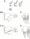
Bovine brain extract–induced rapid actin polymerization in vitro. Aliquots of the bovine brain extracts were treated with anti-WISH antibody, anti–N-WASP antibody, or GST-CA protein to deplete WISH, N-WASP, and Arp2/3 complex, respectively. As a control, mock-treated extracts were used. Actin polymerization activity was measured using pyrene-labeled actin in a total volume of 100 μl. In all assay systems, 100 μM GTPγS was added. When WISH and Cdc42 proteins were added, 4.5 μg of WISH and the equal amounts of WISH SH3, Fyn SH3, or 2 μg of Cdc42 were used. Results shown are means of triplicate measurements. (A) Western blots of WISH-depleted (WISH dep.), N-WASP–depleted (NW dep.), and Arp2/3 complex–depleted (Arp2/3 dep.) extracts. Degree of depletion was checked by scanning Western blots with a densitometer as described in the text. (B) Mock treatment. Mock-treated extract (MOCK), mock-treated extracts + 0.2 μM WISH (MOCK + WISH full), mock-treated extracts + 0.2 μM WISH SH3 (MOCK + WISH SH3), and mock-treated extracts + 0.2 μM Fyn SH3 (MOCK + Fyn SH3). Ba, Effect of Cdc42, WISH, WISH SH3, and Fyn SH3 on actin polymerization was examined using mock-treated bovine brain extracts. Bb, Initial rates of actin polymerization. Results shown are the ratio to the mock. Mean values from three experiments are indicated. Error bars represent SD. Asterisks indicate significant difference by Student's t test (P < 0.01) from mock-treated extracts (*). (C) Arp2/3 complex is essential for a rapid actin polymerization. Mock-treated extract (MOCK), mock-treated extracts + 0.2 μM WISH (MOCK + WISH full), mock-treated extracts + 0.5 μM Cdc42 (MOCK + Cdc42), Arp2/3 complex–depleted extracts (Arp2/3 dep.), Arp2/3 complex–depleted extracts + 0.5 μM Cdc42 (Arp2/3 dep. + Cdc42), and Arp2/3 complex–depleted extracts + WISH (Arp dep. + WISH full). Ca, actin polymerization curves; Cb, initial rates of actin polymerization. Results shown are the ratio to the mock. Mean values from three experiments are indicated. Error bars represent SD. Asterisks indicate significant difference by Student's t test (P < 0.01) from mock-treated extracts (*) and Arp2/3 complex–depleted extracts (**). (D) WISH depletion experiments. Mock-treated extracts (MOCK), mock-treated extracts + 0.2 μM WISH (MOCK + WISH), WISH-depleted extracts (WISH dep.), and WISH-depleted extracts + 0.2 μM WISH addback (WISH dep. + WISH full). Da, actin polymerization curves in WISH-depleted extracts; Db, initial rates of actin polymerization in WISH-depleted extracts. Results shown are the ratio to the mock. Mean values from three experiments are indicated. Error bars represent SD. Asterisks indicate significant difference by Student's t test (P < 0.01) from mock-treated extracts (*) and WISH-depleted extracts (**). (E) WISH activates Arp2/3 complex in N-WASP–depleted extracts. Mock-treated extracts (MOCK), N-WASP–depleted extracts (NW dep.), N-WASP–depleted extracts + 0.2 μM WISH (NW dep. + WISH full), N-WASP–depleted extracts + 0.5 μM Cdc42 (NW dep. + Cdc42), N-WASP–depleted extracts + 0.2 μM WISH SH3 (NW dep. + WISH SH3), and N-WASP–depleted extracts + Ash/Grb2 (NW dep. + Ash/Grb2). Ea, actin polymerization curves in N-WASP–depleted extracts; Eb, initial rates of actin polymerization in N-WASP–depleted extracts. Results shown are the ratio to the mock. Mean values from three experiments are indicated. Error bars represent SD. Asterisks indicate significant difference by Student's t test (P < 0.01) from mock-treated extracts (*) and N-WASP–depleted extracts (**).
Figure 6
Figure 6
Bovine brain extract–induced rapid actin polymerization in vitro. Aliquots of the bovine brain extracts were treated with anti-WISH antibody, anti–N-WASP antibody, or GST-CA protein to deplete WISH, N-WASP, and Arp2/3 complex, respectively. As a control, mock-treated extracts were used. Actin polymerization activity was measured using pyrene-labeled actin in a total volume of 100 μl. In all assay systems, 100 μM GTPγS was added. When WISH and Cdc42 proteins were added, 4.5 μg of WISH and the equal amounts of WISH SH3, Fyn SH3, or 2 μg of Cdc42 were used. Results shown are means of triplicate measurements. (A) Western blots of WISH-depleted (WISH dep.), N-WASP–depleted (NW dep.), and Arp2/3 complex–depleted (Arp2/3 dep.) extracts. Degree of depletion was checked by scanning Western blots with a densitometer as described in the text. (B) Mock treatment. Mock-treated extract (MOCK), mock-treated extracts + 0.2 μM WISH (MOCK + WISH full), mock-treated extracts + 0.2 μM WISH SH3 (MOCK + WISH SH3), and mock-treated extracts + 0.2 μM Fyn SH3 (MOCK + Fyn SH3). Ba, Effect of Cdc42, WISH, WISH SH3, and Fyn SH3 on actin polymerization was examined using mock-treated bovine brain extracts. Bb, Initial rates of actin polymerization. Results shown are the ratio to the mock. Mean values from three experiments are indicated. Error bars represent SD. Asterisks indicate significant difference by Student's t test (P < 0.01) from mock-treated extracts (*). (C) Arp2/3 complex is essential for a rapid actin polymerization. Mock-treated extract (MOCK), mock-treated extracts + 0.2 μM WISH (MOCK + WISH full), mock-treated extracts + 0.5 μM Cdc42 (MOCK + Cdc42), Arp2/3 complex–depleted extracts (Arp2/3 dep.), Arp2/3 complex–depleted extracts + 0.5 μM Cdc42 (Arp2/3 dep. + Cdc42), and Arp2/3 complex–depleted extracts + WISH (Arp dep. + WISH full). Ca, actin polymerization curves; Cb, initial rates of actin polymerization. Results shown are the ratio to the mock. Mean values from three experiments are indicated. Error bars represent SD. Asterisks indicate significant difference by Student's t test (P < 0.01) from mock-treated extracts (*) and Arp2/3 complex–depleted extracts (**). (D) WISH depletion experiments. Mock-treated extracts (MOCK), mock-treated extracts + 0.2 μM WISH (MOCK + WISH), WISH-depleted extracts (WISH dep.), and WISH-depleted extracts + 0.2 μM WISH addback (WISH dep. + WISH full). Da, actin polymerization curves in WISH-depleted extracts; Db, initial rates of actin polymerization in WISH-depleted extracts. Results shown are the ratio to the mock. Mean values from three experiments are indicated. Error bars represent SD. Asterisks indicate significant difference by Student's t test (P < 0.01) from mock-treated extracts (*) and WISH-depleted extracts (**). (E) WISH activates Arp2/3 complex in N-WASP–depleted extracts. Mock-treated extracts (MOCK), N-WASP–depleted extracts (NW dep.), N-WASP–depleted extracts + 0.2 μM WISH (NW dep. + WISH full), N-WASP–depleted extracts + 0.5 μM Cdc42 (NW dep. + Cdc42), N-WASP–depleted extracts + 0.2 μM WISH SH3 (NW dep. + WISH SH3), and N-WASP–depleted extracts + Ash/Grb2 (NW dep. + Ash/Grb2). Ea, actin polymerization curves in N-WASP–depleted extracts; Eb, initial rates of actin polymerization in N-WASP–depleted extracts. Results shown are the ratio to the mock. Mean values from three experiments are indicated. Error bars represent SD. Asterisks indicate significant difference by Student's t test (P < 0.01) from mock-treated extracts (*) and N-WASP–depleted extracts (**).
Figure 2
Figure 2
Tissue distribution of WISH mRNA and protein. (A) Northern hybridization analysis of WISH mRNA. The WISH mRNA (2.8 kb) is indicated by the arrow. (B) Western blot analysis with anti-WISH antibody. Various rat tissues were homogenized and cleared by ultracentrifugation. WISH (90 kD) is indicated by the arrow.
Figure 3
Figure 3
Association of WISH with Ash/Grb2. (A) Ectopically expressed WISH associates with Ash/Grb2 in vivo. Myc-tagged full-length WISH expressing plasmid (Myc-WISH) and empty vector (Myc-vec) alone were transfected into Cos7 cells. Anti-Myc immunoprecipitates (left) and whole cell lysates (right) from these cells were immunoblotted with anti-Myc antibody (top) or anti-Ash/Grb2 antibody (bottom). (B) Endogenous WISH associates with Ash/Grb2 in vivo. N1E-115 cell lysates were immunoprecipitated with preimmune serum (Pre) or anti-WISH antibody (α-WISH). The immunoprecipitates (I.P.) and whole cell lysates were immunoblotted with anti-WISH antibody (top) or anti-Ash/Grb2 antibody (bottom). (C) In vitro association of WISH with Ash/Grb2. Binding of the His-tagged WISH proline-rich sequence (His-Pro) was investigated. GST fusion proteins of full-length Ash/Grb2 (GST-Ash), the NH2-terminal SH3 domain of Ash/Grb2 (GST-AshN), and the COOH-terminal SH3 of Ash/Grb2 (GST-AshC) were immobilized on glutathione-agarose beads and incubated with (right) or without (left) His-Pro. Bound proteins were analyzed by Western blotting with anti–His-tag antibody (α-His).
Figure 4
Figure 4
Association of WISH with N-WASP. (A) WISH binding proteins. Cell lysates from [35S]methionine-prelabeled N1E-115 cells were immunoprecipitated with anti-WISH antibody (α-WISH) or preimmune rabbit serum (Pre). The precipitates (I.P.) were subjected to SDS-PAGE and autoradiography. (B) Association of WISH with proline-rich proteins. N1E-115 cell lysates were immunoprecipitated as described in A. The immunoprecipitates and whole cell lysates were immunoblotted with anti-WISH antibody, anti-Sos antibody, anti–c-Cbl antibody, antisynaptojanin antibody (α-S.J.), anti-WAVE antibody, and anti–N-WASP antibody. (C) Various deletion constructs of WISH GST fusion proteins: full-length (Full), SH3 domain (SH3), proline-rich sequence (Pro), middle region (Mid), leucine-rich region (Leu), and COOH-terminal region (C). (D) WISH associates with N-WASP through its SH3 domain. The ability to bind GST fusion proteins of various deletion constructions of WISH was examined. These proteins were immobilized on glutathione-agarose beads and mixed with N-WASP protein. Bound proteins were immunoblotted with anti–N-WASP antibody. (E) WISH SH3 domain binds to the proline-rich region of N-WASP. GST fusion protein of the N-WASP proline-rich region (GST–NW-Pro) was immobilized on glutathione-agarose beads and mixed with His-tagged WISH SH3 (His-WISH–SH3). Bound proteins were analyzed by Western blotting with anti–His-tag antibody (α-His).
Figure 5
Figure 5
WISH stimulates N-WASP–induced Arp2/3 complex activation. (A) Proteins used in actin polymerization assays. Purified proteins were subjected to SDS-PAGE and stained with Coomassie blue. (B) Effect of WISH on N-WASP–dependent Arp2/3 complex–induced actin polymerization. The pyrene-labeled actin assay was used to monitor the polymerization of 2.2 μM G-actin (2 μM unlabeled actin + 0.2 μM pyrene-labeled actin) in the presence of 60 nM Arp2/3 complex alone (actin+Arp2/3), Arp2/3 complex + 0.2 μM GST-VCA (VCA), Arp2/3 complex + 0.2 μM N-WASP (NW), Arp2/3 complex + N-WASP + 0.5 μM Cdc42 (NW+Cdc42), Arp2/3 complex + 0.2 μM WISH (WISH), Arp2/3 complex + N-WASP + WISH (NW+WISH), or Arp2/3 complex + N-WASP + WISH + Cdc42 (NW+WISH+Cdc42). Experiments were repeated three times. Results shown are means of triplicate measurements. (C) Effect of various SH3 domains on N-WASP–induced Arp2/3 complex activation. The pyrene-labeled actin assay was carried out with 0.2 μM WISH, Ash/Grb2 (Ash), myelin basic protein (MBP), or SH3s of WISH (SH3), Nck, Ash/Grb2 NH2 terminus (AshN), PI 3-kinase 85-kD subunit (p85), PLCγ1 (PLCγ), or Fyn in the presence of 60 nM Arp2/3 complex + 0.2 μM N-WASP (NW). Results shown are means of triplicate measurements. (D) Concentration-dependent effect on initial actin polymerization rate. Effects of Cdc42, WISH, WISH SH3 (SH3), Ash/Grb2 (Ash), Ash/Grb2 NH2-terminal SH3 (AshN), and Fyn SH3 (Fyn) in the presence of 60 nM Arp2/3 complex + 0.2 μM N-WASP (NW). Initial rates of actin polymerization were calculated by differentiating the plot curves. Error bars represent the standard deviation of three different experiments.
Figure 7
Figure 7
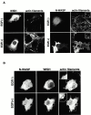
WISH enhances N-WASP–induced microspike formation in vivo. (A) Microspike formation in Cos7 cells expressing N-WASP or Myc-WISH. Transfected cells were treated with or without EGF for 5 min, and then fixed, immunostained with anti–N-WASP (N-WASP) or anti-Myc (WISH) antibodies, and stained with rhodamine-phalloidin (actin filaments). (B) Microspike formation in Cos7 cells expressing N-WASP and Myc-WISH. Transfected cells were treated with or without EGF for 5 min, and then fixed, immunostained with anti–N-WASP (N-WASP) and anti-Myc (WISH) antibodies, and stained with rhodamine-phalloidin (actin filaments). (C) Activation of N-WASP by WISH is independent of Cdc42. Cos7 cells expressing the N-WASP mutant H208D only or together with Myc-WISH were observed for microspike formation. Cells were incubated with or without EGF for 5 min, and then fixed, immunostained with anti–N-WASP (H208D) and anti-Myc (WISH) antibodies, and stained with rhodamine-phalloidin (actin filaments). (D) Real-time observation of microspike formation. Cos7 cells were microinjected with purified N-WASP and/or GST-WISH proteins. After microinjection, cells were observed with a phase–contrast microscope. Membrane protrusions (filopodia) are indicated with arrows. (E) Quantification of microspike formation. Transfected cells were serum-starved and then stimulated with or without EGF for 5 min. The percentage of cells forming microspikes among transfected cells was calculated. Error bars represent the SD of three different measurements. At least 50 cells were counted in each determination.
Figure 7
Figure 7
WISH enhances N-WASP–induced microspike formation in vivo. (A) Microspike formation in Cos7 cells expressing N-WASP or Myc-WISH. Transfected cells were treated with or without EGF for 5 min, and then fixed, immunostained with anti–N-WASP (N-WASP) or anti-Myc (WISH) antibodies, and stained with rhodamine-phalloidin (actin filaments). (B) Microspike formation in Cos7 cells expressing N-WASP and Myc-WISH. Transfected cells were treated with or without EGF for 5 min, and then fixed, immunostained with anti–N-WASP (N-WASP) and anti-Myc (WISH) antibodies, and stained with rhodamine-phalloidin (actin filaments). (C) Activation of N-WASP by WISH is independent of Cdc42. Cos7 cells expressing the N-WASP mutant H208D only or together with Myc-WISH were observed for microspike formation. Cells were incubated with or without EGF for 5 min, and then fixed, immunostained with anti–N-WASP (H208D) and anti-Myc (WISH) antibodies, and stained with rhodamine-phalloidin (actin filaments). (D) Real-time observation of microspike formation. Cos7 cells were microinjected with purified N-WASP and/or GST-WISH proteins. After microinjection, cells were observed with a phase–contrast microscope. Membrane protrusions (filopodia) are indicated with arrows. (E) Quantification of microspike formation. Transfected cells were serum-starved and then stimulated with or without EGF for 5 min. The percentage of cells forming microspikes among transfected cells was calculated. Error bars represent the SD of three different measurements. At least 50 cells were counted in each determination.
Figure 8
Figure 8
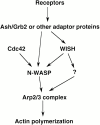
Possible signalings of WISH to Arp2/3 complex activation. When cells are stimulated, an adaptor protein, such as WISH, is recruited to the plasma membrane. At the membrane, WISH transmits signals to Arp2/3 complex through both N-WASP–dependent and –independent pathways. Cdc42 is not required in either pathway.

Similar articles

Cited by

References

    1. Arvidsson A.K., Rupp E., Nanberg E., Downward J., Ronnstrand L., Wennstrom S., Schlessinger J., Heldin C.H., Claesson-Welsh L. Tyr-716 in the platelet-derived growth factor beta-receptor kinase insert is involved in GRB2 binding and Ras activation. Mol. Cell. Biol. 1994;10:6715–6726. - PMC - PubMed
    1. Baltensperger K., Kozma L.M., Cherniack A.D., Klarlund J.K., Chawla A., Banerjee U., Czech M.P. Binding of the Ras activator son of sevenless to insulin receptor substrate-1 signaling complexes. Science. 1993;260:1950–1952. - PubMed
    1. Carlier M.F., Nioche P., Broutin-L'Hermite I., Boujemaa R., Le Clainche R.C., Egile C., Garbay C., Ducruix A., Sansonetti P., Pantaloni D. GRB2 links signaling to actin assembly by enhancing interaction of neural Wiskott-Aldrich syndrome protein (N-WASp) with actin-related protein (ARP2/3) complex. J. Biol. Chem. 2000;275:21946–21952. - PubMed
    1. Fukuoka M.H., Miki H., Takenawa T. Identification of N-WASP homologs in human and rat brain. Gene. 1997;196:43–48. - PubMed
    1. Feller S.M., Ren R., Hanafusa H., Baltimore D. SH2 and SH3 domains as molecular adhesivesthe interactions of Crk and Abl. Trends Biochem. Sci. 1994;19:453–458. - PubMed

Publication types

MeSH terms

Substances