. 2000 Dec 25;151(7):1435-48.
doi: 10.1083/jcb.151.7.1435.

Actopaxin, a new focal adhesion protein that binds paxillin LD motifs and actin and regulates cell adhesion

Affiliations

Actopaxin, a new focal adhesion protein that binds paxillin LD motifs and actin and regulates cell adhesion

S N Nikolopoulos et al. J Cell Biol. .

Abstract

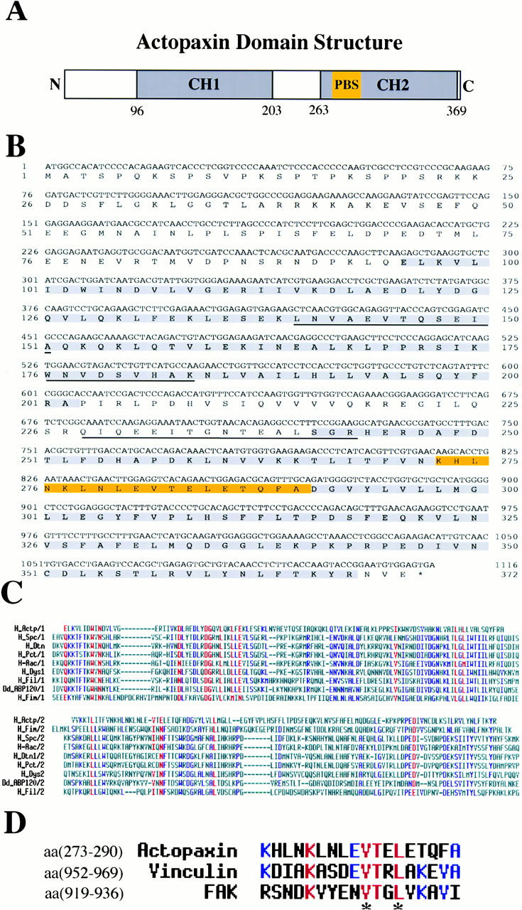
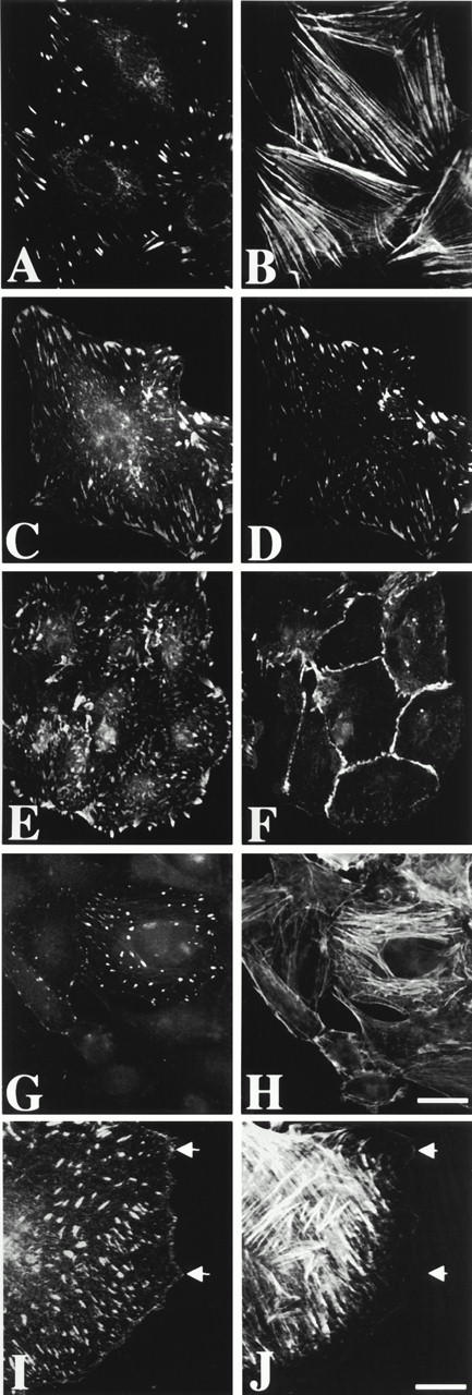
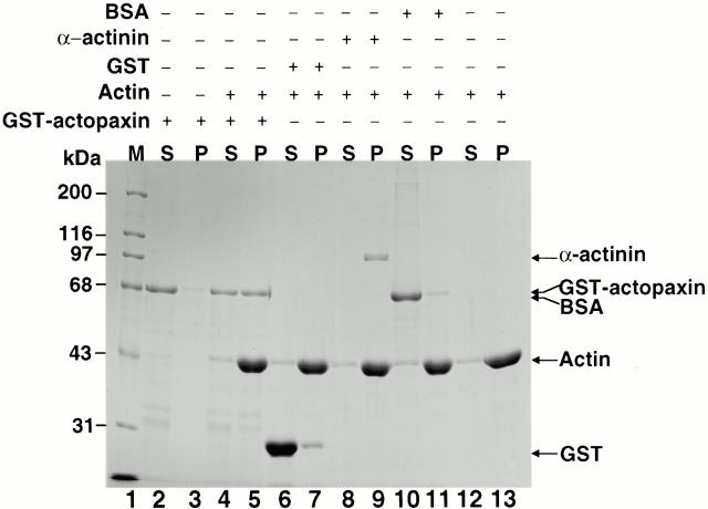
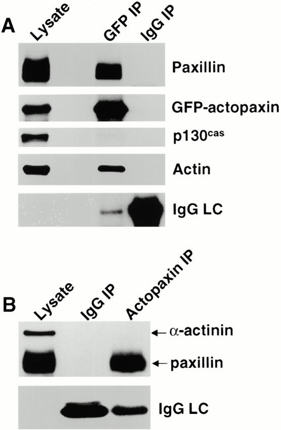
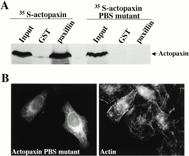
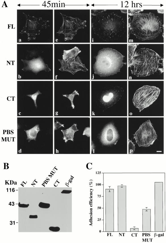
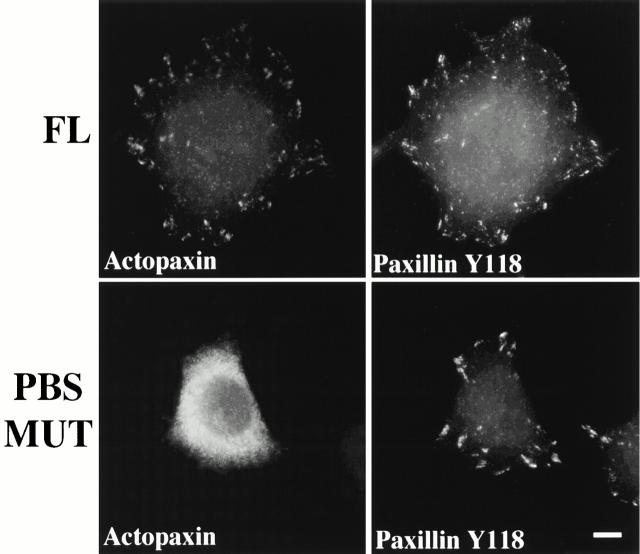
Paxillin is a focal adhesion adapter protein involved in the integration of growth factor- and adhesion-mediated signal transduction pathways. Paxillin LD motifs have been demonstrated to bind to several proteins associated with remodeling of the actin cytoskeleton including the focal adhesion kinase, vinculin, and a complex of proteins comprising p95PKL, PIX, and PAK (Turner, C.E., M. C. Brown, J.A. Perrotta, M.C. Riedy, S.N. Nikolopoulos, A.R. McDonald, S. Bagrodia, S. Thomas, and P.S. Leventhal. 1999. J. Cell Biol. 145:851-863). In this study, we report the cloning and initial characterization of a new paxillin LD motif-binding protein, actopaxin. Analysis of the deduced amino acid sequence of actopaxin reveals a 42-kD protein with two calponin homology domains and a paxillin-binding subdomain (PBS). Western blotting identifies actopaxin as a widely expressed protein. Actopaxin binds directly to both F-actin and paxillin LD1 and LD4 motifs. It exhibits robust focal adhesion localization in several cultured cell types but is not found along the length of the associated actin-rich stress fibers. Similar to paxillin, it is absent from actin-rich cell-cell adherens junctions. Also, actopaxin colocalizes with paxillin to rudimentary focal complexes at the leading edge of migrating cells. An actopaxin PBS mutant incapable of binding paxillin in vitro cannot target to focal adhesions when expressed in fibroblasts. In addition, ectopic expression of the PBS mutant and/or the COOH terminus of actopaxin in HeLa cells resulted in substantial reduction in adhesion to collagen. Together, these results suggest an important role for actopaxin in integrin-dependent remodeling of the actin cytoskeleton during cell motility and cell adhesion.


Figures

Figure 1
Figure 1
Sequence analysis of actopaxin. (A) Schematic representation of the domain structure of actopaxin showing the relative positions of CH domains (CH1 and CH2) and PBS sequence (PBS). (B) Nucleotide and deduced aa sequence of human actopaxin cDNA. The gray boxes highlight the CH domains. The aa sequences of the three peptides derived from the microsequencing of the 42-kD band are underlined. The PBS is highlighted in yellow. The sequence data are available from GenBank/EMBL/DDBJ databases under accession no. AF264765. (C) Sequence alignment of CH domains from different actin-binding proteins. The CH1 domains are aligned on the top and the CH2 domains on the bottom. Residues conserved in >90% of the sequences are colored red; residues conserved in >50% of the sequences are colored blue. The names, database accession numbers (SWISSPROT [SW] or SPTREMBL [SE]), and sequence residues are as follows: H_Actp, human actopaxin aa 96–202 and aa 262–368; H_Spc, human β spectrin, SW Q01082, aa 53–163 and aa 172–281; H-Aac, human α-actinin1 SW P12814, aa 30–140 and aa 143–253; H_Dtn_l, human dystonin isoform 1, SE Q13266, aa 34–143 and aa 150–258; H_Pct, human plectin, SE Q15149, aa 178–287 and aa 294–403; H_Dys, human dystrophin, SW P11532, aa 14–124 and aa 133–243; H_Fil, human nonmuscle filamin (ABP-280), SW P21333, aa 42–154 and aa 165–272; Dd_ABP-120, D. discoideum ABP-120, SW P13466, aa 11–122 and aa 124–230; H_Fim, human fimbrin, SW P13797, aa 119–241 and aa 260–379. The figure was produced with MULTALIN software (Corpet 1988). (D) Sequence alignment of the PBS domains of actopaxin, vinculin, and FAK. Identical residues are colored red; residues conserved in at least two of the sequences are colored blue. Asterisks indicate the aa that have been reported previously to be critical for FAK binding to paxillin and which are mutated in the actopaxin PBS mutant.
Figure 2
Figure 2
Expression of actopaxin protein in various tissues and cell lines. Organs were dissected from a healthy adult rat, homogenized, and lysed in SDS-PAGE sample buffer. 50 μg of each lysate was loaded per lane. (A) Coomassie blue staining of the gel. (B) Western blotting of an identical gel shown in A probed with the actopaxin antibody. (C) The same blot was stripped and reprobed with an actin antibody to indicate relative loading among lanes. (D) Western blot of total lysates (50 μg) from HISM cells, rat embryo fibroblasts (REF-52), and rat intestinal epithelial cells (IEC-18) probed with the actopaxin antibody.
Figure 4
Figure 4
Actopaxin binds to the LD1 and LD4 motifs of paxillin. (A) Coomassie blue staining of the GST-paxillin LD motif fusion proteins. (B) Each paxillin LD motif fusion protein was incubated with 200 μg total lysate from REF-52 cells. Coprecipitating proteins were resolved by SDS-PAGE and transferred to nitrocellulose. The membrane was probed with actopaxin antibody (top). The same blot was stripped and probed with α-actinin antibody (bottom). (C) Individual GST-LD domains of paxillin were incubated with in vitro–translated 35S-labeled actopaxin and binding proteins, precipitated using glutathione-agarose beads, resolved by SDS-PAGE, and visualized by fluorography.
Figure 5
Figure 5
The second CH domain of actopaxin binds directly to paxillin and hic-5 in vitro. (A) Coomassie blue–stained gel of GST-actopaxin fusion proteins wild-type actopaxin (1–372), NH2 terminus actopaxin (1–222), COOH terminus actopaxin (223–372). (B) GST-actopaxin fusion proteins shown in A were incubated with 300 μg of total lysate from REF-52 cells. Coprecipitated proteins were resolved by SDS-PAGE and transferred to nitrocellulose. The membrane was probed with paxillin antibody (clone 349) that recognizes paxillin and hic-5 (top). The same blot was stripped and reprobed with vinculin antibody (bottom). (C) Paxillin was 35S-metabolically labeled in vitro by coupled transcription/translation and tested for its ability to bind directly to GST-actopaxin fusion proteins.
Figure 3
Figure 3
Subcellular distribution of actopaxin. Immunofluorescence staining with actopaxin antibody of (A) rat embryo fibroblasts (REF-52), (C) HISM cells, and (E) rat intestinal epithelial cells (IEC-18). Cells were stained simultaneously with rhodamine phalloidin to visualize actin stress fibers (B), paxillin antibody to demonstrate colocalization of actopaxin with paxillin at focal adhesions (D), and β-catenin antibody to demonstrate that actopaxin is absent from cell–cell contacts (F). Xpress-tagged actopaxin was transfected in CHO-K1 cells (G and H). The cells were stained simultaneously with anti-Xpress antibody to detect the transfected actopaxin (G) and rhodamine phalloidin (H) to visualize actin stress fibers. Xpress-actopaxin was enriched at focal adhesions at the ends of actin stress fibers (G and H). Actopaxin is recruited to the focal complexes (arrows) at the leading edge of migrating fibroblasts (I and J). HISM cells were stimulated to migrate by scrape wounding a confluent monolayer of cells. 3 h after wounding, the cells were stained for immunofluorescence microscopy with actopaxin antibody (I) and rhodamine phalloidin (J) to visualize actin stress fibers. Bars: (A–H) 5 μm; (I and J) 2 μm.
Figure 6
Figure 6
Actin-binding activity of GST-actopaxin. The ability of actopaxin to associate with F-actin in vitro was examined by actin cosedimentation assays using GST-actopaxin fusion protein. The following recombinant proteins were incubated with skeletal muscle F-actin: GST-actopaxin (lanes 4–5), GST (lanes 6–7), α-actinin (lanes 8–9), and BSA (lanes 10–11). GST-actopaxin (lanes 2–3) and F-actin (lanes 12–13) were also incubated in the absence of F-actin and GST-actopaxin, respectively. The supernatant (S) and pellet (P) fractions were analyzed by SDS-PAGE on 10% gel stained with Coomassie blue. Molecular weight markers (M) are shown (lane 1).
Figure 7
Figure 7
Actopaxin binds to paxillin and actin in vivo. (A) GFP-actopaxin was transfected into Cos-7 cells. After lysis, proteins were immunoprecipitated from 400 μg of total lysate with an antibody to either GFP or IgG control. 20 μg of total lysate and the immunoprecipitates (IP) were subjected to SDS-PAGE followed by Western blotting with antibodies to paxillin, GFP, actin, and p130Cas. (B) Coimmunoprecipitation of paxillin with endogenous actopaxin. Asynchronously growing adherent HISM cells were lysed in coimmunoprecipitation buffer. Actopaxin and IgG immunoprecipitations were performed from 500 μg lysates and subjected to SDS-PAGE followed by Western blotting with antibodies to paxillin and α-actinin. Paxillin was coimmunoprecipitated with the actopaxin antibody in contrast to the control IgG. No binding of α-actinin to either actopaxin or IgG coimmunoprecipitates was observed. IgG LC, immunoglobulin light chain.
Figure 8
Figure 8
Actopaxin-PBS mutant does not bind paxillin in vitro and does not localize to focal adhesions. (A) GST or GST-paxillin was incubated with either in vitro–translated 35S-labeled Xpress-actopaxin or 35S-labeled Xpress-actopaxin PBS mutant, and binding proteins were resolved by SDS-PAGE and visualized by fluorography. (B) Xpress-actopaxin PBS mutant was transfected into HeLa, and the cells were stained simultaneously with anti-Xpress antibody to detect the transfected actopaxin and rhodamine phalloidin to visualize actin stress fibers. There was no evidence of focal adhesion localization for the actopaxin PBS mutant, although some perinuclear staining was detected. Bar, 5 μm.
Figure 9
Figure 9
Actopaxin mediates adhesion and spreading of HeLa cells. (A) HeLa cells transfected with Xpress-actopaxin constructs were plated on collagen type I–coated slips for 45 min or for 12 h. For each time point, slips were fixed and processed for immunofluorescence with Xpress antibody. The wild-type full-length actopaxin (FL) localizes efficiently to focal adhesions at both time points (a and i). The COOH terminus actopaxin (CT) localizes weakly to focal adhesions at later time points (c and k). Both the NH2 terminus (NT) and the PBS mutant (PBS MUT) failed to target to focal adhesions (b–j and d–l). Dual labeling of actin in these cells with rhodamine phalloidin indicates that overexpression of the CT or the PBS mutants results in a partial reduction in actin filament content during the initial phases of cell spreading (g and h). (B) Protein expression of the actopaxin constructs and β-galactosidase control. 50 μg of total cell lysates for each construct of actopaxin transfectants were run on SDS-PAGE, transferred to nitrocellulose, and probed with Xpress antibody. (C) Adhesion and spreading efficiency of HeLa cells expressing wild-type full-length, NH2 terminus, COOH terminus, PBS mutant actopaxin, or β-galactosidase. Data represent the mean ± SD of three experiments. Bar, 5 μm.
Figure 10
Figure 10
Ectopic expression of wild-type full-length actopaxin or actopaxin PBS mutant has no effect on paxillin localization to focal adhesions during spreading on collagen. HeLa cells transfected with Xpress-actopaxin constructs were plated on collagen type I–coated coverslips. Coverslips were fixed and stained for immunofluorescence with Xpress and paxillin Y118 antibodies. FL, full-length actopaxin; PBSMUT, actopaxin PBS mutant. Bar, 5 μm.

Similar articles

Cited by

References

    1. Bagrodia S., Cerione R.A. PAK to the future. Trends Cell Biol. 1999;9:350–355. - PubMed
    1. Bagrodia S., Taylor S.J., Jordon K.A., Van Aelst L., Cerione R.A. A novel regulator of p21-activated kinases. J. Biol. Chem. 1998;273:23633–23636. - PubMed
    1. Banuelos S., Saraste M., Carugo K.D. Structural comparisons of calponin homology domainsimplications for actin binding. Structure. 1998;6:1419–1431. - PubMed
    1. Brown M.C., Perrotta J.A., Turner C.E. Identification of LIM3 as the principal determinant of paxillin focal adhesion localization and characterization of a novel motif on paxillin directing vinculin and focal adhesion kinase binding. J. Cell Biol. 1996;135:1109–1123. - PMC - PubMed
    1. Brown M.C., Curtis M.S., Turner C.E. Paxillin LD motifs may define a new family of protein recognition domains Nat. Struct. Biol 5 1998. 677 678a - PubMed

Publication types

MeSH terms

Associated data