. 2000 Dec 11;151(6):1131-40.
doi: 10.1083/jcb.151.6.1131.

Coordinate control of muscle cell survival by distinct insulin-like growth factor activated signaling pathways

Affiliations

Coordinate control of muscle cell survival by distinct insulin-like growth factor activated signaling pathways

M A Lawlor et al. J Cell Biol. .

Abstract

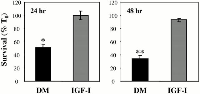
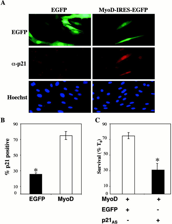
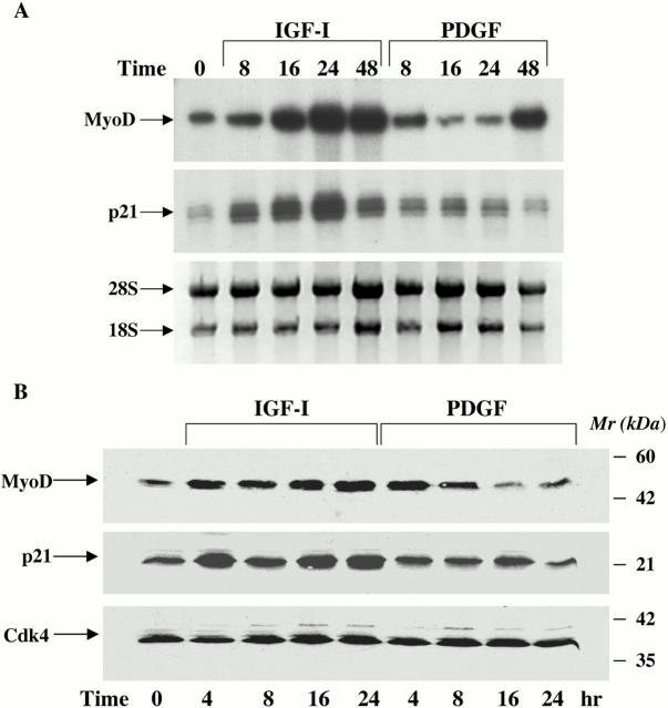
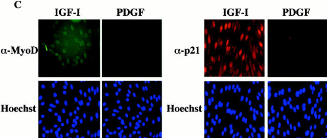
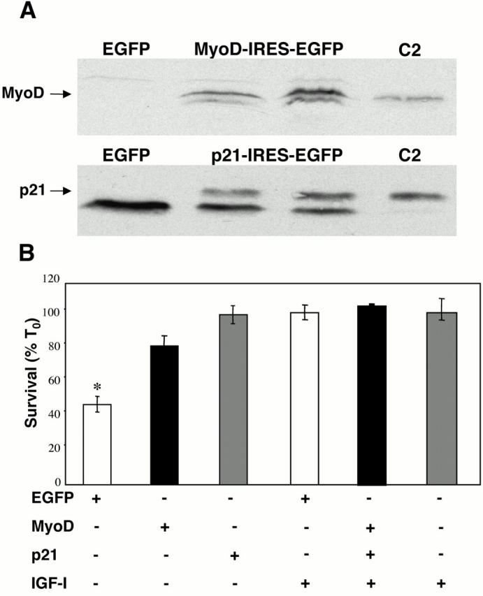
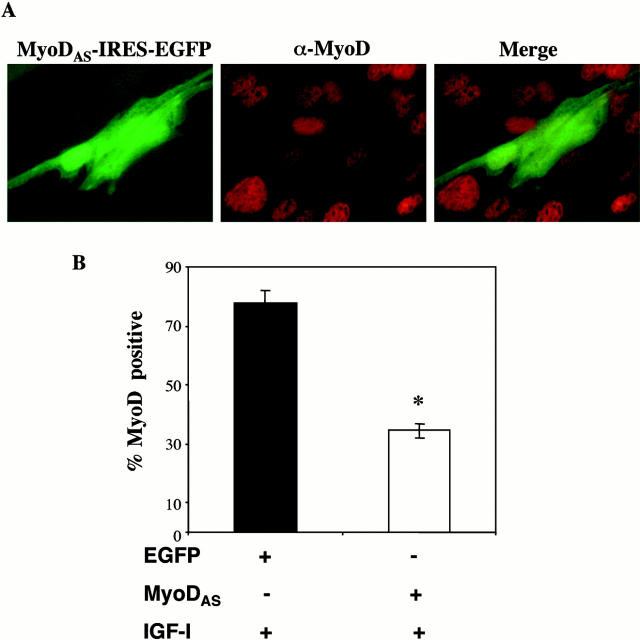
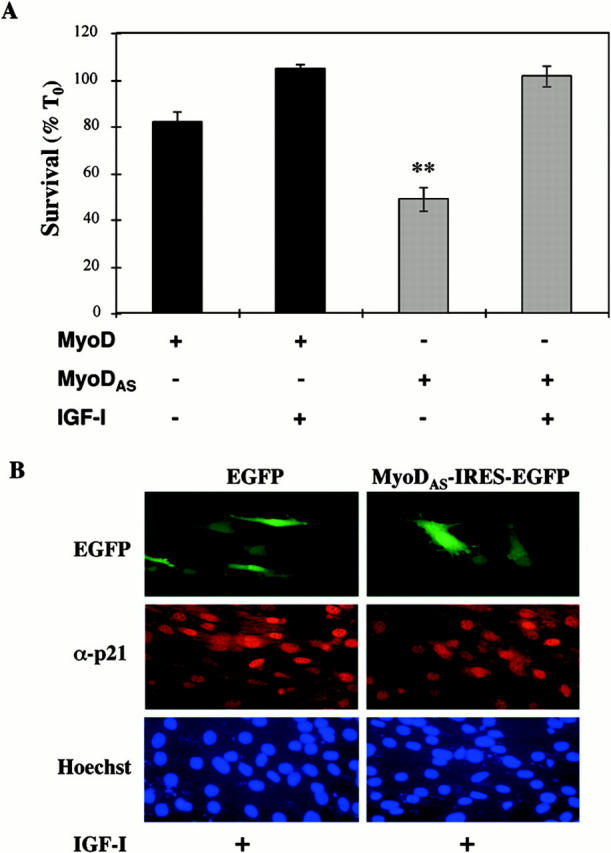
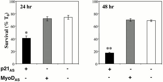
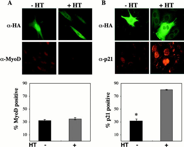
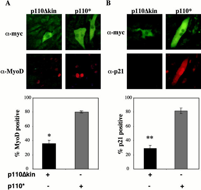
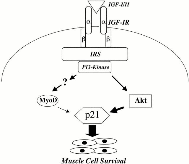
Peptide growth factors control diverse cellular functions by regulating distinct signal transduction pathways. In cultured myoblasts, insulin-like growth factors (IGFs) stimulate differentiation and promote hypertrophy. IGFs also maintain muscle cell viability. We previously described C2 skeletal muscle lines lacking expression of IGF-II. These cells did not differentiate, but underwent progressive apoptotic death when incubated in differentiation medium. Viability could be sustained and differentiation enabled by IGF analogues that activated the IGF-I receptor; survival was dependent on stimulation of phosphatidylinositol 3-kinase (PI3-kinase). We now find that IGF action promotes myoblast survival through two distinguishable PI3-kinase-regulated pathways that culminate in expression of the cyclin-dependent kinase inhibitor, p21. Incubation with IGF-I or transfection with active PI3-kinase led to rapid induction of MyoD and p21, and forced expression of either protein maintained viability in the absence of growth factors. Ectopic expression of MyoD induced p21, and inhibition of p21 blocked MyoD-mediated survival, thus defining one PI3-kinase-dependent pathway as leading first to MyoD, and then to p21 and survival. Unexpectedly, loss of MyoD expression did not impede IGF-mediated survival, revealing a second pathway involving activation by PI3-kinase of Akt, and subsequent induction of p21. Since inhibition of p21 caused death even in the presence of IGF-I, these results establish a central role for p21 as a survival factor for muscle cells. Our observations also define a MyoD-independent pathway for regulating p21 in muscle, and demonstrate that distinct mechanisms help ensure appropriate expression of this key protein during differentiation.


Figures

Figure 1
Figure 1
IGF-I treatment maintains myoblast survival. Results are shown for cell counts (expressed as percent at time 0, % T0) of C2AS12 myoblasts after a 24 or 48 h incubation in DM with or without R3IGF-I (2 nM). Asterisks indicate that significantly fewer cells survived (*P < 0.005, **P < 0.001) in untreated than in growth factor–treated cells (mean ± SEM of three experiments, each performed in duplicate).
Figure 4
Figure 4
p21 is required for MyoD-stimulated muscle cell survival. (A) MyoD induces p21 protein expression. C2AS12 cells were transfected with either EGFP or MyoD-IRES-EGFP expression plasmids, and following a 24-h incubation in DM, fixed, and stained with an antibody to p21. (Top) Expression of EGFP, (middle) immunostaining for p21, and (bottom) nuclear staining with Hoechst dye. (B) The graph records the percent of transfected cells expressing p21. Significantly fewer cells are positive for p21 after transfection with EGFP than with MyoD-IRES-EGFP (*P < 0.005; mean ± SEM of three experiments, 100 transfected cells counted per experiment). (C) Inhibition of p21 prevents MyoD-stimulated muscle cell survival. C2AS12 cells were cotransfected with MyoD-IRES-EGFP (MyoD) and either EGFP or p21AS-IRES-EGFP (p21AS), as outlined in Materials and Methods. Cell counts of transfected myoblasts were performed after a 24-h incubation in DM. Results are presented as percent survival compared with T0 (mean ± SEM of three experiments, each performed in duplicate). *Survival was significantly less in myoblasts cotransfected with p21AS-IRES-EGFP than with EGFP (P < 0.014).
Figure 2
Figure 2
Treatment with IGF-I stimulates expression of MyoD and p21. (A) Induction of MyoD and p21 mRNAs by IGF-I. The autoradiographs show results of representative ribonuclease protection assays performed using total RNA isolated from C2AS12 cells incubated with either R3IGF-I (2 nM) or PDGF-BB (0.4 nM) for the indicated times (top, MyoD; middle, p21). (Bottom) Photograph of an ethidium bromide–stained gel of the RNA used in these studies. Similar results were seen in three independent experiments. (B) Induction of MyoD and p21 proteins by IGF-I. Representative immunoblots are shown using whole-cell protein extracts from C2AS12 cells incubated with either R3IGF-I (2 nM) or PDGF-BB (0.4 nM) for the indicated times, and antibodies to MyoD (top), p21 (middle), and CDK-4 (bottom). Similar results were seen in three independent experiments. (C) Representative fluorescence micrographs of C2AS12 myoblasts treated with either R3IGF-I (2 nM) or PDGF-BB (0.4 nM) for 24 h, as described in Materials and Methods, and immunostained with antibodies to MyoD (top left), or p21 (top right), and incubated with Hoechst nuclear dye (bottom).
Figure 2
Figure 2
Treatment with IGF-I stimulates expression of MyoD and p21. (A) Induction of MyoD and p21 mRNAs by IGF-I. The autoradiographs show results of representative ribonuclease protection assays performed using total RNA isolated from C2AS12 cells incubated with either R3IGF-I (2 nM) or PDGF-BB (0.4 nM) for the indicated times (top, MyoD; middle, p21). (Bottom) Photograph of an ethidium bromide–stained gel of the RNA used in these studies. Similar results were seen in three independent experiments. (B) Induction of MyoD and p21 proteins by IGF-I. Representative immunoblots are shown using whole-cell protein extracts from C2AS12 cells incubated with either R3IGF-I (2 nM) or PDGF-BB (0.4 nM) for the indicated times, and antibodies to MyoD (top), p21 (middle), and CDK-4 (bottom). Similar results were seen in three independent experiments. (C) Representative fluorescence micrographs of C2AS12 myoblasts treated with either R3IGF-I (2 nM) or PDGF-BB (0.4 nM) for 24 h, as described in Materials and Methods, and immunostained with antibodies to MyoD (top left), or p21 (top right), and incubated with Hoechst nuclear dye (bottom).
Figure 3
Figure 3
MyoD and p21 promote myoblast survival. (A) Expression of MyoD and p21 in transfected Cos7 cells. Cos7 cells were transiently transfected with expression plasmids encoding MyoD-IRES-EGFP (MyoD), p21-IRES-EGFP (p21), or EGFP alone, as outlined in Materials and Methods. Whole-cell protein extracts were isolated 48 h after transfection and used for immunoblotting with antibodies to MyoD (top) or p21 (bottom). Whole-cell extracts from differentiating C2 myoblasts (C2) were used as a positive control. (B) MyoD and p21 promote muscle cell survival. C2AS12 myoblasts were transfected with plasmids encoding either MyoD-IRES-EGFP (MyoD), p21-IRES-EGFP (p21), or EGFP alone. Counts of transfected cells were performed after a 24-h incubation in DM without or with R3IGF-I (2 nM). Results are presented as percent survival compared with T0 (mean ± SEM of four experiments, each performed in duplicate). *Survival was significantly lower in myoblasts transfected with EGFP than in cells transfected with MyoD or p21, or treated with IGF-I (P < 0.001).
Figure 5
Figure 5
Forced expression of a MyoD antisense plasmid blocks IGF-I–stimulated MyoD expression. (A) C2AS12 cells were transiently transfected with the MyoDAS-IRES-EGFP expression plasmid, as described in Materials and Methods, and incubated in DM plus R3IGF-I (2 nM) for 24 h, followed by fixation and staining for MyoD. (Left) Expression of EGFP, (center) immunostaining for MyoD, and (right) merged image. (B) The graph indicates the percentage of transfected cells expressing MyoD, as assessed by immunocytochemistry following IGF-I treatment. Significantly fewer cells are positive for MyoD after transfection with MyoDAS-IRES-EGFP than with EGFP (*P < 0.01; mean ± SEM of three experiments, counting 100 cells per experiment).
Figure 6
Figure 6
MyoD is not required for IGF-mediated survival of C2AS12 myoblasts. (A) C2AS12 cells were transiently transfected with MyoD-IRES-EGFP or MyoDAS-IRES-EGFP expression plasmids, as described in Materials and Methods. Cell counts of transfected myoblasts were performed after a 24-h incubation in DM or in DM supplemented with R3IGF-I (2 nM). Results are expressed as the mean ± SEM of three independent experiments, each performed in duplicate. **Survival was significantly less in myoblasts transfected with MyoDAS than in cells transfected with MyoD or treated with IGF-I (P < 0.001). (B) Inhibition of MyoD expression does not block IGF-mediated induction of p21. Representative fluorescence micrographs of C2AS12 myoblasts transiently transfected with expression plasmids for MyoDAS-IRES-EGFP or EGFP alone, and treated with IGF-I (2 nM) in DM for 24 h. (Top) Expression of EGFP, (middle) immunostaining for p21, and (bottom) nuclei stained with Hoechst dye. As determined by cell counting, ∼80% of cells transfected with either plasmid expressed p21 after incubation with IGF-I.
Figure 7
Figure 7
Inhibition of MyoD does not block survival of C2 myoblasts. C2 myoblasts were transiently transfected with MyoDAS-IRES-EGFP or p21AS-IRES-EGFP expression plasmids, as described in Materials and Methods. Cell counts of transfected cells were performed after a 24 or 48 h incubation in DM. Results are expressed as the mean ± SEM of three independent experiments, each performed in duplicate. The asterisks denote that viability was significantly less in myoblasts transfected with p21AS than in cells transfected with MyoDAS or untransfected cells (*P < 0.003, **P < 0.00002).
Figure 9
Figure 9
Induction of p21 but not MyoD after activation of transfected Akt. (A) Akt does not induce MyoD. Pictured are representative fluorescence micrographs of C2AS12 cells transiently transfected with hydroxytamoxifen-inducible HA-tagged Akt (iAkt), as described in Materials and Methods. After a 24-h incubation with DM in the absence or presence of hydroxytamoxifen, cells were fixed and immunostained using antibodies to the HA tag (top) and MyoD (bottom). The graph shows that the fraction of transfected cells expressing MyoD was not significantly different in cells incubated without or with HT (mean ± SEM of three experiments, counting 100 cells per experiment). (B) Akt induces p21. Illustrated are representative fluorescence micrographs of C2AS12 cells transiently transfected with iAkt, as described in Materials and Methods. After a 24-h incubation in DM without or with HT, the cells were immunostained using antibodies to the HA tag (top) and to p21 (bottom). The graph shows that the percentage of transfected cells expressing p21 was significantly greater after incubation with HT (mean ± SEM of three experiments, counting 100 cells per experiment, *P < 0.01).
Figure 8
Figure 8
Induction of MyoD and p21 after transfection of active PI3-kinase. (A) Induction of MyoD by PI3-kinase. Pictured are representative fluorescence micrographs of C2AS12 muscle cells transiently transfected with plasmids encoding active (p110*) or inert (p110Δkin) PI3-kinase, as described in Materials and Methods. After a 24-h incubation in DM, cells were fixed and immunostained for expression of either PI-3 kinase using an antibody directed against the c-myc epitope tag (top) or for MyoD (bottom). The graph shows that the percent of transfected cells expressing MyoD was significantly less after transfection with p110Δkin than with p110* (mean ± SEM of three experiments, counting 100 cells per experiment, *P < 0.01). (B) Induction of p21 by PI3-kinase. Illustrated are representative fluorescence micrographs of C2AS12 muscle cells transiently transfected with the p110* or p110Δkin expression plasmids, as described in A. After a 24-h incubation in DM cells were immunostained for expression of either protein (top), and for p21 (bottom). The graph shows that the percentage of transfected cells expressing p21 was significantly smaller after transfection with p110Δkin than with p110* (mean ± SEM of three experiments, counting 100 cells per experiment, **P < 0.001).
Figure 10
Figure 10
Model for IGF-regulated muscle cell survival. Ligand-induced activation of the IGF-I receptor (IGF-IR) leads to stimulation of PI3-kinase enzymatic activity through the adapter molecules, IRS-1 and -2. PI3-kinase subsequently activates Akt, which stimulates expression of p21 mRNA and protein, which then promotes muscle cell survival. PI3-kinase through unidentified steps also stimulates expression of MyoD, which then induces p21. MyoD plays a secondary role in the induction of p21 and in IGF-mediated cell survival.

Similar articles

Cited by

References

    1. Andres V., Walsh K. Myogenin expression, cell cycle withdrawal, and phenotypic differentiation are temporally separable events that precede cell fusion upon myogenesis. J. Cell Biol. 1996;132:657–666. - PMC - PubMed
    1. Arnold H.H., Winter B. Muscle differentiationmore complexity to the network of myogenic regulators. Curr. Opin. Genet. Dev. 1998;8:539–544. - PubMed
    1. Ball K.L. p21structure and functions associated with cyclin-CDK binding. Prog. Cell Cycle Res. 1997;3:125–134. - PubMed
    1. Barton-Davis E.R., Shoturma D.I., Musaro A., Rosenthal N., Sweeney H.L., Kurabayashi M., Dutta S., Kedes L. Viral mediated expression of insulin-like growth factor I blocks the aging-related loss of skeletal muscle function. Proc. Natl. Acad. Sci. USA. 1998;95:5603–15607. - PMC - PubMed
    1. Baserga R., Resnicoff M., D'Ambrosio C., Valentinis B. The role of the IGF-I receptor in apoptosis. Vitam. Horm. 1997;53:65–98. - PubMed

Publication types