. 2000 Dec 15;19(24):6742-50.
doi: 10.1093/emboj/19.24.6742.

Epstein-Barr virus-encoded poly(A)(-) RNA supports Burkitt's lymphoma growth through interleukin-10 induction

Affiliations

Epstein-Barr virus-encoded poly(A)(-) RNA supports Burkitt's lymphoma growth through interleukin-10 induction

N Kitagawa et al. EMBO J. .

Abstract

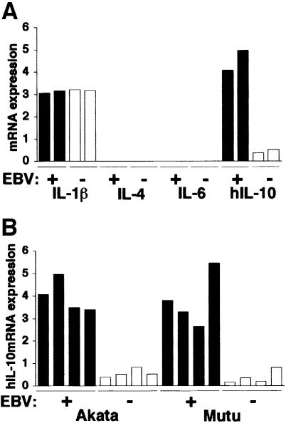
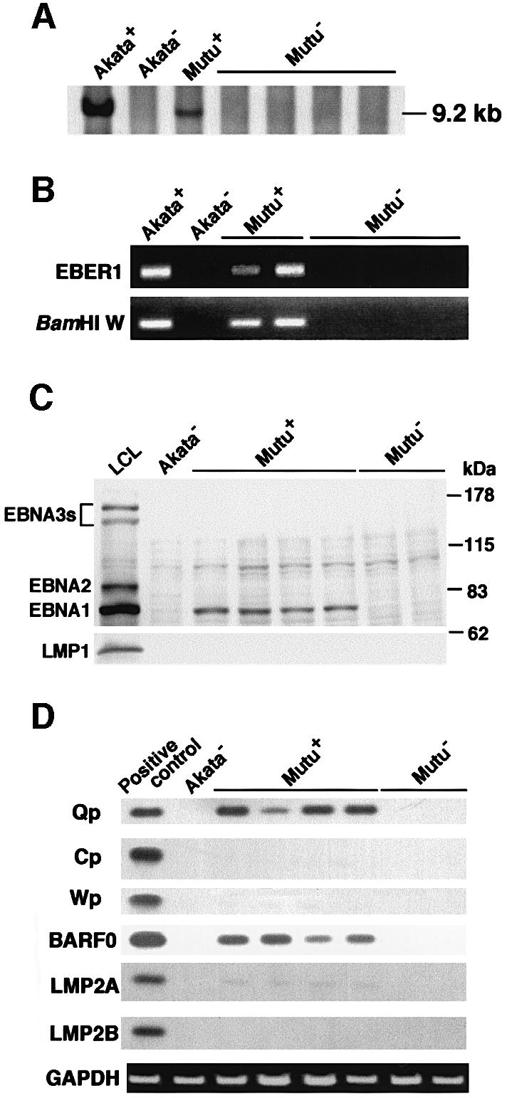
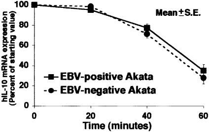
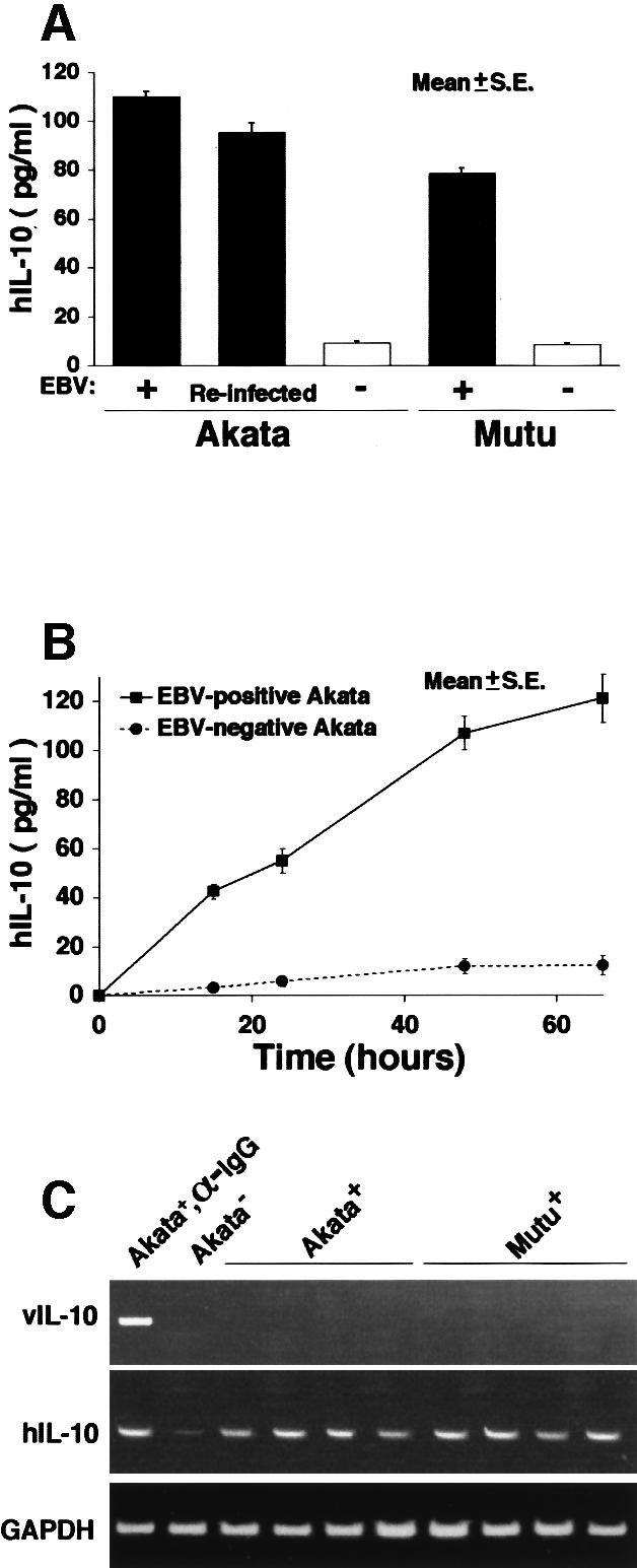
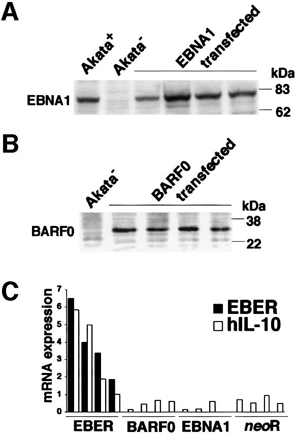
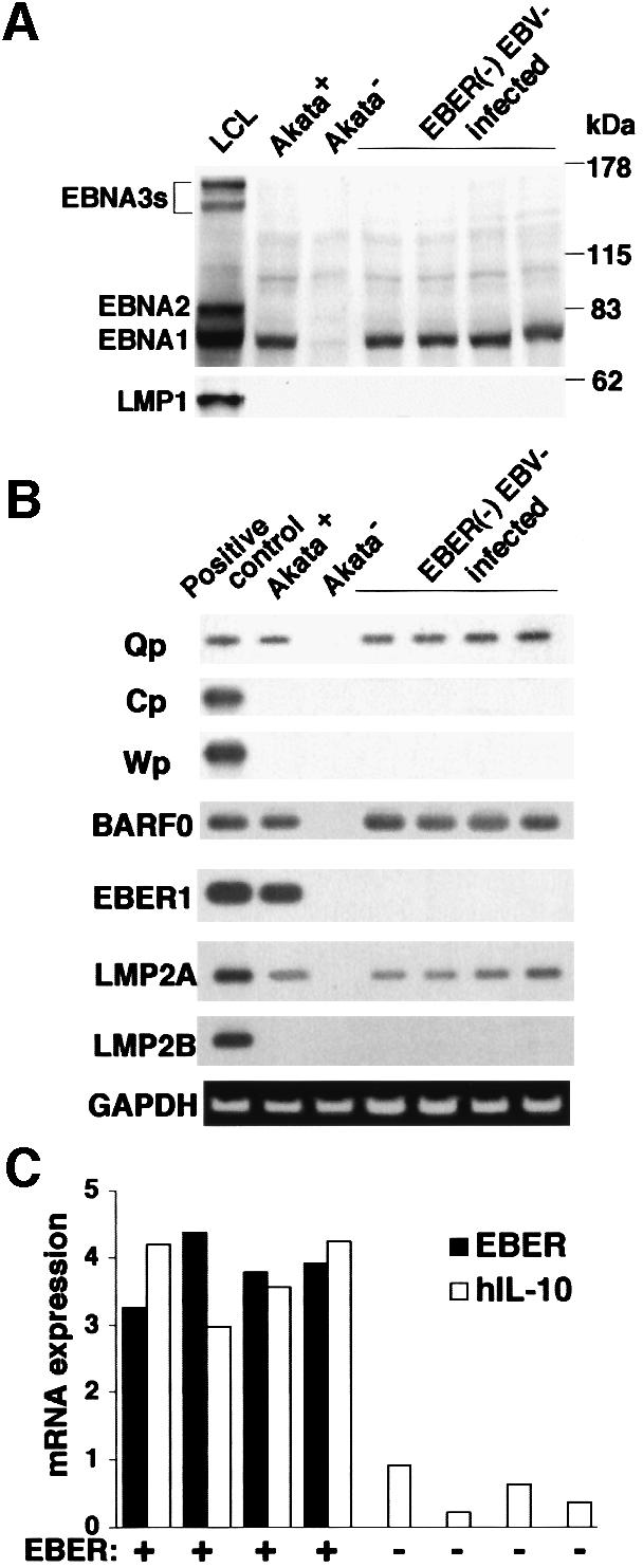
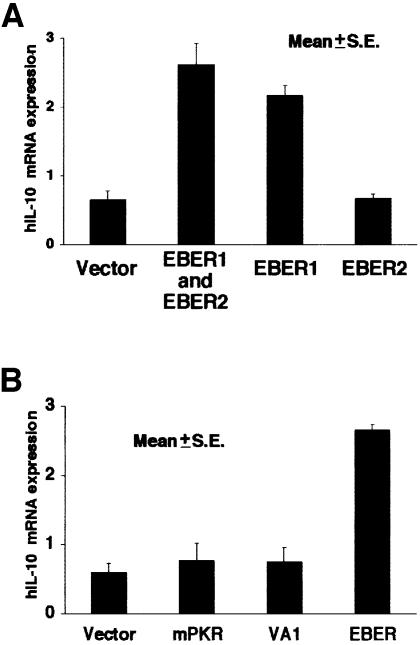
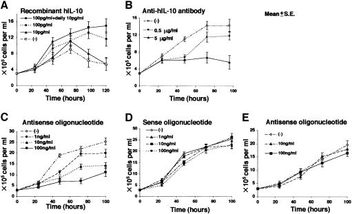
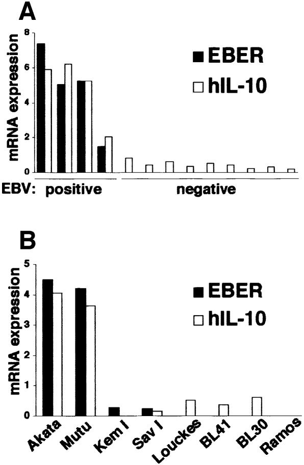
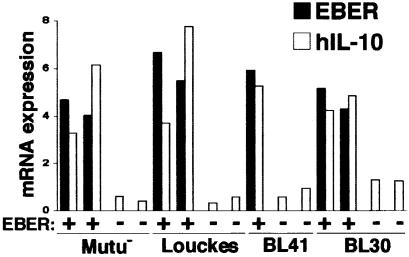
Akata and Mutu cell lines are derived from Burkitt's lymphoma (BL) and retain the in vivo phenotype of Epstein-Barr virus (EBV) expression that is characterized by expression of EBV-determined nuclear antigen 1 (EBNA1), EBV-encoded RNAs (EBERs) and transcripts from the BAM:HI A region (BARF0). We found that EBV-positive Akata and Mutu cell clones expressed higher levels of interleukin (IL)-10 than their EBV-negative subclones at the transcriptional level. Transfection of an individual EBV latent gene into EBV-negative Akata cells revealed that EBERs were responsible for IL-10 induction. Recombinant IL-10 enabled EBV-negative Akata cells to grow in low (0.1%) serum conditions. On the other hand, growth of EBV-positive Akata cells was blocked by treatment either with an anti-IL-10 antibody or antisense oligonucleotide against IL-10. EBV-positive BL biopsies consistently expressed IL-10, but EBV-negative BL biopsies did not. These results suggest that IL-10 induced by EBERs acts as an autocrine growth factor for BL. EBERs, EBER1 and EBER2, are non-polyadenylated RNAs and are 166 and 172 nucleotides long, respectively. The present findings indicate that RNA molecules could regulate cell growth.


Figures

None
Fig. 1. Cytokine expression in BL-derived Akata and Mutu cell lines. Both cell lines were originally 100% EBV positive, and EBV-negative subclones were isolated by limiting dilution from the parental cultures. All analyses were performed by the real-time quantitative RT–PCR assay using a LightCycler™ (Fink et al., 1998). The results are expressed as the ratio to the value of GAPDH (K × cytokine copies/5 × 103 copies GAPDH; K = constant). (A) Cytokine mRNA expression in EBV-positive and -negative Akata cell clones (two clones each). (B) IL-10 mRNA expression in EBV-positive and -negative Akata and Mutu cell clones (four clones each).
None
Fig. 2. Detection of EBV in EBV-positive and -negative Mutu cell clones. (A) Southern blot analysis of EBV genomes. DNA samples (5 µg each) were digested with BamHI, and the blot was probed with the BamHI C fragment of EBV DNA. (B) PCR analysis of EBV genomes. DNA samples (100 ng each) were amplified with primers for EBER1 and BamHI W regions. (C) Immunoblot analysis for detection of EBNAs and LMP1. The blots were probed with EBNA-positive human serum (upper blot) and an anti-LMP1 monoclonal antibody (lower blot). Protein samples extracted from 105 cells were loaded per slot. (D) RT–PCR analysis of EBNA promoter usage and EBV latent gene expression. Akata cells were used as a positive control for detection of Qp-initiated EBNA mRNA, and a lymphoblastoid cell line immortalized by Akata EBV (LCL) was used as a positive control for detection of Cp- or Wp-initiated EBNA mRNAs and BARF0, LMP2A and LMP2B mRNAs.
None
Fig. 3. IL-10 mRNA degradation in EBV-positive and -negative Akata cells. Actinomycin D (5 µg/ml) was added to the culture to block mRNA synthesis. At varying times thereafter, RNA was isolated and the amount of IL-10 mRNA remaining was determined. A representative result of several experiments is shown. In EBV-positive cells, t1/2 = 51.1 min; it was 49.3 min in EBV-negative cells. There was no significant difference between EBV-positive and -negative cells.
None
Fig. 4. (A) IL-10 production in EBV-positive and -negative Akata and Mutu cell clones. After 3 days of culture of 106 cells in 1 ml of RPMI with 10% FCS, the amount of IL-10 in the culture supernatant was measured with a high-sensitivity IL-10 human ELISA system (Amersham Pharmacia Biotech). (B) Time course of IL-10 production in EBV-positive and -negative Akata cells. (C) RT–PCR analysis of viral IL-10 expression in EBV-positive Akata and Mutu cell clones. EBV-positive Akata cells treated with anti-Ig antibody were used as a positive control for detection of viral IL-10.
None
Fig. 5. IL-10 expression in EBV-negative Akata cell clones transfected with an individual EBV latent gene expressed in BL. An EBV-negative Akata cell clone was transfected with an individual EBV gene, and cell clones that stably expressed similar levels of EBV-positive Akata cells were selected and analyzed. (A) EBNA1 expression in EBV-negative Akata cell clones transfected with the EBNA1 gene. EBNA1 was detected by immunoblotting using EBNA1-positive human serum. (B) BARF0 expression in EBV-negative Akata cell clones transfected with the FLAG epitope-tagged BARF0 gene. BARF0 was detected by immunoblotting using an anti-FLAG M2 antibody (Upstate Technology Inc.). (C) IL-10 expression. Four clones each were examined for the expression of IL-10 and EBER by the real-time quantitative RT–PCR assay. The results are expressed as the ratio to the value of GAPDH (K × IL-10 copies/5 × 103 copies GAPDH; K × EBER copies/1 copy GAPDH; K = constant).
None
Fig. 6. IL-10 expression in EBER-knockout EBV-infected Akata cells. An EBV-negative Akata cell clone was reinfected with EBER-positive or -negative EBV, and 100% EBV-positive cell clones were isolated. (A) Immunoblot analysis for detection of EBNAs and LMP1. The blots were probed with EBNA-positive human serum (upper blot) and anti-LMP1 monoclonal antibody (lower blot). Protein samples extracted from 105 cells were loaded per slot. (B) RT–PCR analysis of EBNA promoter usage and EBV latent gene expression. Akata cells were used as a positive control for detection of Qp-initiated EBNA mRNA, and a lymphoblastoid cell line immortalized by Akata EBV (LCL) was used as a positive control for detection of Cp- or Wp-initiated EBNA mRNAs and BARF0, EBER1, LMP2A and LMP2B mRNAs. (C) IL-10 expression. Four clones each were examined for the expression of IL-10 and EBER by the real-time quantitative RT–PCR assay. The results are expressed as the ratio to the value of GAPDH (K × IL-10 copies/5 × 103 copies GAPDH; K × EBER copies/1 copy GAPDH; K = constant).
None
Fig. 7. (A) IL-10 induction by transient expression of EBER1 or EBER2 in EBV-negative Akata cells. Cells (5 × 10⁶) were transfected with the EBER plasmid (50 µg) by the electroporation method, harvested after 48 h of incubation, and subjected to the real-time quantitative RT–PCR assay. The results are expressed as the ratio to the value of GAPDH (K × IL-10 copies/5 × 103 copies GAPDH; K = constant). (B) Effect of adenovirus VA1 and a mutant form of PKR (mPKR) on expression of IL-10 in EBV-negative Akata cells. Cells (5 × 10⁶) were transfected with the VA1 or mPKR plasmid (50 µg) by the electroporation method. Cells were harvested after 48 h of incubation and subjected to the real-time quantitative RT–PCR assay. The results are expressed as the ratio to the value of GAPDH (K × IL-10 copies/5 × 103 copies GAPDH; K = constant).
None
Fig. 8. (A) Effect of recombinant IL-10 on growth of EBV-negative Akata cells in low (0.1%) FCS conditions. Recombinant IL-10 (Endogen) was added to the culture medium in various concentrations. (B) Effect of anti-IL-10 antibody on growth of EBV-positive Akata cells. Purified rat anti-human IL-10 antibody (PharMingen), at various concentrations, was added to the culture medium containing 0.1% FCS. (C) Effect of antisense oligonucleotide against IL-10 on growth of EBV-positive Akata cells. Antisense oligonucleotide, at various concentrations, was added to the culture medium containing 10% FCS. (D) Effect of sense oligonucleotide against IL-10 on growth of EBV-positive Akata cells. Sense oligonucleotide, at various concentrations, was added to the culture medium containing 10% FCS. (E) Effect of antisense oligonucleotide against IL-10 on growth of K562 cells. Cell proliferation was determined by counting the viable cells.
None
Fig. 9. IL-10 mRNA expression in BL biopsies (A), type I BL cell lines and EBV-negative BL cell lines (B). IL-10 mRNA was quantified by the real-time RT–PCR assay. The results are expressed as the ratio to the value of GAPDH (K × IL-10 copies/5 × 103 copies GAPDH; K × EBER copies/1 copy GAPDH; K = constant).
None
Fig. 10. IL-10 mRNA expression in EBER-transfected cell clones of EBV-negative BL cell lines. EBV-negative Mutu, Louckes, BL41 and BL30 cells were transfected with the EBER plasmid, and cell clones that stably expressed EBERs were selected and analyzed. Cell clones transfected with the neoR gene were used as controls. IL-10 mRNA was quantified by the real-time RT–PCR assay. The results are expressed as the ratio to the value of GAPDH (K × IL-10 copies/5 × 103 copies GAPDH; K × EBER copies/1 copy GAPDH; K = constant).

Similar articles

Cited by

References

    1. Baer R. et al. (1984) DNA sequence and expression of the B95-8 Epstein–Barr virus genome. Nature, 310, 207–211. - PubMed
    1. Benjamin D., Knobloch,T.J. and Dayton,M.A. (1992) Human B-cell interleukin-10: B-cell lines derived from patients with acquired immunodeficiency syndrome and Burkitt’s lymphoma constitutively secrete a large quantity of interleukin-10. Blood, 80, 1289–1298. - PubMed
    1. Bhat R.A. and Thimmappaya,B. (1983) Two small RNAs encoded by Epstein–Barr virus can functionally substitute for the virus-associated RNAs in the lytic growth of adenovirus 5. Proc. Natl Acad. Sci. USA, 80, 4789–4793. - PMC - PubMed
    1. Burdin N., Peronne,C., Banchereau,J. and Rousset,F. (1993) Epstein–Barr virus transformation induces B lymphocytes to produce human interleukin 10. J. Exp. Med., 177, 295–304. - PMC - PubMed
    1. Calender A., Billaud,M., Aubry,J.P., Banchereau,J., Vuillaume,M. and Lenoir,G. (1987) Epstein–Barr virus (EBV) induces expression of B-cell activation markers on in vitro infection of EBV-negative B-lymphoma cells. Proc. Natl Acad. Sci. USA, 84, 8060–8064. - PMC - PubMed

Publication types