. 2001 Jan;21(1):330-42.
doi: 10.1128/MCB.21.1.330-342.2001.

Inhibition of the Wnt signaling pathway by Idax, a novel Dvl-binding protein

Affiliations

Inhibition of the Wnt signaling pathway by Idax, a novel Dvl-binding protein

S Hino et al. Mol Cell Biol. 2001 Jan.

Abstract

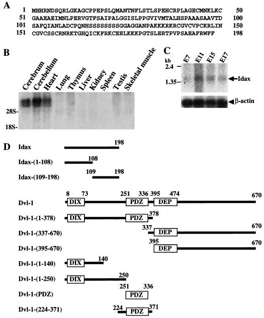
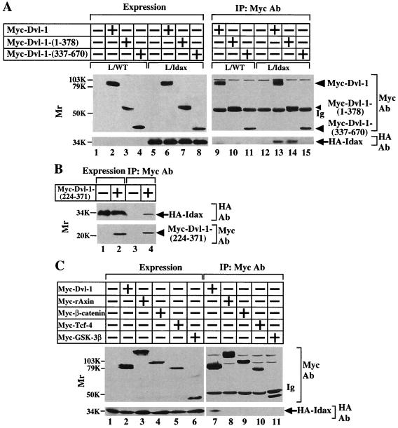
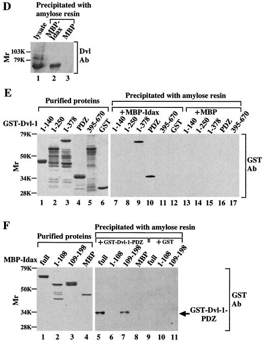
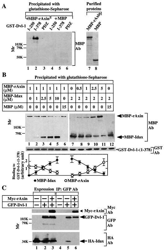
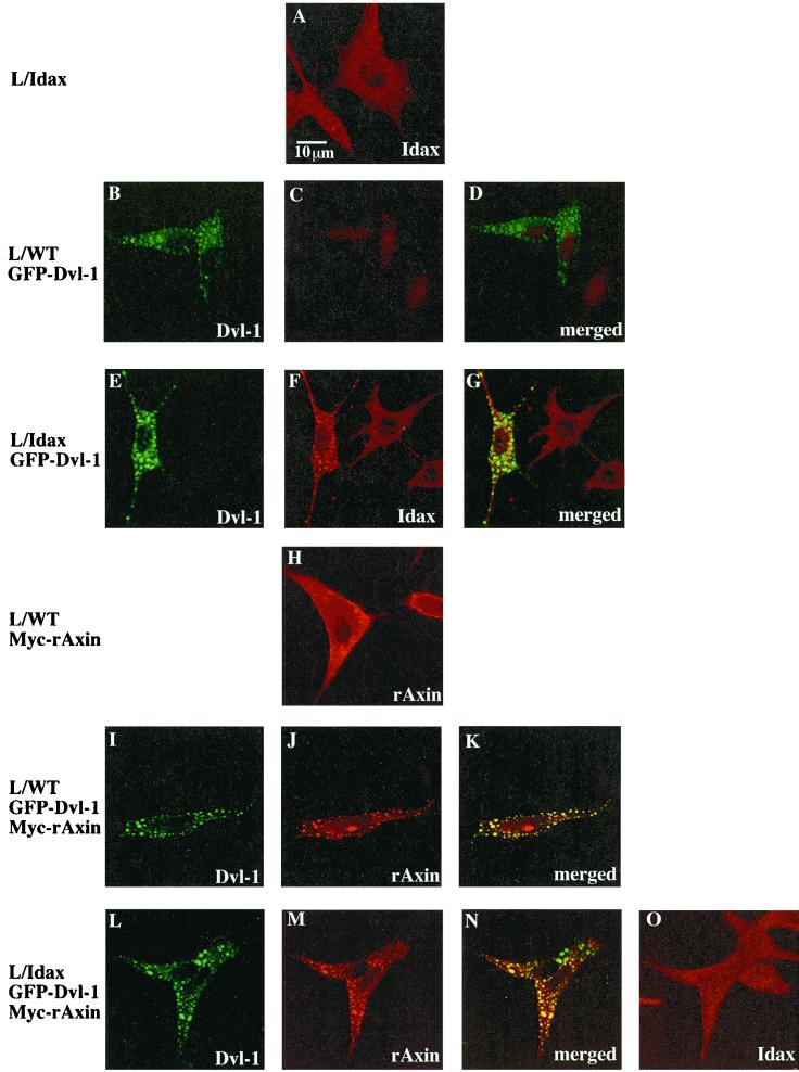
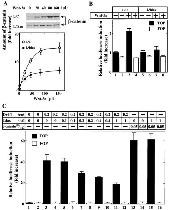
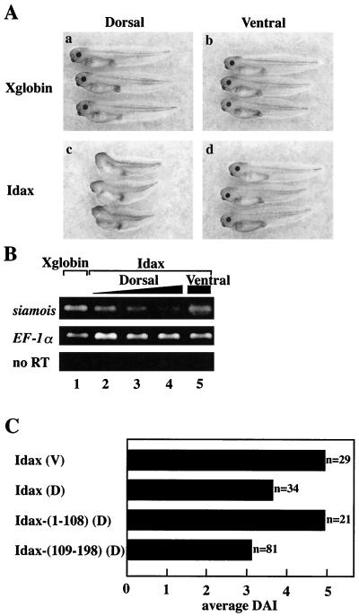
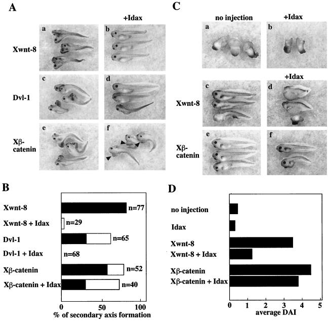
In attempting to clarify the roles of Dvl in the Wnt signaling pathway, we identified a novel protein which binds to the PDZ domain of Dvl and named it Idax (for inhibition of the Dvl and Axin complex). Idax and Axin competed with each other for the binding to Dvl. Immunocytochemical analyses showed that Idax was localized to the same place as Dvl in cells and that expression of Axin inhibited the colocalization of Dvl and Idax. Further, Wnt-induced accumulation of beta-catenin and activation of T-cell factor in mammalian cells were suppressed by expression of Idax. Expression of Idax in Xenopus embryos induced ventralization with a reduction in the expression of siamois, a Wnt-inducible gene. Idax inhibited Wnt- and Dvl- but not beta-catenin-induced axis duplication. It is known that Dvl is a positive regulator in the Wnt signaling pathway and that the PDZ domain is important for this activity. Therefore, these results suggest that Idax functions as a negative regulator of the Wnt signaling pathway by directly binding to the PDZ domain of Dvl.


Figures

FIG. 1
FIG. 1
Structure and gene expression of Idax. (A) Amino acid sequence of Idax. (B) Northern blot analysis of Idax in rat tissues. Total RNA (20 μg per lane) from the indicated rat tissues was probed with full-length cDNA of rat Idax. The positions of 18S and 28S rRNA are indicated. (C) Northern blot analysis of Idax in mouse embryos. Poly(A) RNA (2 μg per lane) from the indicated stages of mouse embryos was probed with the cDNAs of mouse β-actin and rat Idax-(369-495), which is identical with mouse Idax. E, embryonic day. (D) Schematic representation of deletion mutants of rat Idax and human Dvl-1 used in this study.
FIG. 2
FIG. 2
Interaction of Idax with Dvl. (A) Interaction of Idax with Dvl in intact cells. The lysates (20 μg of protein) of wild-type L cells (lanes 1 to 4) or L-Idax cells (lanes 5 to 8) expressing Myc–Dvl-1 (lanes 2 and 6), Myc–Dvl-1-(1-378) (lanes 3 and 7), or Myc–Dvl-1-(337-670) (lanes 4 and 8) were probed with the anti-Myc and anti-HA antibodies. The same lysates (150 μg of protein) prepared in lanes 2 to 8 were immunoprecipitated with the anti-Myc antibody, and the immunoprecipitates were probed with the anti-Myc and anti-HA antibodies (lanes 9 to 15). IP, immunoprecipitation; Ab, antibody. (B) Interaction of Idax with the PDZ domain of Dvl. The lysates of L-Idax cells with (lane 2) or without (lane 1) expression of Myc–Dvl-1-(224-371) were probed with the anti-Myc and anti-HA antibodies. The same lysates (150 μg of protein) prepared in lanes 1 and 2 were immunoprecipitated with the anti-Myc antibody, and the immunoprecipitates were probed with the anti-Myc and anti-HA antibodies (lanes 3 and 4). (C) Inability of Idax to bind to other Wnt signaling molecules. The lysates (20 μg of protein) of L-Idax cells expressing Myc–Dvl-1 (lane 2), Myc-rAxin (lane 3), Myc–β-catenin (lane 4), Myc–Tcf-4 (lane 5), or Myc–GSK-3β (lane 6) were probed with the anti-Myc and anti-HA antibodies. The same lysates (150 μg of protein) prepared in lanes 2 to 6 were immunoprecipitated with the anti-Myc antibody, and the immunoprecipitates were probed with the anti-Myc and anti-HA antibodies (lanes 7 to 11). (D) Complex formation of Idax with Dvl in rat brain. Rat brain cytosol (30 μg of protein) was probed with the anti-Dvl antibody (lane 1). The cytosol (100 μg of protein) was incubated with MBP-Idax (lane 2) or MBP (lane 3) (30 pmol) immobilized to amylose resin, and MBP fusion proteins were precipitated by centrifugation and the precipitates were probed with the anti-Dvl antibody. (E) Direct interaction of Idax with Dvl. GST–Dvl-1-(1-140), GST–Dvl-1-(1-250), GST–Dvl-1-(1-378), GST–Dvl-1-PDZ, GST–Dvl-1-(395-670), and GST (1 μg of protein) were stained with Coomassie brilliant blue (lanes 1 to 6). GST–Dvl-1-(1-140) (lanes 7 and 13), GST–Dvl-1-(1-250) (lanes 8 and 14), GST–Dvl-1-(1-378) (lanes 9 and 15), GST–Dvl-1-PDZ (lanes 10 and 16), GST–Dvl-1-(395-670) (lanes 11 and 17), or GST (lane 12) (1 μM) was incubated with MBP-Idax (lanes 7 to 12) or MBP (lanes 13 to 17) (30 pmol) immobilized on amylose resin, and MBP fusion proteins were precipitated by centrifugation and the precipitates were probed with the anti-GST antibody. (F) The region of Idax which binds to Dvl. MBP-Idax, MBP-Idax-(1-108), MBP-Idax-(109-198), and MBP (0.5 μg of protein) were stained with Coomassie brilliant blue (lanes 1 to 4). GST–Dvl-1-PDZ (lanes 5 to 8) or GST (lanes 9 to 11) (1 μM) was incubated with MBP-Idax (lanes 5 and 9), MBP-Idax-(1-108) (lanes 6 and 10), MBP-Idax-(109-198) (lanes 7 and 11), or MBP (lanes 8) (30 pmol) immobilized on amylose resin. MBP fusion proteins were precipitated by centrifugation, and the precipitates were probed with the anti-GST antibody. The results shown are representative of three independent experiments.
FIG. 2
FIG. 2
Interaction of Idax with Dvl. (A) Interaction of Idax with Dvl in intact cells. The lysates (20 μg of protein) of wild-type L cells (lanes 1 to 4) or L-Idax cells (lanes 5 to 8) expressing Myc–Dvl-1 (lanes 2 and 6), Myc–Dvl-1-(1-378) (lanes 3 and 7), or Myc–Dvl-1-(337-670) (lanes 4 and 8) were probed with the anti-Myc and anti-HA antibodies. The same lysates (150 μg of protein) prepared in lanes 2 to 8 were immunoprecipitated with the anti-Myc antibody, and the immunoprecipitates were probed with the anti-Myc and anti-HA antibodies (lanes 9 to 15). IP, immunoprecipitation; Ab, antibody. (B) Interaction of Idax with the PDZ domain of Dvl. The lysates of L-Idax cells with (lane 2) or without (lane 1) expression of Myc–Dvl-1-(224-371) were probed with the anti-Myc and anti-HA antibodies. The same lysates (150 μg of protein) prepared in lanes 1 and 2 were immunoprecipitated with the anti-Myc antibody, and the immunoprecipitates were probed with the anti-Myc and anti-HA antibodies (lanes 3 and 4). (C) Inability of Idax to bind to other Wnt signaling molecules. The lysates (20 μg of protein) of L-Idax cells expressing Myc–Dvl-1 (lane 2), Myc-rAxin (lane 3), Myc–β-catenin (lane 4), Myc–Tcf-4 (lane 5), or Myc–GSK-3β (lane 6) were probed with the anti-Myc and anti-HA antibodies. The same lysates (150 μg of protein) prepared in lanes 2 to 6 were immunoprecipitated with the anti-Myc antibody, and the immunoprecipitates were probed with the anti-Myc and anti-HA antibodies (lanes 7 to 11). (D) Complex formation of Idax with Dvl in rat brain. Rat brain cytosol (30 μg of protein) was probed with the anti-Dvl antibody (lane 1). The cytosol (100 μg of protein) was incubated with MBP-Idax (lane 2) or MBP (lane 3) (30 pmol) immobilized to amylose resin, and MBP fusion proteins were precipitated by centrifugation and the precipitates were probed with the anti-Dvl antibody. (E) Direct interaction of Idax with Dvl. GST–Dvl-1-(1-140), GST–Dvl-1-(1-250), GST–Dvl-1-(1-378), GST–Dvl-1-PDZ, GST–Dvl-1-(395-670), and GST (1 μg of protein) were stained with Coomassie brilliant blue (lanes 1 to 6). GST–Dvl-1-(1-140) (lanes 7 and 13), GST–Dvl-1-(1-250) (lanes 8 and 14), GST–Dvl-1-(1-378) (lanes 9 and 15), GST–Dvl-1-PDZ (lanes 10 and 16), GST–Dvl-1-(395-670) (lanes 11 and 17), or GST (lane 12) (1 μM) was incubated with MBP-Idax (lanes 7 to 12) or MBP (lanes 13 to 17) (30 pmol) immobilized on amylose resin, and MBP fusion proteins were precipitated by centrifugation and the precipitates were probed with the anti-GST antibody. (F) The region of Idax which binds to Dvl. MBP-Idax, MBP-Idax-(1-108), MBP-Idax-(109-198), and MBP (0.5 μg of protein) were stained with Coomassie brilliant blue (lanes 1 to 4). GST–Dvl-1-PDZ (lanes 5 to 8) or GST (lanes 9 to 11) (1 μM) was incubated with MBP-Idax (lanes 5 and 9), MBP-Idax-(1-108) (lanes 6 and 10), MBP-Idax-(109-198) (lanes 7 and 11), or MBP (lanes 8) (30 pmol) immobilized on amylose resin. MBP fusion proteins were precipitated by centrifugation, and the precipitates were probed with the anti-GST antibody. The results shown are representative of three independent experiments.
FIG. 3
FIG. 3
Competition of Idax and Axin for their binding to Dvl. (A) Requirement of the DIX and PDZ domains of Dvl for its binding to Axin. After GST–Dvl-1-(1-250) (lanes 1 and 4), GST–Dvl-1-(1-378) (lanes 2 and 5), or GST–Dvl-1-PDZ (lanes 3 and 6) (0.5 μM) was incubated with MBP-rAxin (lanes 1 to 3) or MBP (lanes 4 to 6) (0.5 μM), the GST fusion proteins were precipitated by centrifugation and the precipitates were probed with the anti-MBP antibody. MBP-rAxin (lane 7) and MBP (lane 8) (0.5 μg) were stained with Coomassie brilliant blue. (B) Competition of Idax and Axin for their binding to Dvl in vitro. MBP-rAxin (1 μM) was incubated with GST–Dvl-1-(1-378) (5 pmol) immobilized on glutathione Sepharose 4B in the presence of the indicated concentrations of MBP-Idax (lanes 1 to 5) or MBP (lane 6). GST–Dvl-1-(1-378) was precipitated by centrifugation, and the precipitates were probed with the anti-MBP antibody (upper panel). The amounts of GST–Dvl-1-(1-378) precipitated in each binding assay are shown (middle panel). In reciprocal experiments, MBP-Idax (2 μM) was incubated with GST–Dvl-1-(1-378) (5 pmol) immobilized on glutathione Sepharose 4B in the presence of the indicated concentrations of MBP-rAxin (lanes 7 to 11) or MBP (lane 12). The results shown in the upper and middle panels are representative of three independent experiments. The amounts of MBP-rAxin and MBP-Idax were analyzed using the NIH Image system and expressed as arbitrary units. The results shown are the mean ± the standard error of three independent experiments (lower panel). (C) Inhibition of the binding of Idax to Dvl by Axin in intact cells. The lysates (20 μg of protein) of L-Idax cells transfected with empty vector (lane 1), pEGFPC1-Dvl-1 (lane 2), or pEGFPC1-Dvl-1 and pBJ-Myc/rAxin (lane 3) were probed with the anti-Myc, anti-GFP, and anti-HA antibodies. The same lysates (150 μg of protein) were immunoprecipitated with the anti-GFP antibody, and the immunoprecipitates were probed with the anti-Myc, anti-GFP, and anti-HA antibodies (lanes 4 to 6). IP, immunoprecipitation; Ab, antibody. The results shown are representative of three independent experiments.
FIG. 4
FIG. 4
Subcellular localization of Idax, Dvl-1, and rAxin. L-Idax cells (A), wild-type L cells expressing GFP–Dvl-1 (B, C, and D), L-Idax cells expressing GFP–Dvl-1 (E, F, and G), wild-type L cells expressing Myc-rAxin (H), wild-type L cells coexpressing GFP–Dvl-1 and Myc-rAxin (I, J, and K), and L-Idax cells coexpressing GFP–Dvl-1 and Myc-rAxin (L, M, N, and O) were fixed and permeabilized. Some of them were directly viewed with a confocal laser-scanning microscope to detect GFP–Dvl-1 (B, E, I, and L), and the others were stained with the anti-HA antibody to detect HA-Idax (A, C, F, and O) or with the anti-Axin antibody to observe Myc-rAxin (H, J, and M). Note that nuclei were stained nonspecifically with the anti-HA antibody. Merged pictures of panels B and C, E and F, I and J, and L and M are shown in panels D, G, K, and N, respectively. GFP–Dvl-1, Cy5-labeled HA-Idax, and Alexa 546-labeled Myc-rAxin produced no cross staining. The results shown are representative of three independent experiments.
FIG. 5
FIG. 5
Effects of Idax on β-catenin signaling. (A) Inhibition of Wnt-3a-induced accumulation of β-catenin by Idax. After L cells stably expressing Neo (control L cells [L/C]) (upper panel) and L-Idax cells (middle panel) were treated with the indicated amounts of Wnt-3a conditioned medium for 40 min, the cells were lysed and probed with the anti-β-catenin antibody. The results shown in the upper and middle panels are representative of three independent experiments. The amounts of β-catenin were analyzed using the NIH Image system and expressed as fold increase compared with the level observed in L/C cells treated with control medium. The results shown are the mean ± the standard error (SE) of three independent experiments (lower panel). ○, L/C cells; ●, L-Idax cells. (B) Inhibition of Wnt-3a-induced Tcf activation by Idax. L/C cells (lanes 1 to 4) and L-Idax cells (lanes 5 to 8) transfected with pTOPFLASH (black bars) or pFOPFLASH (white bars) were treated with Wnt-3a-conditioned medium (lanes 3, 4, 7, and 8) or control medium (lanes 1, 2, 5, and 6) for 6 h. Luciferase activity was assayed and expressed as fold increase compared with the level observed in cells transfected with pTOPFLASH and treated with control medium. These results represent the mean ± SE of five independent experiments. (C) Inhibition of Dvl- and β-catenin-dependent Tcf activation by Idax. pCGN-Dvl-1 and pEF-BOS-HA/Idax (lanes 3 to 12) or pUC/EF-1α/β-cateninSA and pEF-BOS-HA/Idax (lanes 13 to 16) were transfected into 293 cells with pTOPFLASH (black bars) or pFOPFLASH (white bars). After 46 h, luciferase activity was assayed and expressed as fold increase compared with the level observed in cells transfected with pTOPFLASH. These results represent the mean ± SE of four independent experiments.
FIG. 6
FIG. 6
Effects of Idax on axis formation in Xenopus embryos. (A) Ventralizing activity of Idax. (a) Dorsal injection of Xglobin (1 ng). (b) Ventral injection of Xglobin. (c) Dorsal injection of Idax (200 pg). (d) Ventral injection of Idax. (B) Inhibition of siamois expression by Idax. siamois expression in embryos was detected with RT-PCR analysis. Shown are dorsal injection with Xglobin (lane 1, 1 ng); dorsal injection with Idax (lane 2, 50 pg; lane 3, 100 pg; lane 4, 200 pg); and ventral injection with Idax (lane 5, 200 pg). The amounts of cDNA were standardized with EF-1α. no RT, experiments without RT-PCR. (C) Ventralizing activity of Idax deletion mutants. Embryos were injected ventrally (V) or dorsally (D) with Idax (full length) or dorsally with Idax-(1-108) or Idax-(109-198). The phenotypes were expressed as DAI. DAI 0, completely ventralized; DAI 5, normal.
FIG. 7
FIG. 7
Effects of Idax on Wnt signal-dependent axis formation. (A) Effects of Idax on Wnt signal-dependent secondary axis formation. Embryos were injected ventrally with Xwnt-8 (40 pg) (a), Xwnt-8 and Idax (200 pg) (b), Dvl-1 (1 ng) (c), Dvl-1 and Idax (d), Xβ-catenin (1 ng) (e), or Xβ-catenin and Idax (f). Arrowheads in panel f indicate secondary axes. (B) The results from panel A are expressed as percentage of secondary axis formation. Black bars show complete axis duplication, which includes eyes and cement glands. White bars indicate incomplete axis duplication characterized by a lack of head structures but with a distinct branched axis. (C) Effects of Idax on Wnt-dependent rescue of axis formation. UV-treated embryos were injected with nothing (a), Idax (200 pg) (b), Xwnt-8 (40 pg) (c), Xwnt-8 and Idax (d), Xβ-catenin (1 ng) (e), or Xβ-catenin and Idax (f). (D) The results from panel C are expressed as DAI.

Similar articles

Cited by

References

    1. Axelrod J D, Miller J R, Shulman J M, Moon R T, Perrimon N. Differential recruitment of Dishevelled provides signaling specificity in the planar cell polarity and Wingless signaling pathways. Genes Dev. 1998;12:2610–2622. - PMC - PubMed
    1. Behrens J, Jerchow B-A, Würtele M, Grimm J, Asbrand C, Wirtz R, Kühl M, Wedlich D, Birchmeier W. Functional interaction of an Axin homolog, conductin, with β-catenin, APC, and GSK3β. Science. 1998;280:596–599. - PubMed
    1. Behrens J, von Kries J P, Kühl M, Bruhn L, Wedlich D, Grosschedl R, Birchmeier W. Functional interaction of β-catenin with the transcription factor LEF-1. Nature. 1996;382:638–642. - PubMed
    1. Boutros M, Paricio N, Strutt D I, Mlodzik M. Dishevelled activates JNK and discriminates between JNK pathways in planar polarity and wingless signaling. Cell. 1998;94:109–118. - PubMed
    1. Bradford M M. A rapid and sensitive method for the quantitation of microgram quantities of protein utilizing the principle of protein-dye binding. Anal Biochem. 1976;72:248–254. - PubMed

Publication types

MeSH terms

Substances

Associated data