. 2000 Nov 2;19(46):5270-80.
doi: 10.1038/sj.onc.1203906.

Interactions of the PDZ-protein MAGI-1 with adenovirus E4-ORF1 and high-risk papillomavirus E6 oncoproteins

Affiliations

Interactions of the PDZ-protein MAGI-1 with adenovirus E4-ORF1 and high-risk papillomavirus E6 oncoproteins

B A Glaunsinger et al. Oncogene. .

Abstract

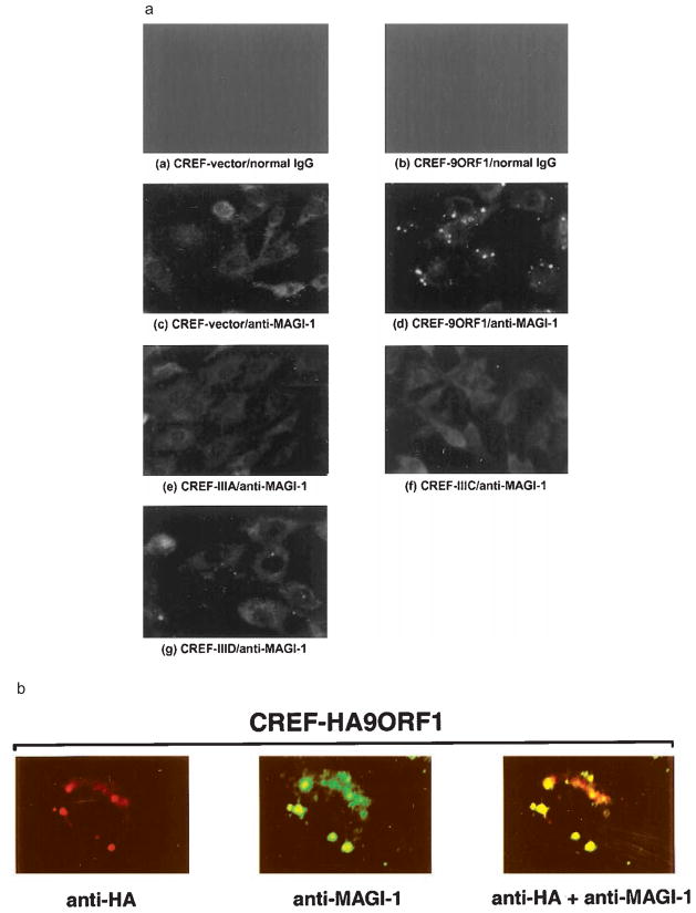
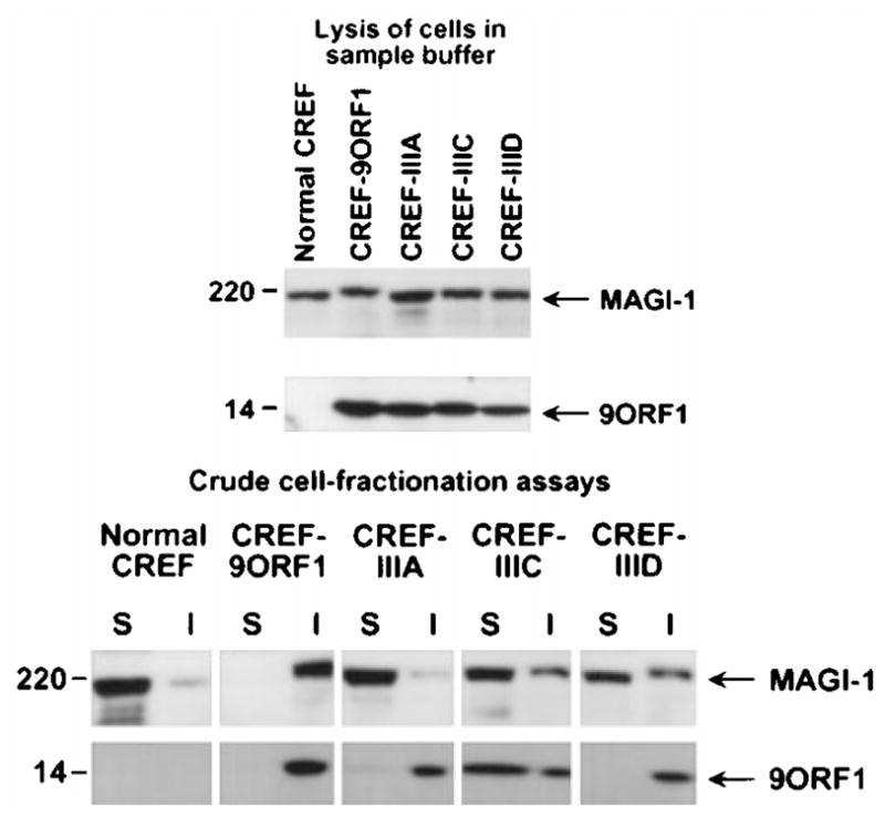
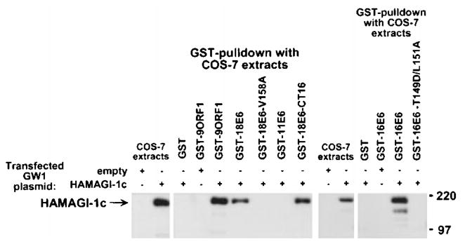
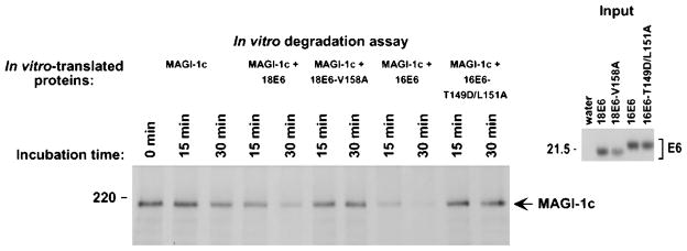
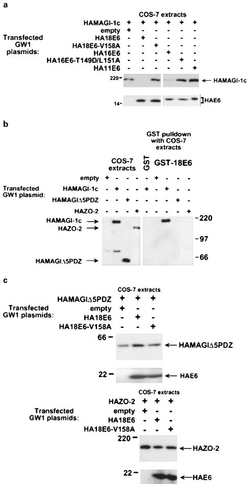
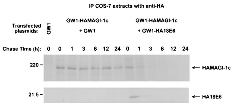
The oncoproteins of small DNA tumor viruses promote tumorigenesis by complexing with cellular factors intimately involved in the control of cell proliferation. The major oncogenic determinants for human adenovirus type 9 (Ad9) and high-risk human papillomaviruses (HPV) are the E4-ORF1 and E6 proteins, respectively. These seemingly unrelated viral oncoproteins are similar in that their transforming activities in cells depend, in part, on a carboxyl-terminal PDZ domain-binding motif which mediates interactions with the cellular PDZ-protein DLG. Here we demonstrated that both Ad9 E4-ORF1 and high-risk HPV E6 proteins also bind to the DLG-related PDZ-protein MAGI-1. These interactions resulted in MAGI-1 being aberrantly sequestered in the cytoplasm by the Ad9 E4-ORF1 protein or being targeted for degradation by high-risk HPV E6 proteins. Transformation-defective mutant viral proteins, however, were deficient for these activities. Our findings indicate that MAGI-1 is a member of a select group of cellular PDZ proteins targeted by both adenovirus E4-ORF1 and high-risk HPV E6 proteins and, in addition, suggest that the tumorigenic potentials of these viral oncoproteins depend, in part, on an ability to inhibit the function of MAGI-1 in cells.


Figures

Figure 1
Figure 1
Three isoforms of MAGI-1. MAGI-1 has an inverted MAGUK domain structure with a guanylate kinase-homology domain (GuK) at its amino-terminus and PDZ domains at its carboxyl-terminus. MAGI-1a, -1b, and -1c isoforms are identical except that their sequences diverge carboxyl-terminal to PDZ5. WW, WW domain; NLS, putative bipartite nuclear localization signal
Figure 2
Figure 2
Binding of 9ORF1 to MAGI-1 in vitro. (a) 9ORF1 protein binding to mouse MAGI-1c detected in GST-pulldown assays. Extracts of RIPA buffer-lysed COS-7 cells transfected with 5 μg of empty GW1 or 5 μg of GWI-HAMAGI-1c plasmid were used in GST-pulldown reactions with the indicated GST fusion protein, and recovered proteins were immunoblotted with HA antibodies. Upper panel, MAGI-1 binding to wild-type and mutant 9ORF1 proteins (see Table 1). Lower panel, MAGI-1 binding to the wild-type E4-ORF1 proteins of Ad9, Ad5 (5ORF1), and Ad12 (12ORF1). COS-7 extracts representing one-half the amount used in GST pulldown reactions were also directly immunoblotted with HA antibodies as a control. (b) 9ORF1 binding to endogenous rat MAGI-1 of CREF cells using GST-pulldown assays. CREF cell extracts in RIPA buffer were subjected to GST-pulldown assays and then immunoblotted with MAGI-1 antibodies
Figure 3
Figure 3
Co-migration of MAGI-1 and 9ORF1-associated protein p180 in a protein gel. GST-pulldown reactions using GST or GST-9ORF1 protein were performed with extracts of human TE85 cells in RIPA buffer. Recovered proteins from duplicate GST-pulldown reactions were separated in parallel by SDS–PAGE and transferred to a membrane, and membranes were either blotted with a radiolabeled 9ORF1 protein probe (left) or with MAGI-1 antibodies (right)
Figure 4
Figure 4
Binding of 9ORF1 to MAGI-1 in vivo. Co-immuno-precipitation assays were performed with extracts of COS-7 cells transfected with 4 μg of GW1-HAMAGI-1 plasmid and either 4 μg empty GW1 or 4 μg of GW1 plasmid expressing wild-type or the indicated mutant 9ORF1 protein. COS-7 extracts in RIPA buffer were subjected to immunoprecipitation with either HA antibodies (upper panel) or 9ORF1 antibodies (lower panel), and recovered proteins were separately immunoblotted with the same two antibodies. In the lower panel, immunoprecipitation of the COS-7 extract with pre-immune serum (pre) was included as a negative control
Figure 5
Figure 5
Binding of 9ORF1 to MAGI-1 PDZ1 and PDZ3. (a) Strong binding of 9ORF1 to two of five MAGI-1 PDZ domains. GST proteins fused to individual MAGI-1 PDZ domains were separated by SDS– PAGE, immobilized on duplicate membranes, and either stained with coomassie or protein blotted with the indicated wild-type or mutant 9ORF1 fusion protein probe. (b) Illustration of MAGI-1 deletion mutants. (c) A MAGI-1 mutant missing PDZ1 and PDZ3 fails to interact with 9ORF1 in GST pulldown assays. Extracts of RIPA buffer-lysed COS-7 cells transfected with 5 μg of GW1 plasmid expressing either HA-tagged wild-type or the indicated mutant MAGI-1 protein were subjected to GST-pulldown reactions with either GST or GST-9ORF1 fusion protein, and recovered proteins were immunoblotted with HA antibodies. COS-7 extracts representing one-tenth the amount used in GST pulldown reactions were also directly immunoblotted with HA antibodies as a control. (d) A MAGI-1 mutant missing PDZ1 and PDZ3 fails to interact with 9ORF1 in co-immunoprecipitation assays. Co-immunoprecipitation assays were performed with extracts of COS-7 cells transfected with 4 μg of wild-type or mutant GW1-HAMAGI-1 plasmid and either 4 μg empty GW1 or 4 μg of GW1-9ORF1 plasmid. COS-7 extracts in RIPA buffer were subjected to immunoprecipitation with 9ORF1 antibodies, and recovered proteins were separately immunoblotted with either HA antibodies (upper panels) or 9ORF1 antibodies (lower panels)
Figure 6
Figure 6
Aberrant sequestration of MAGI-1 in the cytoplasm of 9ORF1-expressing CREF cells. (a) Localization of MAGI-1 in normal CREF cells or CREF cells expressing wild-type or mutant 9ORF1 proteins. Indirect immunofluorescence assays were performed with normal CREF cells (panels a and c) or CREF cells stably expressing wild-type 9ORF1 (panels b and d), mutant IIIA (panel e), mutant IIIC (panel f), or mutant IIID (panel g). Cells were reacted with either normal rabbit IgG (panels a–b) or MAGI-1 antibodies (panels c–g) and visualized by fluorescence microscopy. (b) Co-localization of 9ORF1 and MAGI-1 proteins in CREF cells. Double-label indirect immunofluorescence assays were performed by reacting CREF cells stably expressing HA epitope-tagged 9ORF1 (CREF-HA9ORF1) with both HA and MAGI-1 antibodies. Each of the three panels represents the same field containing a single representative cell stained for 9ORF1 (left panel), MAGI-1 (center panel), or the merged images (right panel)
Figure 7
Figure 7
Aberrant sequestration of MAGI-1 within RIPA buffer-insoluble complexes in 9ORF1-expressing CREF cells. Normal CREF cells or CREF cells stably expressing wild-type or mutant 9ORF1 protein were lysed either in sample buffer (upper panel) or in RIPA buffer and subsequently centrifuged to yield RIPA buffer-soluble supernatant (S) and RIPA buffer-insoluble pellet (I) fractions (lower panel). Extracts of sample buffer-lysed cells or equal volumes of the S and I fractions from RIPA buffer-lysed cells were separately immunoblotted with MAGI-1 antibodies or 9ORF1 antiserum
Figure 8
Figure 8
Binding of high-risk HPV E6 oncoproteins to MAGI-1 in vitro. Extracts of COS-7 cells transfected with 5 μg of empty GW1 or 5 μg of GWI-HAMAGI-1c plasmid were subjected to GST-pulldown reactions with the indicated GST fusion protein, and recovered proteins were immunoblotted with HA antibodies. GST pulldown assays with the indicated GST fusion protein were performed with COS-7 extracts in RIPA buffer or NETN buffer. COS-7 extracts representing one-tenth the amount used in GST pulldown reactions were also directly immunoblotted with HA antibodies as a control
Figure 9
Figure 9
HPV-18 E6-induced degradation of MAGI-1 in vitro. In vitro-translated FLAG epitope-tagged MAGI-1c (MAGI-1c) was mixed with in vitro-translated wild-type 18E6, mutant 18E6-V158A, wild-type 16E6, mutant 16E6-T149D/L151A, or control water-primed lysates and incubated at 30°C for the indicated times. At each time point, reactions were immunoprecipitated with FLAG antibodies, and recovered proteins were separated by SDS–PAGE and visualized by autoradiography (left panel). The amount of in vitro-translated E6 protein used in each assay is shown (right panel)
Figure 10
Figure 10
Selective reduction in MAGI-1 protein levels in vivo by high-risk HPV E6 proteins. (a) Decrease in the steady-state protein levels of MAGI-1 induced by high-risk HPV E6 oncoproteins. Extracts of RIPA buffer-lysed COS-7 cells transfected with 0.5 μg of GW1-HAMAGI-1c plasmid alone or in combination with 4 μg of a GW1 plasmid expressing the indicated wild-type or mutant HPV E6 protein were immunoblotted with HA antibodies. (b) Failure of 18E6 to bind a MAGI-1 deletion mutant missing all five PDZ domains (MAGI-1Δ5PDZ) or the wild-type ZO-2 protein. Extracts of RIPA buffer-lysed COS-7 cells transfected with 5 μg of the indicated GW1 expression plasmid were subjected to GST-pulldown reactions with either GST or GST-18E6 protein. Recovered proteins were immunoblotted with HA antibodies. COS-7 extracts representing one-tenth the amount used in GST pulldown reactions were also directly immunoblotted with HA antibodies as a control. (c) Inability of 18E6 to reduce the steady-state protein levels of MAGI-1Δ5PDZ or ZO-2 in cells. Extracts of RIPA buffer-lysed COS-7 cells transfected with 0.1 μg of GWI-HAMAGIΔ5PDZ plasmid (upper panel) or with 0.01 μg of GW1-HAZO-2 plasmid (lower panel), alone or in combination with 4 μg of GW1 plasmid expressing wild-type or mutant HA18E6, were immunoblotted with HA antibodies
Figure 11
Figure 11
Decrease in MAGI-1 protein half-life induced by the high-risk HPV-18 E6 protein. COS-7 cells transfected with 0.5 μg of GW1-HAMAGI-1c plasmid in combination with 4 μg of empty GW1 or GW1-HA18E6 plasmid were pulse-labeled for 15 min with [35S] EXPRESS protein label and subsequently chased with unlabeled culture medium for the indicated times. At each time point, cell extracts were immunoprecipitated with HA antibodies. Recovered MAGI-1 protein was visualized by autoradiography and quantified by phosphorimager analysis

Similar articles

Cited by

References

    1. Bosch FX, Manos MM, Munoz N, Sherman M, Jansen AM, Peto J, Schiffman MH, Moreno V, Kurman R, Shah KV. J Natl Cancer Inst. 1995;87:796–802. - PubMed
    1. Bradford MM. Anal Biochem. 1976;72:248–254. - PubMed
    1. Craven SE, Bredt DS. Cell. 1998;93:495–498. - PubMed
    1. Dobrosotskaya I, Guy RK, James GL. J Biol Chem. 1997;272:31589–31597. - PubMed
    1. Fanning AS, Anderson JM. J Clin Invest. 1999;103:767–772. - PMC - PubMed

Publication types

MeSH terms

LinkOut - more resources