Corticolimbic interactions associated with performance on a short-term memory task are modified by age
- PMID: 11069948
- PMCID: PMC6773167
- DOI: 10.1523/JNEUROSCI.20-22-08410.2000
Corticolimbic interactions associated with performance on a short-term memory task are modified by age
Abstract
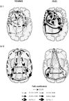
Aging has been associated with a decline in memory abilities dependent on hippocampal processing. We investigated whether the functional interactions between the hippocampus and related cortical areas were modified by age. Young and old subjects' brain activity was measured using positron emission tomography (PET) while they performed a short-term memory task (delayed visual discrimination) in which they determined which of two successively presented sine-wave gratings had the highest spatial frequency. Behavioral performance was equal for the two groups. Partial least squares (PLS) analysis of PET images identified a hippocampal voxel whose activity was similarly correlated with performance across groups. Using this voxel as a seed, a second PLS analysis identified cortical regions functionally connected to the hippocampus. Quantification of the neural interactions with structural equation modeling suggested that a different hippocampal network supported performance in the elderly. Unlike the neural network engaged by the young, which included prefrontal cortex Brodmann's area (BA) 10, fusiform gyrus, and posterior cingulate gyrus, the network recruited by the old included more anterior areas, i.e., dorsolateral prefrontal cortex (BA 9/46), middle cingulate gyrus, and caudate nucleus. Recruitment of a distinct corticolimbic network for visual memory in the elderly suggests that age-related neurobiological deterioration not only results in focal changes but also in the modification of large-scale network operations.
Figures


Similar articles
-
The cognitive control network: Integrated cortical regions with dissociable functions.Neuroimage. 2007 Aug 1;37(1):343-60. doi: 10.1016/j.neuroimage.2007.03.071. Epub 2007 Apr 25. Neuroimage. 2007. PMID: 17553704
-
Changes in limbic and prefrontal functional interactions in a working memory task for faces.Cereb Cortex. 1996 Jul-Aug;6(4):571-84. doi: 10.1093/cercor/6.4.571. Cereb Cortex. 1996. PMID: 8670683 Clinical Trial.
-
Aging gracefully: compensatory brain activity in high-performing older adults.Neuroimage. 2002 Nov;17(3):1394-402. doi: 10.1006/nimg.2002.1280. Neuroimage. 2002. PMID: 12414279
-
Mapping cognition to the brain through neural interactions.Memory. 1999 Sep-Nov;7(5-6):523-48. doi: 10.1080/096582199387733. Memory. 1999. PMID: 10659085 Review.
-
Hemispheric asymmetry reduction in older adults: the HAROLD model.Psychol Aging. 2002 Mar;17(1):85-100. doi: 10.1037//0882-7974.17.1.85. Psychol Aging. 2002. PMID: 11931290 Review.
Cited by
-
Aging-Related Alterations to Persistent Firing in the Lateral Entorhinal Cortex Contribute to Deficits in Temporal Associative Memory.Front Aging Neurosci. 2022 Mar 11;14:838513. doi: 10.3389/fnagi.2022.838513. eCollection 2022. Front Aging Neurosci. 2022. PMID: 35360205 Free PMC article. Review.
-
Multivariate analyses suggest genetic impacts on neurocircuitry in schizophrenia.Neuroreport. 2008 Apr 16;19(6):603-7. doi: 10.1097/WNR.0b013e3282fa6d8d. Neuroreport. 2008. PMID: 18382271 Free PMC article.
-
II. Temporal patterns of longitudinal change in aging brain function.Neurobiol Aging. 2008 Apr;29(4):497-513. doi: 10.1016/j.neurobiolaging.2006.11.011. Epub 2006 Dec 18. Neurobiol Aging. 2008. PMID: 17178430 Free PMC article.
-
Prominent hippocampal CA3 gene expression profile in neurocognitive aging.Neurobiol Aging. 2011 Sep;32(9):1678-92. doi: 10.1016/j.neurobiolaging.2009.10.005. Epub 2009 Nov 13. Neurobiol Aging. 2011. PMID: 19913943 Free PMC article.
-
Age-related neural changes in autobiographical remembering and imagining.Neuropsychologia. 2011 Nov;49(13):3656-69. doi: 10.1016/j.neuropsychologia.2011.09.021. Epub 2011 Sep 19. Neuropsychologia. 2011. PMID: 21945808 Free PMC article.
References
-
- Arikuni T, Sako H, Murata A. Ipsilateral connections of the anterior cingulate cortex with the frontal and medial temporal cortices in the macaque monkey. Neurosci Res. 1994;21:19–39. - PubMed
-
- Bach ME, Barad M, Son H, Zhuo M, Lu YF, Shih R, Mansuy I, Hawkins RD, Kandel ER. Age-related defects in spatial memory are correlated with defects in the late phase of hippocampal long-term potentiation in vitro and are attenuated by drugs that enhance the cAMP signaling pathway. Proc Natl Acad Sci USA. 1999;96:5280–5285. - PMC - PubMed
-
- Bachevalier J, Meunier M, Lu MX, Ungerleider LG. Thalamic and temporal cortex input to medial prefrontal cortex in rhesus monkeys. Exp Brain Res. 1997;115:430–444. - PubMed
-
- Barnes CA. Memory deficits associated with senescence: a neurophysiological and behavioral study in the rat. J Comp Physiol. 1979;931:74–104. - PubMed
Publication types
MeSH terms
LinkOut - more resources
Full Text Sources
Medical
