. 2000 Nov;106(9):1127-37.
doi: 10.1172/JCI9914.

Cathepsin B contributes to TNF-alpha-mediated hepatocyte apoptosis by promoting mitochondrial release of cytochrome c

Affiliations

Cathepsin B contributes to TNF-alpha-mediated hepatocyte apoptosis by promoting mitochondrial release of cytochrome c

M E Guicciardi et al. J Clin Invest. 2000 Nov.

Abstract

TNF-alpha-induced apoptosis is thought to involve mediators from acidic vesicles. Cathepsin B (cat B), a lysosomal cysteine protease, has recently been implicated in apoptosis. To determine whether cat B contributes to TNF-alpha-induced apoptosis, we exposed mouse hepatocytes to the cytokine in vitro and in vivo. Isolated hepatocytes treated with TNF-alpha in the presence of the transcription inhibitor actinomycin D (AcD) accumulated cat B in their cytosol. Further experiments using cell-free systems indicated that caspase-8 caused release of active cat B from purified lysosomes and that cat B, in turn, increased cytosol-induced release of cytochrome c from mitochondria. Consistent with these observations, the ability of TNF-alpha/AcD to induce mitochondrial release of cytochrome c, caspase activation, and apoptosis of isolated hepatocytes was markedly diminished in cells from CatB(-/-) mice. Deletion of the CatB gene resulted in diminished liver injury and enhanced survival after treatment in vivo with TNF-alpha and an adenovirus construct expressing the IkappaB superrepressor. Collectively, these observations suggest that caspase-mediated release of cat B from lysosomes enhances mitochondrial release of cytochrome c and subsequent caspase activation in TNF-alpha-treated hepatocytes.


Figures
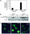
Figure 1
Figure 1
Cat B contributes to TNF-α/AcD–induced hepatocyte apoptosis. Isolated hepatocytes from catB+/+ and catB–/– mice were incubated in the absence (control) or presence of TNF-α (28 ng/ml) and AcD (0.2 μg/ml) for up to 12 hours. (a) Intracellular cat B activity was measured fluorometrically in the cells after 4 hours of treatment using the fluorogenic substrate VLK-CMAC and digitized video microscopy, as described in Methods. (b) At the indicated time points, cytosolic extracts were prepared by selective permeabilization with digitonin as described in Methods and subjected to immunoblot analysis using an anti–cat B antiserum. Locations of 30-kDa (p30) and 27-kDa (p27) active fragments of cat B are indicated. Immunoblot analysis of β-actin was performed as a control for protein loading. Cont., control. (c) Cultured McNtcp.24 cells grown on collagen-coated glass coverslips were transfected with the plasmid construct encoding the cat B-GFP fusion protein (control, TNF-α) or double-transfected with the cat B-GFP plasmid and the plasmid encoding the viral protein CrmA (CrmA + TNF-α). Forty-eight hours later, cells were incubated in the absence (control) or presence of TNF-α/AcD at 37°C for 2 hours and transferred to the stage of an inverted confocal microscope. Cat B-GFP fluorescence was imaged as described in Methods.
Figure 2
Figure 2
Caspase-8 induces release of cat B from lysosomes. Isolated lysosomes from catB+/+ mouse liver (10 μg protein) were incubated at 37°C with either active recombinant human caspase-8 (20 ng) (a) or rabbit m-calpain (10–25 ng) (b), in the absence or presence of S-100 cytosol fraction (50 μg protein) and the caspase inhibitor Z-VAD-fmk (20 μM). Lysosomes were also treated with 0.1% Triton-X 100 to induce maximal release of cat B (positive control). After 1 hour, lysosomes were pelleted by centrifugation at 15,000 g for 30 minutes. Supernatants were subjected SDS-PAGE on gels containing 10% acrylamide, transferred to nitrocellulose, and probed with anti–cat B antiserum.
Figure 3
Figure 3
catB–/– mouse hepatocytes are more resistant to TNF-α–induced apoptosis. Isolated hepatocytes from catB+/+ and catB–/– mice were incubated in the absence (control) or presence of TNF-α and AcD. (a) Apoptosis was quantitated in catB+/+ and catB–/– hepatocytes at different times of incubation after staining with both FITC-annexin V (dotted lines) and DAPI (solid lines). Cells were considered apoptotic if either externalization of phosphatidylserine residues on the plasma membrane or chromatin condensation and nuclear fragmentation occurred. At least 300 cells in six high-power fields were counted by an individual blinded to the experimental conditions. (b) Apoptosis was quantitated in catB+/+ and catB–/– hepatocytes by DAPI staining after 24 hours of incubation in medium lacking (control) or containing either TNF-α and AcD (TNF-α) or AcD alone (AcD). catB+/+ hepatocytes were also treated with TNF-α and AcD after a 30-minute preincubation with CA-074, a pharmacological inhibitor of cat B. Results are representative of at least three independent experiments using cells from three separate isolations and are expressed as mean ± SEM. Data were compared using a one-tail t test. AP < 0.05, catB–/– vs. catB+/+; BP < 0.05, catB+/+ + CA-074 vs. catB+/+. (c) Isolated mouse hepatocytes from catB+/+ (filled bars) and catB–/– (open bars) mice were infected with an adenovirus expressing the IκB-superrepressor (Ad5IκB) or with an empty adenovirus (Ad5ΔE1) as a negative control, and treated with TNF-α (28 ng/ml) for 12 hours. Apoptosis was quantitated after staining with DAPI. Results are representative of three independent experiments performed in triplicate from separate isolations and are expressed as mean ± SEM.
Figure 4
Figure 4
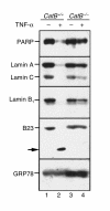
Cleavage of PARP, lamins, and B23 after TNF-α/AcD treatment is diminished in catB–/– hepatocytes. Hepatocytes from catB+/+ and catB–/– mice were incubated for 24 hours with medium lacking or containing TNF-α + AcD. Whole-cell lysates were then prepared as described in Methods. Aliquots containing 50 μg protein were subjected to SDS-PAGE on gradient gels containing 5–15% acrylamide, transferred to nitrocellulose, and sequentially blotted for PARP, lamins A and C, lamin B1, or B23. Cleavage of the substrates was detected by the loss of the bands corresponding to the molecular weight of the native protein, and, in the case of B23, by the appearance of a new band (arrow). GRP78 served as a control for protein loading.
Figure 5
Figure 5
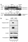
(a) TNF-α–induced release of cytochrome c into the cytosol is reduced in catB–/– mouse hepatocytes. At the indicated times after addition of medium lacking (control) or containing TNF-α + AcD, cytosol fractions were prepared by selective permeabilization with digitonin as described in Methods. Aliquots containing 20 μg of protein were subjected to SDS-PAGE on gels containing 15% acrylamide, transferred to nitrocellulose, and probed for cytochrome c. Samples from catB+/+ hepatocytes were also probed for cytochrome c oxidase, to exclude a possible mitochondrial contamination in the cytosol. (b) Caspase-9 and caspase-3 are processed in TNF-α/AcD–treated catB+/+ hepatocytes but not in catB–/– hepatocytes. Cells were incubated for 24 hours in medium lacking or containing TNF-α + AcD. After whole-cell lysates were prepared as described in Methods, aliquots containing 50 μg protein were subjected to SDS-PAGE, transferred to nitrocellulose membrane, and analyzed by immunoblot using antisera that recognize only active caspase-9 or active caspase-3 (36). The same blot was probed with sera that recognize procaspase-8 and β-actin to confirm loading and transfer of samples from catB–/– mice.
Figure 6
Figure 6
Caspase-8 activation after TNF-α/AcD treatment is reduced in catB–/– cells. After cells were incubated in medium without (cont.) or with TNF-α + AcD for the indicated lengths of time, cytosolic fractions were prepared. Aliquots containing 40 μg protein were subjected to SDS-PAGE on gels containing 15% acrylamide, transferred to nitrocellulose, and immunoblotted for caspase-8. Processing of caspase-8 was detected by the appearance of the 18- to 20-kDa (p20) and 10-kDa (p10) active fragments. β-Actin served as a control for protein loading. Results are representative of three independent experiments.
Figure 7
Figure 7
Cat B–induced release of cytochrome c from mitochondria is enhanced by cytosol and is not due to a nonspecific proteolytic effect. Isolated mitochondria from catB+/+ mouse liver (25 μg protein) were incubated at 37°C with increasing concentrations of purified recombinant cat B (5–50 ng) (a) or m-calpain (10 ng) (b), in the presence or in the absence of S-100 cytosol fraction (50 μg) as described in Methods. Mitochondria were also treated with 0.1% Triton X-100 to induce maximum release of cytochrome c (positive control). After 1 hour, mitochondria were pelleted by centrifugation at 12,000 g for 5 min. Supernatants were subjected to SDS-PAGE on 15% acrylamide gels, transferred to nitrocellulose, and immunoblotted for cytochrome c. Blots were also probed for cytochrome c oxidase (subunit IV) to exclude mitochondria contamination in the supernatant. (c) Active cat B induces release of cytochrome c from isolated catB–/– mouse liver mitochondria in the presence of cytosol. Isolated mitochondria from catB–/– mouse liver (25 μg protein) were incubated at 37°C with purified recombinant cat B (25 ng), in the presence or in the absence of S-100 cytosol fraction (50 μg) obtained from the same animal. Mitochondria were also treated with 0.1% Triton X-100 to induce maximum release of cytochrome c (positive control). After 1 hour, mitochondria were pelleted by centrifugation at 12,000 g for 5 minutes, and the resulting supernatants were subjected to SDS-PAGE and subsequent immunoblot analysis for cytochrome c as already above. Immunoblot for cytochrome c oxidase (subunit IV) was also performed to exclude mitochondria contamination in the supernatant.
Figure 8
Figure 8
catB–/– mice are more resistant to TNF-α–induced liver damage. catB–/– and catB+/+ were injected via tail vein with the adenovirus Ad5IκB (0.35 × 10⁹ pfu/mouse) encoding for an IκB superrepressor. In control experiments, mice were injected with the adenovirus Ad5ΔE1 (0.35 × 10⁹ pfu/mouse in 0.22 ml sterile saline) or with sterile saline (0.22 ml).Twenty-four hours later, each mouse received a dose of 0.5 μg of recombinant mouse TNF-α intravenously. Mice were sacrificed after 2- and 4-hour treatment with TNF-α. (a) Serum alanine aminotransferase (ALT) levels were measured and expressed as mean ± SEM (n = 3). AP < 0.01. ALT values in control samples were < 20 IU/L, except in the Ad5ΔE1-injected mice, in which they were < 750 IU/L at 2 hours and < 1850 IU/L at 4 hours (data not shown). (b) H&E staining of Ad5IκB-injected catB+/+ (left) and catB–/– (right) mouse liver harvested 4 hours after treatment with TNF-α.
Figure 9
Figure 9
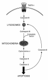
Model of TNF-α signaling pathway through acidic compartment. Triggering of TNFR-1 leads to activation of a small amount of caspase-8, sufficient to induce the release of cat B from the lysosomes. Active cat B in turn promotes the release of cytochrome c from mitochondria by cleaving one or more still unidentified cytosolic substrates. Release of cytochrome c results in cleavage of caspase-9 and caspase-3 followed by further apoptotic changes. Strong activation of caspase-8 occurs downstream of mitochondria, possibly by effector caspases. An amplification loop might generate more active caspase-8, inducing further release of cat B from lysosomes.

Comment in

Similar articles

Cited by

References

    1. Wallach D, et al. Tumor necrosis factor receptor and Fas signaling mechanisms. Annu Rev Immunol. 1999;17:331–367. - PubMed
    1. Colletti LM, et al. Role of tumor necrosis factor-alpha in the pathophysiologic alterations after hepatic ischemia/reperfusion injury in the rat. J Clin Invest. 1990;85:1936–1943. - PMC - PubMed
    1. McClain CJ, Cohen DA. Increased tumor necrosis factor production by monocytes in alcoholic hepatitis. Hepatology. 1989;9:349–351. - PubMed
    1. Gonzalez-Amaro R, et al. Induction of tumor necrosis factor alpha production by human hepatocytes in chronic viral hepatitis. J Exp Med. 1994;179:841–848. - PMC - PubMed
    1. Leist M, et al. Tumor necrosis factor-induced apoptosis during the poisoning of mice with hepatotoxins. Gastroenterology. 1997;112:923–934. - PubMed

Publication types

MeSH terms