Evolution of bovine respiratory syncytial virus
- PMID: 11044116
- PMCID: PMC110946
- DOI: 10.1128/jvi.74.22.10714-10728.2000
Evolution of bovine respiratory syncytial virus
Abstract
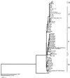
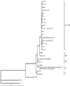
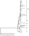
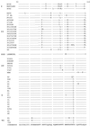
Until now, the analysis of the genetic diversity of bovine respiratory syncytial virus (BRSV) has been based on small numbers of field isolates. In this report, we determined the nucleotide and deduced amino acid sequences of regions of the nucleoprotein (N protein), fusion protein (F protein), and glycoprotein (G protein) of 54 European and North American isolates and compared them with the sequences of 33 isolates of BRSV obtained from the databases, together with those of 2 human respiratory syncytial viruses and 1 ovine respiratory syncytial virus. A clustering of BRSV sequences according to geographical origin was observed. We also set out to show that a continuous evolution of the sequences of the N, G, and F proteins of BRSV has been occurring in isolates since 1967 in countries where vaccination was widely used. The exertion of a strong positive selective pressure on the mucin-like region of the G protein and on particular sites of the N and F proteins is also demonstrated. Furthermore, mutations which are located in the conserved central hydrophobic part of the ectodomain of the G protein and which result in the loss of four Cys residues and in the suppression of two disulfide bridges and an alpha helix critical to the three-dimensional structure of the G protein have been detected in some recent French BRSV isolates. This conserved central region, which is immunodominant in BRSV G protein, thus has been modified in recent isolates. This work demonstrates that the evolution of BRSV should be taken into account in the rational development of future vaccines.
Figures


Similar articles
-
Sequence conservation in the attachment glycoprotein and antigenic diversity among bovine respiratory syncytial virus isolates.Vet Microbiol. 1997 Mar;54(3-4):201-21. doi: 10.1016/s0378-1135(96)01288-6. Vet Microbiol. 1997. PMID: 9100323
-
A bovine respiratory syncytial virus strain with mutations in subgroup-specific antigenic domains of the G protein induces partial heterologous protection in cattle.Vet Microbiol. 1998 Oct;63(2-4):159-75. doi: 10.1016/s0378-1135(98)00244-2. Vet Microbiol. 1998. PMID: 9850996
-
Serological and genetic characterisation of bovine respiratory syncytial virus (BRSV) indicates that Danish isolates belong to the intermediate subgroup: no evidence of a selective effect on the variability of G protein nucleotide sequence by prior cell culture adaption and passages in cell culture or calves.Vet Microbiol. 1998 Aug 15;62(4):265-79. doi: 10.1016/s0378-1135(98)00226-0. Vet Microbiol. 1998. PMID: 9791873
-
[Immunobiology of bovine respiratory syncytial virus infections].Tijdschr Diergeneeskd. 1998 Nov 15;123(22):658-62. Tijdschr Diergeneeskd. 1998. PMID: 9836385 Review. Dutch.
-
Bovine respiratory syncytial virus infection: immunopathogenic mechanisms.Anim Health Res Rev. 2007 Dec;8(2):207-13. doi: 10.1017/S1466252307001405. Anim Health Res Rev. 2007. PMID: 18218161 Review.
Cited by
-
Bovine Respiratory Syncytial Virus Enhances the Adherence of Pasteurella multocida to Bovine Lower Respiratory Tract Epithelial Cells by Upregulating the Platelet-Activating Factor Receptor.Front Microbiol. 2020 Jul 31;11:1676. doi: 10.3389/fmicb.2020.01676. eCollection 2020. Front Microbiol. 2020. PMID: 32849350 Free PMC article.
-
Evaluating the potential of whole-genome sequencing for tracing transmission routes in experimental infections and natural outbreaks of bovine respiratory syncytial virus.Vet Res. 2022 Dec 12;53(1):107. doi: 10.1186/s13567-022-01127-9. Vet Res. 2022. PMID: 36510312 Free PMC article.
-
Prevalence and Molecular Characteristics of Bovine Respiratory Syncytial Virus in Beef Cattle in China.Animals (Basel). 2022 Dec 12;12(24):3511. doi: 10.3390/ani12243511. Animals (Basel). 2022. PMID: 36552433 Free PMC article.
-
Recombinant bovine respiratory syncytial virus with deletion of the SH gene induces increased apoptosis and pro-inflammatory cytokines in vitro, and is attenuated and induces protective immunity in calves.J Gen Virol. 2014 Jun;95(Pt 6):1244-1254. doi: 10.1099/vir.0.064931-0. Epub 2014 Apr 3. J Gen Virol. 2014. PMID: 24700100 Free PMC article.
-
Pre-fusion RSV F strongly boosts pre-fusion specific neutralizing responses in cattle pre-exposed to bovine RSV.Nat Commun. 2017 Oct 20;8(1):1085. doi: 10.1038/s41467-017-01092-4. Nat Commun. 2017. PMID: 29057917 Free PMC article.
References
-
- Anderson L J, Heirholzer J C, Tson C, Hendry R M, Fernie B N, Stone Y, McIntosh K. Antigenic characterization of respiratory syncytial virus strains with monoclonal antibodies. J Infect Dis. 1985;151:626–633. - PubMed
-
- Beem M. Repeated infections with respiratory syncytial virus. J Immunol. 1987;98:1115–1122. - PubMed
-
- Bourhy H, Kissi B, Audry L, Smreczak M, Sadkowska-Todys M, Kulonen K, Tordo N, Zmudzinski J F, Holmes E C. Ecology and evolution of rabies virus in Europe. J Gen Virol. 1999;99:2545–2557. - PubMed
-
- Bracho M A, Moya A, Barrio E. Contribution of Taq polymerase-induced errors to the estimation of RNA virus diversity. J Gen Virol. 1998;79:2921–2928. - PubMed
Publication types
MeSH terms
Substances
LinkOut - more resources
Full Text Sources
Medical

