. 2000 Oct 15;14(20):2650-63.
doi: 10.1101/gad.836400.

Different human TFIIIB activities direct RNA polymerase III transcription from TATA-containing and TATA-less promoters

Affiliations

Different human TFIIIB activities direct RNA polymerase III transcription from TATA-containing and TATA-less promoters

L Schramm et al. Genes Dev. .

Abstract

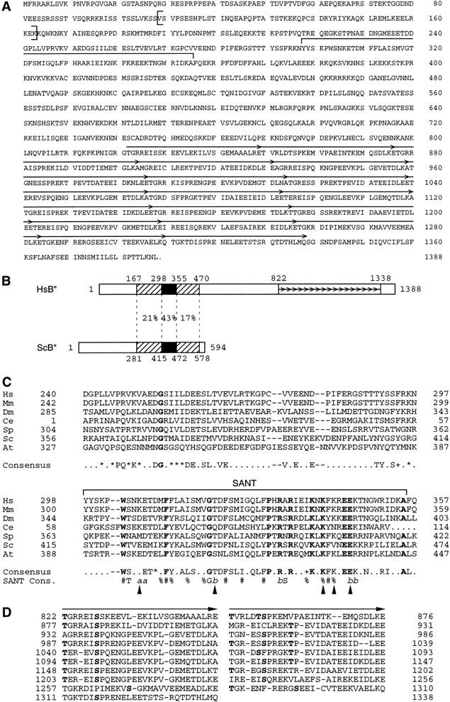
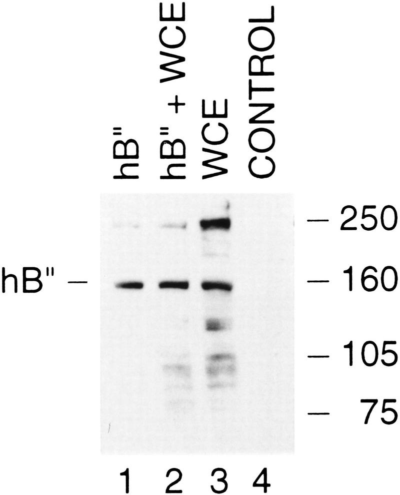
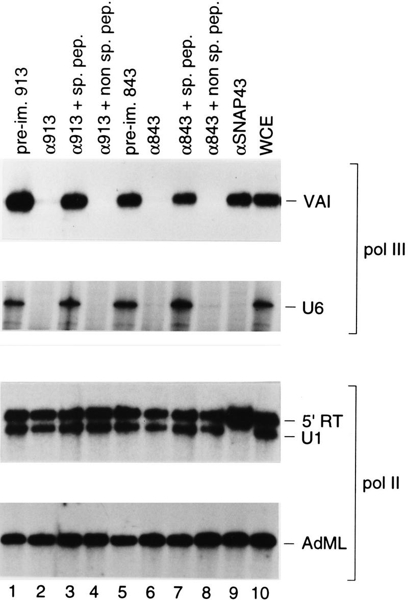
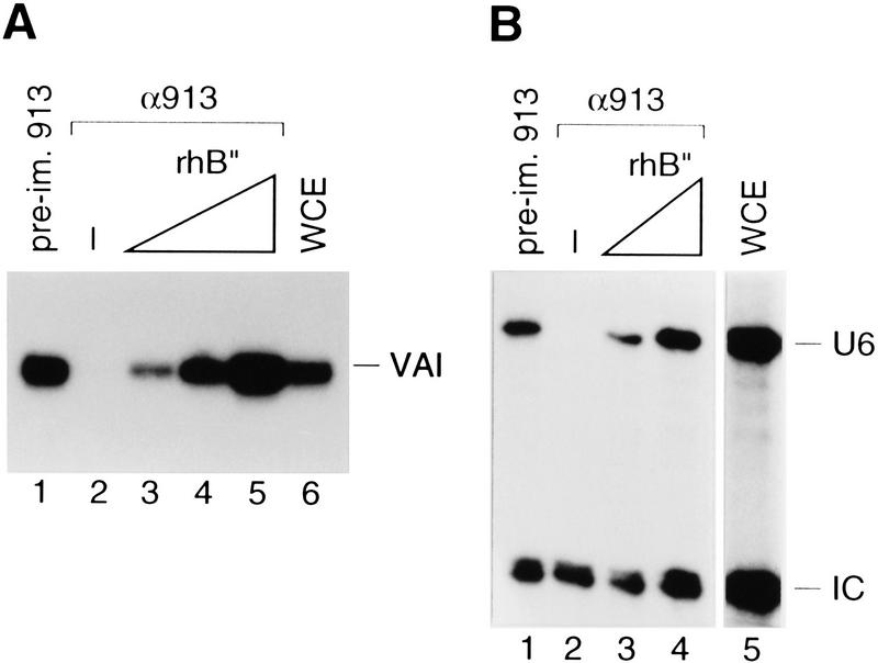
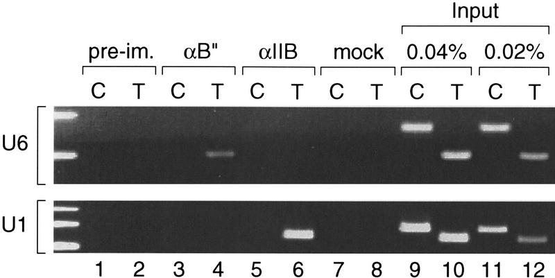
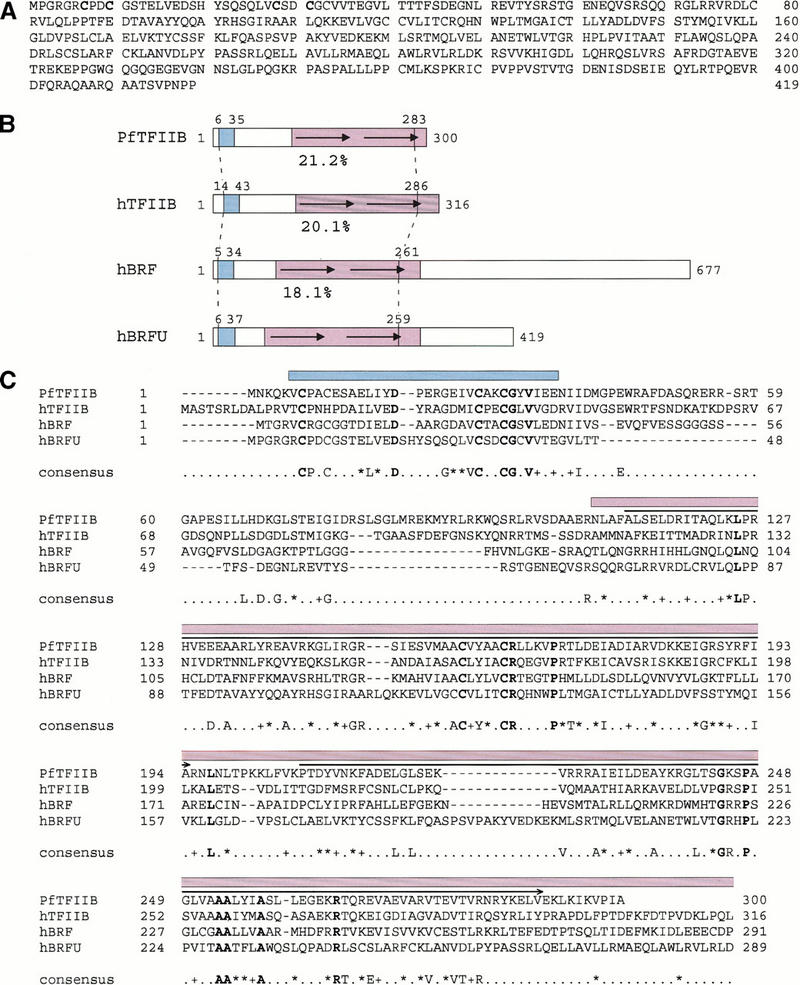
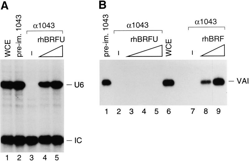
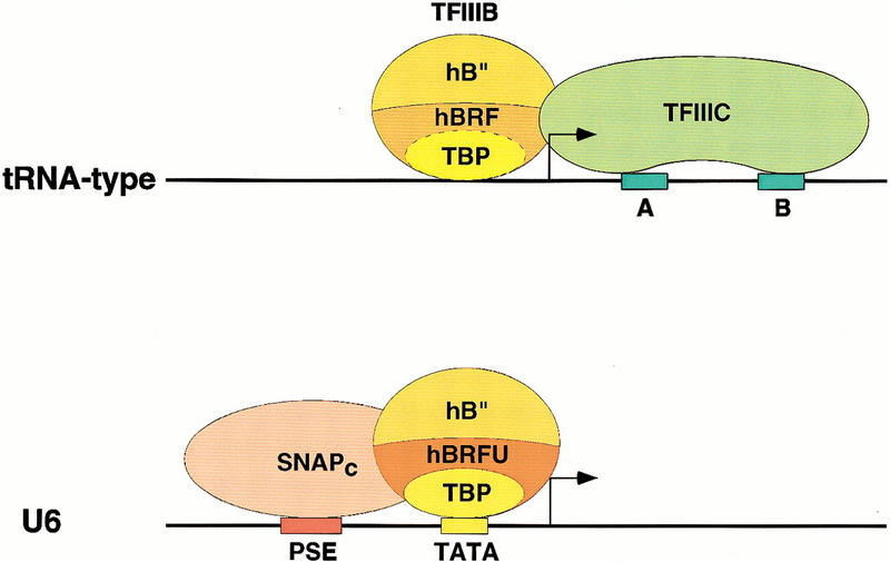
Transcription initiation at RNA polymerase III promoters requires transcription factor IIIB (TFIIIB), an activity that binds to RNA polymerase III promoters, generally through protein-protein contacts with DNA binding factors, and directly recruits RNA polymerase III. Saccharomyces cerevisiae TFIIIB is a complex of three subunits, TBP, the TFIIB-related factor BRF, and the more loosely associated polypeptide beta("). Although human homologs for two of the TFIIIB subunits, the TATA box-binding protein TBP and the TFIIB-related factor BRF, have been characterized, a human homolog of yeast B(") has not been described. Moreover, human BRF, unlike yeast BRF, is not universally required for RNA polymerase III transcription. In particular, it is not involved in transcription from the small nuclear RNA (snRNA)-type, TATA-containing, RNA polymerase III promoters. Here, we characterize two novel activities, a human homolog of yeast B("), which is required for transcription of both TATA-less and snRNA-type RNA polymerase III promoters, and a factor equally related to human BRF and TFIIB, designated BRFU, which is specifically required for transcription of snRNA-type RNA polymerase III promoters. Together, these results contribute to the definition of the basal RNA polymerase III transcription machinery and show that two types of TFIIIB activities, with specificities for different classes of RNA polymerase III promoters, have evolved in human cells.


Figures

Figure 1
Figure 1
Structure of a hB′′. (A) Predicted hB′′ protein sequence (GenBank accession number AF298151). The SANT domain is indicated by a large horizontal bracket above the sequence. The repeated sequences are indicated by arrows. The sequence between the vertical brackets (amino acids 109–163) is lacking in some cDNAs that probably correspond to splicing variants. (B) Regions of similarity in the Homo sapiens and S. cerevisiae B′′ sequences (HsB′′ and ScB′′, respectively). The black boxes correspond to the SANT domain; the hatched boxes indicate regions of lower but still significant similarity on either side of the SANT domain. The percentages indicate identities between the two proteins in the regions delimited by the dotted lines. The numbers above the boxes indicate aa numbering. The small arrows indicate the repeats in hB′′. (C) Alignment of the SANT domain and region immediately upstream from hB′′ (Hs) and ScB′′ (Sc), as well as putative mouse (Mm), Drosophila melanogaster (Dm), Caenorhabditis elegans (Ce), Schizosaccharomyces pombe (Sp), and Arabidopsis thaliana (At) B′′ homologs. The SANT domain is indicated by the horizontal bracket above the sequence. The region N-terminal of the SANT domain was shown, in ScB′′, to be required for efficient transcription of linear yeast U6 templates in vitro (Kassavetis et al. 1998). The amino acids in boldface are identical in all sequences. The amino acids indicated in the consensus are identical in at least four of the seven sequences. (+) similar amino acids in all sequences; (*) similar amino acids in at least five of the seven sequences. The following amino acids were considered similar: V, I, L, and M; D and E; N and Q; R, K, and H; S and T; and W, F, and Y. The SANT consensus sequence (Aasland et al. 1996) is also shown. In the SANT consensus: (%) semiconserved hydrophobicity; (#) strongly conserved hydrophobicity; (a) conserved acidic residues; and (b) conserved basic residues. The positions marked with arrowheads indicate positions not conserved in the B′′ and SANT consensus sequences. The GenBank accession numbers are as follows: Mm ESTs, AI315861, AI315862, and AI787462; Ce genomic sequences U97016 (B0261) (nucleotides 25059–25185 and 25518–25780); Dm protein, AAF53291.1 (CG9305 gene product); Sp protein, T41239; and At protein T08564 (hypothetical protein T22F8.60). The numberings correspond to the sequence shown in (A) for HsB′′, to the sequences published (Kassavetis et al. 1995; Roberts et al. 1996; Rüth et al. 1996) for ScB′′, and to the sequences corresponding to the protein accession numbers indicated for other sequences. For the Mm sequence, the numbering corresponds to a putative mouse B′′ N-terminal region (amino acids 1–377) assembled from an alignment of various ESTs (GenBank accession numbers AI527964, AI317698, AI787358, AI526613, AI316225, AI639984, AA168265, AW456745, AW049390, AI429656, AI430369, AI450927, W91010, AI787462, AI315861, AI315862, AI892036, and AI315863), and for the Ce sequence, the numbering is arbitrary. (D) Alignment of the repeats within the C-terminal half of hB′′. As indicated by the arrows, each repeat can itself be subdivided into two short imperfect repeats. Residues in boldface indicate potential phosphorylation sites for various kinases, including PKC, PKA, CAMII, and CKII kinases.
Figure 2
Figure 2
rhB′′ comigrates with endogenous B′′ from HeLa cells. The immunoblot was performed with an anti-hB′′ antibody (αhB′′-2, CS913). (Lane 1) 5 μL of hB′′ translated in vitro; (lane 2) 5 μL of hB′′ translated in vitro and 2.5 μL of whole HeLa-cell extract (WCE); (lane 3) 2.5 μL of HeLa-cell extract; and (lane 4) 5 μL of unprogrammed rabbit reticulocyte lysates (control). The position of hB′′ and the positions of molecular weight markers are indicated.
Figure 3
Figure 3
Depletion of hB′′ debilitates RNA polymerase III, but not RNA polymerase II, transcription in vitro. Whole-cell extract was treated with beads coated with either the preimmune antibodies (lanes 1 and 5) or the anti-B′′ antibodies (lanes 2–4 and 6–8) indicated above the lanes (913 is αhB′′-2; 843 is αhB′′-3). In lanes 3 and 7, and in lanes 4 and 8, the antibody beads were preincubated either with the specific peptide against which the antibodies were raised or with nonspecific peptide, as indicated. In lane 9, the extract was treated with beads coated with an antibody directed against the SNAP43 subunit of SNAPc. Aliquots of the variously treated extracts were then tested in parallel for their ability to direct transcription from the Ad2 VAI, human U6, human U1, and Ad2 major late (AdML) promoters. The bands corresponding to correctly initiated RNA are indicated in each panel. In the U1 panel, the band labeled 5′ RT corresponds to RNA initiated within the vector and reading through the U1 snRNA promoter (Sadowski et al. 1993). Lane 10 shows transcription in untreated whole-cell extract.
Figure 4
Figure 4
hB′′ is required for RNA polymerase III transcription from the Ad2 VAI and human U6 promoters. (A) Whole-cell extract was treated with beads coated with either preimmune (lane 1) or anti-hB′′ (lanes 2–5) antibodies and tested for transcription from the Ad2 VAI promoter. In lanes 3–5, increasing amounts of rhB′′ (rhB′′) expressed in E. coli were added to the depleted extract. Lane 6 shows VAI transcription in untreated whole-cell extract. (B) Whole-cell extract treated with beads coated with the preimmune (lane 1) or anti-hB′′ (lanes 2–4) antibodies and tested for U6 transcription. In lanes 3 and 4, increasing amounts of rhB′′ expressed in E. coli were added to the depleted extract. Lane 5 shows U6 transcription in untreated whole-cell extract. (IC) internal control signal.
Figure 5
Figure 5
hB′′ is found in the U6 promoter region in vivo. Rapidly growing HeLa cells were treated with formaldehyde; then cross-linked chromatin was extracted, sonicated, and used as starting material for immunoprecipitations with beads coated with either preimmune (lanes 1 and 2), anti-hB′′ (lanes 3 and 4), or anti-TFIIB (lanes 5 and 6) antibodies, or just beads alone (lanes 7 and 8). The immunoprecipitated material was then analyzed by PCR with test (T) primers specific for the U6 (upper panel) or U1 (lower panel) promoters, or control (C) primers hybridizing to a unique region located 4 kb upstream of the human U6 snRNA gene (upper panel) or a unique region within the 45-kb U1 repeat (Bernstein et al. 1985) 7 kb upstream of the U1 gene (see Materials and Methods for details). Lanes 9–12 show PCRs performed with the test (T) or control (C) primers with 0.04% and 0.02% of the input material used for immunoprecipitation, as indicated above the lanes.
Figure 6
Figure 6
Structure of hBRFU. (A) Predicted hBRFU protein sequence (GenBank accession number AF298153). (B) Regions of similarities in the Pyrococcus furiosus TFIIB homolog (PfTFIIB) and the hTFIIB, hBRF, and hBRFU sequences. The location of the structured zinc ribbon as determined in PfTFIIB by nuclear magnetic resonance (Zhu et al. 1996), and as modeled in BRF and TFIIB (Hahn and Roberts 2000), and the location of the corresponding region in hBRFU are indicated in blue. The location of the structured core domain of TFIIB (Bagby et al. 1995; Nikolov et al. 1995) and that of the corresponding regions in the other proteins are indicated in purple. The region conserved between the proteins is indicated by the vertical stripped lines. The percentages below the sequences indicate percentage of identity between hBRFU and PfTFIIB, hTFIIB, or hBRF within the region bracketed by the vertical stripped lines as calculated from the alignment shown in C. (C) Alignment of PfTFIIB, hTFIIB, and the N-terminal regions of hBRF and hBRFU. The alignment was performed with the ClustalW program and the default parameters. The amino acids in boldface are identical in all sequences. The amino acids indicated in the consensus are identical in three of the four sequences. (+) similar amino acids in all sequences; (*) similar amino acids in three of the four sequences. The following amino acids were considered similar: V, I, L, and M; D and E; N and Q; R, K, and H; S and T; and W, F, and Y. The horizontal arrows above the sequences indicate the location of the structural repeats (Nikolov et al. 1995). The location of the structured zinc ribbon is indicated in blue, and that of the core (hTFIIB amino acids 113–316) in purple.
Figure 7
Figure 7
hBRFU is expressed in HeLa cells. The immunoblot was performed with an anti-hBRFU/hBRF antibody (CS1043). (Lane 1) rhBRFU expressed in E. coli; (lane 2) rhBRFU expressed in E. coli and 2 μL of P11 B fraction; and (lane 3) 2 μL of P11 B fraction. rhBRFU migrates as a protein of ∼50 kD. The additional bands seen in the P11 fraction probably correspond to cross-reacting proteins.
Figure 8
Figure 8
hBRFU is specifically required for U6 transcription. (A) A whole-cell extract was treated with beads coated with preimmune (lane 2) or CS1043 (lanes 3–5) antibodies and tested for transcription from the U6 promoter. In lanes 4 and 5, increasing amounts of rhBRFU were added to the depleted extract. Lane 1 shows U6 transcription in untreated whole-cell extract. (B) A whole-cell extract was treated with beads coated with preimmune (lane 1) or CS1043 (lanes 2–5 and 7–9) antibodies and tested for transcription from the VAI promoter. In lanes 3–5, increasing amounts of rhBRFU were added to the depleted extract. In lanes 8 and 9, increasing amounts of rhBRF were added to the depleted extract. Lane 6 shows U6 transcription in untreated whole-cell extract.
Figure 9
Figure 9
Model of the initiation complexes formed on tRNA-type and U6-type promoters. The upper panel shows a tRNA-type promoter with the gene internal A and B box promoter elements recruiting TFIIIC, which in turn can recruit through protein–protein contacts the TFIIIB components TBP, hBRF, and hB′′. The dotted line separating TBP and hBRF indicates that these two factors are tightly associated with each other. The solid line separating hB′′ from the TBP–hBRF complex indicates that hB′′ is only weakly associated with the rest of the complex. The lower panel shows that for the human U6 gene, the external PSE and TATA box core promoter elements recruit SNAPc and the TFIIIB component TBP, respectively. The binding of these factors probably allows the recruitment of hB′′ and a TFIIIB component specific for PSE-containing RNA polymerase III promoters, hBRFU. The solid lines separating TBP, hBRFU, and hB′′ indicate that in this case, the TFIIIB components are either not associated or only weakly associated with each other.

Similar articles

Cited by

References

    1. Aasland R, Stewart AF, Gibson T. The SANT domain: A putative DNA-binding domain in the SWI-SNF and ADA complexes, the transcriptional co-repressor N-CoR and TFIIIB. Trends Biochem Sci. 1996;21:87–88. - PubMed
    1. Bagby S, Kim S, Maldonado E, Tong KI, Reinberg D, Ikura M. Solution structure of the C-terminal core domain of human TFIIB: Similarity to cyclin A and interaction with TATA-binding protein. Cell. 1995;82:857–867. - PubMed
    1. Berger B, Wilson DB, Wolf E, Tonchev T, Milla M, Kim PS. Predicting coiled coils by use of pairwise residue correlations. Proc Natl Acad Sci. 1995;92:8259–8263. - PMC - PubMed
    1. Bernstein LB, Manser T, Weiner AM. Human U1 small nuclear RNA genes: Extensive conservation of flanking sequences suggests cycles of gene amplification and transposition. Mol Cell Biol. 1985;5:2159–2171. - PMC - PubMed
    1. Boyd KE, Wells J, Gutman J, Bartley SM, Farnham PJ. c-Myc target gene specificity is determined by a post–DNA binding mechanism. Proc Natl Acad Sci. 1998;95:13887–13892. - PMC - PubMed

Publication types

MeSH terms