

Evaluation of global DNA hypomethylation in human colon cancer tissues by immunohistochemistry and image analysis
- PMID: 11034586
- PMCID: PMC1728119
- DOI: 10.1136/gut.47.5.689
Evaluation of global DNA hypomethylation in human colon cancer tissues by immunohistochemistry and image analysis
Abstract
Background: Global hypomethylation of DNA is frequently observed in human tumours. This alteration is detected in early adenomas in colorectal tumorigenesis. Information is currently acquired after extraction of DNA from tissues, digestion with nucleases, and analysis by reverse phase chromatography, or treatment with restriction enzymes followed by gel electrophoresis analysis and Southern hybridisation with radiolabelled probes.
Aims: The purpose of our work was to evaluate the global methylation status of DNA in malignant lesions without loosing the histopathological features of the samples.
Patients: The investigation was performed on paired normal-tumour tissues from 13 patients undergoing surgical resection of colorectal adenocarcinomas.
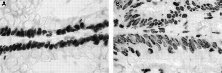
Methods: Antibodies raised against 5-methylcytidine can be used to label methyl rich regions in interphase nuclei. This technique was adapted to the study of paraffin embedded tissues and an immunohistochemical method was developed to assess the global methylation status of individual nuclei while preserving cell morphology and tissue architecture. Computer assisted quantification of the staining intensity was performed on malignant and normal zones of human colon tissues to test the correlation between the immunolabelling signal and the respective histological patterns observed.
Results: Qualitative and quantitative differences were observed and measured between the normal and malignant part of each sample. Morphologically altered nuclei displayed densely labelled spots within faintly labelled areas whereas normal nuclei were darker and uniformly stained. Image analysis allowed calculation of the average integrated optical density of the nuclei in both types of tissues, demonstrating a constant and significantly lower intensity for the former type of cells.
Figures
Similar articles
-
DNA demethylation is directly related to tumour progression: evidence in normal, pre-malignant and malignant cells from uterine cervix samples.Oncol Rep. 2003 May-Jun;10(3):545-9. Oncol Rep. 2003. PMID: 12684621
-
Association of global DNA hypomethylation with clinicopathological variables in colonic tumors of Iraqi patients.Saudi J Gastroenterol. 2016 Mar-Apr;22(2):139-47. doi: 10.4103/1319-3767.178525. Saudi J Gastroenterol. 2016. PMID: 26997221 Free PMC article.
-
Computer-assisted analysis of methylation status of individual interphase nuclei in human cultured cells.Cytometry. 1998 Feb 1;31(2):85-92. doi: 10.1002/(sici)1097-0320(19980201)31:2<85::aid-cyto3>3.3.co;2-8. Cytometry. 1998. PMID: 9482277
-
[DNA content in colorectal carcinomas: application of image analysis to archival material].Pathologica. 1994 Aug;86(4):376-83. Pathologica. 1994. PMID: 7708437 Review. Italian.
-
Methods for DNA methylation analysis and applications in colon cancer.Mutat Res. 2010 Nov 10;693(1-2):84-93. doi: 10.1016/j.mrfmmm.2010.06.010. Epub 2010 Jun 25. Mutat Res. 2010. PMID: 20599551 Review.
Cited by
-
Global DNA methylation evaluation: potential complementary marker in differential diagnosis of thyroid neoplasia.Virchows Arch. 2005 Jul;447(1):18-23. doi: 10.1007/s00428-005-1268-5. Epub 2005 May 13. Virchows Arch. 2005. PMID: 15891902
-
Quantification of Global DNA Methylation in Canine Mammary Gland Tumors via Immunostaining of 5-Methylcytosine: Histopathological and Clinical Correlations.Front Vet Sci. 2021 Feb 25;8:628241. doi: 10.3389/fvets.2021.628241. eCollection 2021. Front Vet Sci. 2021. PMID: 33718471 Free PMC article.
-
PUM1 is upregulated by DNA methylation to suppress antitumor immunity and results in poor prognosis in pancreatic cancer.Transl Cancer Res. 2021 May;10(5):2153-2168. doi: 10.21037/tcr-20-3295. Transl Cancer Res. 2021. PMID: 35116535 Free PMC article.
-
Quantification of Global DNA Methylation in Canine Melanotic and Amelanotic Oral Mucosal Melanomas and Peripheral Blood Leukocytes From the Same Patients With OMM: First Study.Front Vet Sci. 2021 Aug 24;8:680181. doi: 10.3389/fvets.2021.680181. eCollection 2021. Front Vet Sci. 2021. PMID: 34504885 Free PMC article.
-
Epigenomics in Hurthle Cell Neoplasms: Filling in the Gaps Towards Clinical Application.Front Endocrinol (Lausanne). 2021 May 24;12:674666. doi: 10.3389/fendo.2021.674666. eCollection 2021. Front Endocrinol (Lausanne). 2021. PMID: 34108939 Free PMC article. Review.
References
Publication types
MeSH terms
Substances
LinkOut - more resources
Full Text Sources
Other Literature Sources