. 2000 Nov;74(21):10260-8.
doi: 10.1128/jvi.74.21.10260-10268.2000.

Role of the Rous sarcoma virus p10 domain in shape determination of gag virus-like particles assembled in vitro and within Escherichia coli

Affiliations

Role of the Rous sarcoma virus p10 domain in shape determination of gag virus-like particles assembled in vitro and within Escherichia coli

S M Joshi et al. J Virol. 2000 Nov.

Abstract

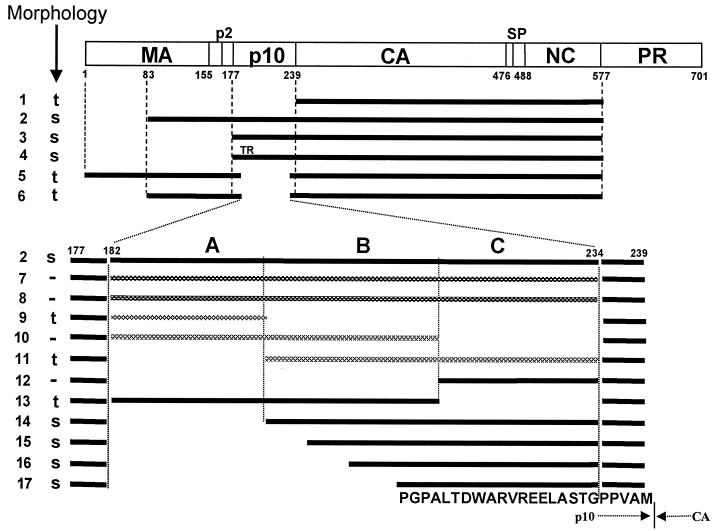
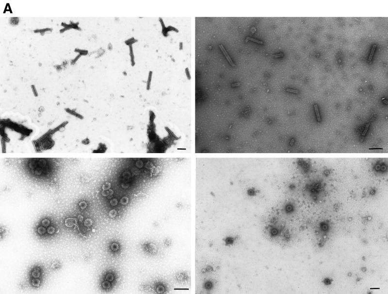
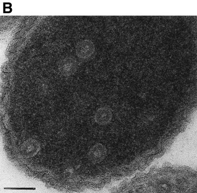
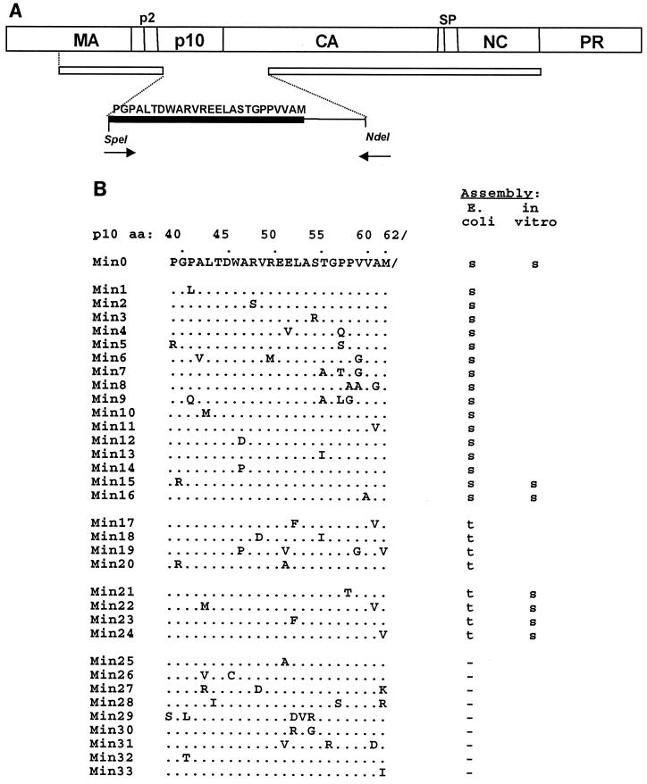
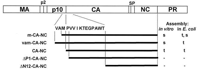
Purified retrovirus Gag proteins can assemble in vitro into virus-like particles (VLPs) in the presence of RNA. It was shown previously that a Rous sarcoma virus Gag protein missing only the protease domain forms spherical particles resembling immature virions lacking a membrane but that a similar protein missing the p10 domain forms tubular particles. Thus, p10 plays a role in spherical particle formation. To further study this shape-determining function, we dissected the p10 domain by mutagenesis and examined VLPs assembled within Escherichia coli or assembled in vitro from purified proteins. The results identified a minimal contiguous segment of 25 amino acid residues at the C terminus of p10 that is sufficient to restore efficient spherical assembly to a p10 deletion mutant. Random and site-directed mutations were introduced into this segment of polypeptide, and the shapes of particles formed in E. coli were examined in crude extracts by electron microscopy. Three phenotypes were observed: tubular morphology, spherical morphology, or no regular structure. While the particle morphology visualized in crude extracts generally was the same as that visualized for purified proteins, some tubular mutants scored as spherical when tested as purified proteins, suggesting that a cellular factor may also play a role in shape determination. We also examined the assembly properties of smaller Gag proteins consisting of the capsid protein-nucleocapsid protein (CA-NC) domains with short N-terminal extensions or deletions. Addition of one or three residues allowed CA-NC to form spheres instead of tubes in vitro, but the efficiency of assembly was extremely low. Deletion of the N-terminal residue(s) abrogated assembly. Taken together, these results imply that the N terminus of CA and the adjacent upstream 25 residues play an important role in the polymerization of the Gag protein.


Figures

FIG. 1
FIG. 1
Diagrammatic representation of assembly results. The rectangle shows the structure of the Gag protein, with vertical lines representing cleavage sites and numbers representing the number of amino acid residues from the N terminus, using the standard numbering for the Prague C strain of RSV (32). Horizontal bars indicate the structures of the proteins studied for their assembly properties from both in vitro and particle extraction experiments. Black bars represent viral sense sequences. Cross-hatched bars indicate either antisense RSV sequences or sequences from other retroviruses. Regions A, B, and C in p10 are segments of approximately 20 aa residues each. All constructs were placed into the pET3XC vector by common cloning techniques, propagated in E. coli DH5α cells, confirmed by sequencing, and transformed into BL21(DE3)/pLysS cells for protein expression and purification. ΔMBDΔPR, dp10 (called Δp10.52 in reference 25), and CA-NC have been described previously (5, 6, 25). ΔMBDdp10 combines the deletions of ΔMBDΔPR and dp10. DNA segments encoding the C-terminal 62 aa residues of M-MuLV p12 or HIV-1 MA or various segments of p10 (AB, BC, and C) were amplified by PCR from the appropriate viral clones, using primers encoding a SpeI site, and inserted into the unique SpeI site in ΔMBDdp10. Lines: 1, CA-NC; 2, ΔMBDΔPR; 3, p10-CA-NC; 4, p10TR-CA-NC; 5, dp10; 6, ΔMBDdp10; 7, C-terminal 62 aa of MMLV p12; 8, C-terminal 62 aa of HIV-1 MA (BH10 strain); 9 to 11, insertions of antisense sequences derived from DNA coding for p10; 12, segment C; 13, segment AB; 14, segment BC; 15, segment C plus the last 15 aa of segment B; 16, segment C plus the last 10 aa of segment B; 17, segment C plus the last 5 aa of segment B. The proteins shown in lines 7 to 14 contain the inserted dipeptide Thr-Ser between the wild-type Gly and Pro residues five residues from the C terminus of p10. The proteins shown in lines 15 to 17 contain the wild-type sequence at this location. The 25-aa sequence corresponding to the minimal insertion sufficient for spherical particle formation is shown at the bottom, with the boundary between p10 and CA marked. s, formation of spheres; t, formation of tubes; −, no regular assembly; TR, Thr-Arg insertion. Assembly for all of the proteins shown was tested both in vitro with purified protein and in E. coli lysates, with the same results, shown in the column marked “Morphology.”
FIG. 2
FIG. 2
Morphology of CA-NC and p10-CA-NC particles assembled in vitro and within E. coli. (A) Electron micrographs of negatively stained particles. Samples were adsorbed onto Formvar- and carbon-coated grids for 2 min and stained with 2% uranyl acetate (pH 5.2) for 20 s. Upper left, CA-NC protein assembled into tubes in vitro; upper right, CA-NC tubes from crude extracts; lower left, p10-CA-NC protein assembled into spheres in vitro; lower right, p10-CA-NC spheres from crude extracts. Bars = 100 nm. (B) Thin-section electron micrograph of VLPs formed within E. coli cells expressing p10-CA-NC protein. Cells were pelleted and fixed for 2 h in 0.1 M sodium maleate (pH 5.2)–3% glutaraldehyde and then washed in 0.1 M sodium cacodylate, pH 7.4. The samples were postfixed for 2 h in 1% OsO4–0.1 M sodium cacodylate (pH 7.4), quickly rinsed in 0.1 M sodium maleate (pH 5.2), and then washed extensively in the same buffer. The samples were then stained with 1% uranyl acetate–0.1 M sodium maleate (pH 6.0), washed in 0.1 M sodium maleate (pH 5.2), and serially dehydrated with 50, 70, 95, and 100% ethanol and 100% propylene oxide. Pellets were embedded in 50% propylene oxide–standard Spurr, and thin sections were stained with 2% uranyl acetate and then Reynold's lead citrate.
FIG. 2
FIG. 2
Morphology of CA-NC and p10-CA-NC particles assembled in vitro and within E. coli. (A) Electron micrographs of negatively stained particles. Samples were adsorbed onto Formvar- and carbon-coated grids for 2 min and stained with 2% uranyl acetate (pH 5.2) for 20 s. Upper left, CA-NC protein assembled into tubes in vitro; upper right, CA-NC tubes from crude extracts; lower left, p10-CA-NC protein assembled into spheres in vitro; lower right, p10-CA-NC spheres from crude extracts. Bars = 100 nm. (B) Thin-section electron micrograph of VLPs formed within E. coli cells expressing p10-CA-NC protein. Cells were pelleted and fixed for 2 h in 0.1 M sodium maleate (pH 5.2)–3% glutaraldehyde and then washed in 0.1 M sodium cacodylate, pH 7.4. The samples were postfixed for 2 h in 1% OsO4–0.1 M sodium cacodylate (pH 7.4), quickly rinsed in 0.1 M sodium maleate (pH 5.2), and then washed extensively in the same buffer. The samples were then stained with 1% uranyl acetate–0.1 M sodium maleate (pH 6.0), washed in 0.1 M sodium maleate (pH 5.2), and serially dehydrated with 50, 70, 95, and 100% ethanol and 100% propylene oxide. Pellets were embedded in 50% propylene oxide–standard Spurr, and thin sections were stained with 2% uranyl acetate and then Reynold's lead citrate.
FIG. 3
FIG. 3
Results of random and site-directed mutagenesis. (A) Mutagenesis strategy. A schematic diagram shows the PCR strategy for the partial-randomization experiment. The top bar represents the Gag polyprotein. The second, narrower bar denotes the ΔMBDdp10 protein; the gap is at the SpeI site. The forward primer starts at the corresponding SpeI site; the reverse primer starts at the natural NdeI site for the CA gene. The sequence that is shown (thick black bar) was partially randomized. The methionine at the end of this sequence is the C-terminal residue of the wild-type p10 protein. (B) Mutant sequences and assembly phenotypes. Dots denote the wild-type sequence, with changes from the wild-type sequence shown by letters. The results are for particles observed in crude lysates and for some particles assembled in vitro from purified proteins. Blanks indicate that the protein was not tested. s, spheres; t, tubes; −, no assembly.
FIG. 4
FIG. 4
Effects of small additions and deletions at the N terminus of CA-NC. Assembly phenotypes of proteins with minimal additions or deletions at the N terminus of CA-NC are shown. ΔP1, deletion of Pro1 residue; ΔN12, deletion of the N-terminal 12 residues of CA. All proteins had an additional initiating Met aside from the sequence shown. VAM/PVVIKTEGPAWT is the sequence of the C-terminal 3 residues of p10 and the N-terminal 12 residues of CA. Mutants with changes in this region are not drawn to scale relative to the rest of the CA-NC protein. s, spheres; t, tubes; −, no assembly.

Similar articles

Cited by

References

    1. Ausubel F M, et al., editors. Current protocols in molecular biology. New York, N.Y: John Wiley & Sons, Inc.; 1994.
    1. Bancroft J B. The self-assembly of spherical plant viruses. Adv Virus Res. 1970;16:99–134. - PubMed
    1. Berthet-Colominas C, Monaco S, Novelli A, Sibal G, Mallet F, Cusack S. Head-to-tail dimers and interdomain flexibility revealed by the crystal structure of HIV-1 capsid protein (p24) complexed with a monoclonal antibody Fab. EMBO J. 1999;18:1124–1136. - PMC - PubMed
    1. Campbell S, Rein A. In vitro assembly properties of human immunodeficiency virus type 1 Gag protein lacking the p6 domain. J Virol. 1999;73:2270–2279. - PMC - PubMed
    1. Campbell S, Vogt V M. Self-assembly in vitro of purified CA-NC proteins from Rous sarcoma virus and human immunodeficiency virus type 1. J Virol. 1995;69:6487–6497. - PMC - PubMed

Publication types

LinkOut - more resources