

Patterning and nuclear beta-catenin expression in the colonic adenoma-carcinoma sequence. Analogies with embryonic gastrulation
- PMID: 11021815
- PMCID: PMC1850184
- DOI: 10.1016/s0002-9440(10)64626-3
Patterning and nuclear beta-catenin expression in the colonic adenoma-carcinoma sequence. Analogies with embryonic gastrulation
Abstract
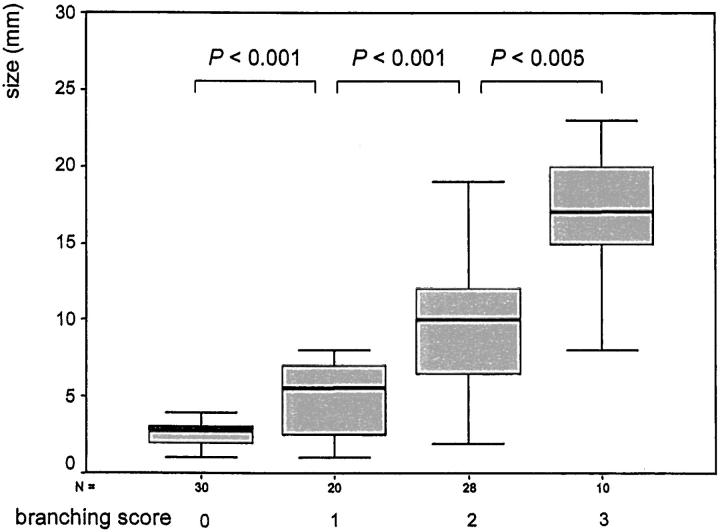
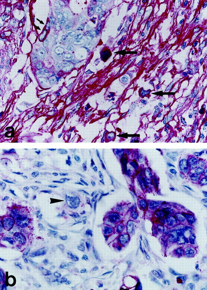
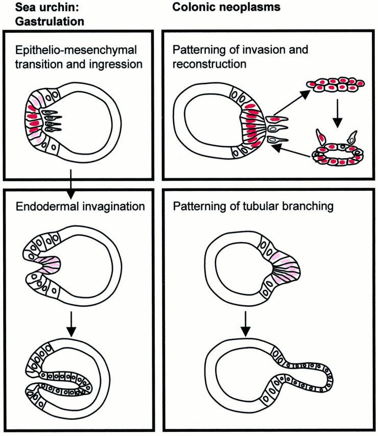
Patterning is a process by which ordered arrangements of cells and tissue structure are attained. The term derived from developmental biology is also useful for the study of colonic carcinogenesis, in which the patterning of neoplastic tubules is necessary for properties of growth, invasion, and metastasis. Interestingly the nuclear expression and transcriptional activity of beta-catenin, a major oncoprotein in colonic carcinogenesis, is decisive for the first patterning of a tubule in embryogenesis, which creates the primitive gut and is called the gastrulation. Thus, basic patternings of embryogenesis and carcinogenesis might be linked. To test this hypothesis we compared morphological patterns and immunohistochemical beta-catenin stainings in colonic adenomas and adenocarcinomas with the gastrulation steps. Two analogies were found: 1) the patterning of invasion with reconstruction in adenocarcinomas corresponded to the epithelio-mesenchymal transition, ingression, and rearrangement of cells during the first phase of gastrulation; and 2) the patterning of tubular branching in adenomas and adenocarcinomas resembled the endodermal invagination during the second phase. The intratumorous distribution and intensity of nuclear beta-catenin expression was significantly correlated with the two patternings, similar to the findings in gastrulation. The results indicate microenvironmental regulations of nuclear beta-catenin expression and a return of neoplastic cells to embryonic transcriptional susceptibilities during colonic carcinogenesis.
Figures


Similar articles
-
Tumor patterning: analogies of neoplastic morphogenesis with embryogenesis.Verh Dtsch Ges Pathol. 2000;84:22-7. Verh Dtsch Ges Pathol. 2000. PMID: 11217443 Review.
-
[Morphogenetic aspects of colorectal cancer].Pathologe. 2003 Feb;24(1):44-8. doi: 10.1007/s00292-002-0603-8. Epub 2003 Jan 14. Pathologe. 2003. PMID: 12601477 Review. German.
-
Nuclear localization of beta-catenin and plakoglobin in primary and metastatic human colonic carcinomas, colonic adenomas, and normal colon.Int J Surg Pathol. 2001 Oct;9(4):273-9. doi: 10.1177/106689690100900403. Int J Surg Pathol. 2001. PMID: 12574842
-
Expression of beta-catenin and full-length APC protein in normal and neoplastic colonic tissues.Carcinogenesis. 2000 Nov;21(11):1935-40. doi: 10.1093/carcin/21.11.1935. Carcinogenesis. 2000. PMID: 11062151
-
Altered expression of beta-catenin during radiation-induced colonic carcinogenesis.Pathol Res Pract. 2002;198(11):717-24. doi: 10.1078/0344-0338-00326. Pathol Res Pract. 2002. PMID: 12530573
Cited by
-
Beta-catenin simultaneously induces activation of the p53-p21WAF1 pathway and overexpression of cyclin D1 during squamous differentiation of endometrial carcinoma cells.Am J Pathol. 2004 May;164(5):1739-49. doi: 10.1016/s0002-9440(10)63732-7. Am J Pathol. 2004. PMID: 15111320 Free PMC article.
-
Nuclear beta-catenin expression is closely related to ulcerative growth of colorectal carcinoma.Br J Cancer. 2002 Apr 8;86(7):1124-9. doi: 10.1038/sj.bjc.6600214. Br J Cancer. 2002. PMID: 11953860 Free PMC article.
-
Hypoxia generates a more invasive phenotype of tumour cells: an in vivo experimental setup based on the chorioallantoic membrane.Pathol Oncol Res. 2009 Sep;15(3):417-22. doi: 10.1007/s12253-008-9140-y. Pathol Oncol Res. 2009. PMID: 19082873
-
Dog as model for down-expression of E-cadherin and beta-catenin in tubular epithelial cells in renal fibrosis.Virchows Arch. 2008 Dec;453(6):617-25. doi: 10.1007/s00428-008-0684-8. Epub 2008 Oct 24. Virchows Arch. 2008. PMID: 18949487
-
The majority of β-catenin mutations in colorectal cancer is homozygous.BMC Cancer. 2020 Oct 28;20(1):1038. doi: 10.1186/s12885-020-07537-2. BMC Cancer. 2020. PMID: 33115416 Free PMC article.
References
-
- Fearon ER, Vogelstein B: A genetic model for colorectal tumorigenesis. Cell 1990, 61:759-767 - PubMed
-
- Korinek V, Barker N, Morin PJ, van Wichen D, de Weger R, Kinzler KW, Vogelstein B, Clevers H: Constitutive transcriptional activation by a beta-catenin-Tcf complex in APC−/− colon carcinoma. Science 1997, 275:1784-1787 - PubMed
-
- Morin PJ, Sparks AB, Korinek V, Barker N, Clevers H, Vogelstein B, Kinzler KW: Activation of beta-catenin-Tcf signaling in colon cancer by mutations in beta-catenin or APC. Science 1997, 275:1787-1790 - PubMed
-
- He TC, Sparks AB, Rago C, Hermeking H, Zawel L, da Costa LT, Morin PJ, Vogelstein B, Kinzler KW: Identification of c-MYC as a target of the APC pathway. Science 1998, 281:1509-1512 - PubMed
-
- Tetsu O, McCormick F: Beta-catenin regulates expression of cyclin D1 in colon carcinoma cells. Nature 1999, 398:422-426 - PubMed
MeSH terms
Substances
LinkOut - more resources
Full Text Sources
Other Literature Sources
Miscellaneous
