Substitutions at the putative receptor-binding site of an encephalitic flavivirus alter virulence and host cell tropism and reveal a role for glycosaminoglycans in entry
- PMID: 10982329
- PMCID: PMC102081
- DOI: 10.1128/jvi.74.19.8867-8875.2000
Substitutions at the putative receptor-binding site of an encephalitic flavivirus alter virulence and host cell tropism and reveal a role for glycosaminoglycans in entry
Abstract
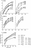
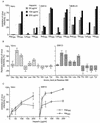
The flavivirus receptor-binding domain has been putatively assigned to a hydrophilic region (FG loop) in the envelope (E) protein. In some flaviviruses this domain harbors the integrin-binding motif Arg-Gly-Asp (RGD). One of us has shown earlier that host cell adaptation of Murray Valley encephalitis virus (MVE) can result in the selection of attenuated variants altered at E protein residue Asp(390), which is part of an RGD motif. Here, a full-length, infectious cDNA clone of MVE was constructed and employed to systematically investigate the impact of single amino acid changes at Asp(390) on cell tropism, virus entry, and virulence. Each of 10 different E protein 390 mutants was viable. Three mutants (Gly(390), Ala(390), and His(390)) showed pronounced differences from an infectious clone-derived control virus in growth in mammalian and mosquito cells. The altered cell tropism correlated with (i) a difference in entry kinetics, (ii) an increased dependence on glycosaminoglycans (determined by inhibition of virus infectivity by heparin) for attachment of the three mutants to different mammalian cells, and (iii) the loss of virulence in mice. These results confirm a functional role of the FG loop in the flavivirus E protein in virus entry and suggest that encephalitic flaviviruses can enter cells via attachment to glycosaminoglycans. However, it appears that additional cell surface molecules are also used as receptors by natural isolates of MVE and that the increased dependence on glycosaminoglycans for entry results in the loss of neuroinvasiveness.
Figures


Similar articles
-
Mechanism of virulence attenuation of glycosaminoglycan-binding variants of Japanese encephalitis virus and Murray Valley encephalitis virus.J Virol. 2002 May;76(10):4901-11. doi: 10.1128/jvi.76.10.4901-4911.2002. J Virol. 2002. PMID: 11967307 Free PMC article.
-
Attenuation of Murray Valley encephalitis virus by site-directed mutagenesis of the hinge and putative receptor-binding regions of the envelope protein.J Virol. 2001 Aug;75(16):7692-702. doi: 10.1128/JVI.75.16.7692-7702.2001. J Virol. 2001. PMID: 11462041 Free PMC article.
-
Common E protein determinants for attenuation of glycosaminoglycan-binding variants of Japanese encephalitis and West Nile viruses.J Virol. 2004 Aug;78(15):8271-80. doi: 10.1128/JVI.78.15.8271-8280.2004. J Virol. 2004. PMID: 15254199 Free PMC article.
-
Japanese encephalitis virus invasion of cell: allies and alleys.Rev Med Virol. 2016 Mar;26(2):129-41. doi: 10.1002/rmv.1868. Epub 2015 Dec 23. Rev Med Virol. 2016. PMID: 26695690 Review.
-
Virus entry and release in polarized epithelial cells.Curr Top Microbiol Immunol. 1995;202:209-19. doi: 10.1007/978-3-642-79657-9_14. Curr Top Microbiol Immunol. 1995. PMID: 7587364 Review. No abstract available.
Cited by
-
The mutation of Japanese encephalitis virus envelope protein residue 389 attenuates viral neuroinvasiveness.Virol J. 2024 Jun 5;21(1):128. doi: 10.1186/s12985-024-02398-8. Virol J. 2024. PMID: 38840203 Free PMC article.
-
Novel approaches for the rapid development of rationally designed arbovirus vaccines.One Health. 2023 May 13;16:100565. doi: 10.1016/j.onehlt.2023.100565. eCollection 2023 Jun. One Health. 2023. PMID: 37363258 Free PMC article.
-
Charge-changing point mutations in the E protein of tick-borne encephalitis virus.Arch Virol. 2023 Mar 5;168(3):100. doi: 10.1007/s00705-023-05728-3. Arch Virol. 2023. PMID: 36871232
-
Glycosaminoglycan binding by arboviruses: a cautionary tale.J Gen Virol. 2022 Feb;103(2):001726. doi: 10.1099/jgv.0.001726. J Gen Virol. 2022. PMID: 35191823 Free PMC article. Review.
-
Murine Trophoblast Stem Cells and Their Differentiated Cells Attenuate Zika Virus In Vitro by Reducing Glycosylation of the Viral Envelope Protein.Cells. 2021 Nov 9;10(11):3085. doi: 10.3390/cells10113085. Cells. 2021. PMID: 34831310 Free PMC article.
References
-
- Bernfield M, Götte M, Park P W, Reizes O, Fitzgerald M L, Lincecum J, Zako M. Functions of cell surface heparan sulfate proteoglycans. Annu Rev Biochem. 1999;68:729–777. - PubMed
-
- Castle E, Nowak T, Leidner U, Wengler G, Wengler G. Sequence analysis of the viral core protein and the membrane-associated proteins V1 and NV2 of the flavivirus West Nile virus and of the genome sequence for these proteins. Virology. 1985;145:227–236. - PubMed
MeSH terms
Substances
LinkOut - more resources
Full Text Sources
Other Literature Sources

