Experimental infection model for Sin Nombre hantavirus in the deer mouse (Peromyscus maniculatus)
- PMID: 10973478
- PMCID: PMC27067
- DOI: 10.1073/pnas.180197197
Experimental infection model for Sin Nombre hantavirus in the deer mouse (Peromyscus maniculatus)
Abstract
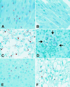
The relationship between hantaviruses and their reservoir hosts is not well understood. We successfully passaged a mouse-adapted strain of Sin Nombre virus from deer mice (Peromyscus maniculatus) by i.m. inoculation of 4- to 6-wk-old deer mouse pups. After inoculation with 5 ID(50), antibodies to the nucleocapsid (N) antigen first became detectable at 14 d whereas neutralizing antibodies were detectable by 7 d. Viral N antigen first began to appear in heart, lung, liver, spleen, and/or kidney by 7 d, whereas viral RNA was present in those tissues as well as in thymus, salivary gland, intestine, white fat, and brown fat. By 14 d nearly all tissues examined displayed both viral RNA and N antigen. We noted no consistent histopathologic changes associated with infection, even when RNA load was high. Viral RNA titers peaked on 21 d in most tissues, then began to decline by 28 d. Infection persisted for at least 90 d. The RNA titers were highest in heart, lung, and brown fat. Deer mice can be experimentally infected with Sin Nombre virus, which now allows provocative examination of the virus-host relationship. The prominent involvement of heart, lung, and brown fat suggests that these sites may be important tissues for early virus replication or for maintenance of the virus in nature.
Figures


Similar articles
-
Maporal Hantavirus Causes Mild Pathology in Deer Mice (Peromyscus maniculatus).Viruses. 2016 Oct 18;8(10):286. doi: 10.3390/v8100286. Viruses. 2016. PMID: 27763552 Free PMC article.
-
Differential lymphocyte and antibody responses in deer mice infected with Sin Nombre hantavirus or Andes hantavirus.J Virol. 2014 Aug;88(15):8319-31. doi: 10.1128/JVI.00004-14. Epub 2014 May 14. J Virol. 2014. PMID: 24829335 Free PMC article.
-
Persistent Sin Nombre virus infection in the deer mouse (Peromyscus maniculatus) model: sites of replication and strand-specific expression.J Virol. 2003 Jan;77(2):1540-50. doi: 10.1128/jvi.77.2.1540-1550.2002. J Virol. 2003. PMID: 12502867 Free PMC article.
-
Seroepidemiologic studies of hantavirus infection among wild rodents in California.Emerg Infect Dis. 1997 Apr-Jun;3(2):183-90. doi: 10.3201/eid0302.970213. Emerg Infect Dis. 1997. PMID: 9204301 Free PMC article. Review.
-
New ecological aspects of hantavirus infection: a change of a paradigm and a challenge of prevention--a review.Virus Genes. 2005 Mar;30(2):157-80. doi: 10.1007/s11262-004-5625-2. Virus Genes. 2005. PMID: 15744574 Review.
Cited by
-
Antiviral immune responses of bats: a review.Zoonoses Public Health. 2013 Feb;60(1):104-16. doi: 10.1111/j.1863-2378.2012.01528.x. Epub 2012 Aug 1. Zoonoses Public Health. 2013. PMID: 23302292 Free PMC article. Review.
-
Elevated cytokines, thrombin and PAI-1 in severe HCPS patients due to Sin Nombre virus.Viruses. 2015 Feb 10;7(2):559-89. doi: 10.3390/v7020559. Viruses. 2015. PMID: 25674766 Free PMC article.
-
Trimeric hantavirus nucleocapsid protein binds specifically to the viral RNA panhandle.J Virol. 2004 Aug;78(15):8281-8. doi: 10.1128/JVI.78.15.8281-8288.2004. J Virol. 2004. PMID: 15254200 Free PMC article.
-
Hantaviruses in the americas and their role as emerging pathogens.Viruses. 2010 Dec;2(12):2559-86. doi: 10.3390/v2122559. Epub 2010 Nov 25. Viruses. 2010. PMID: 21994631 Free PMC article.
-
Amending Koch's postulates for viral disease: When "growth in pure culture" leads to a loss of virulence.Antiviral Res. 2017 Jan;137:1-5. doi: 10.1016/j.antiviral.2016.11.002. Epub 2016 Nov 8. Antiviral Res. 2017. PMID: 27832942 Free PMC article. Review.
References
-
- Nichol S T, Spiropoulou C F, Morzunov S, Rollin P E, Ksiazek T G, Feldmann H, Sanchez A, Childs J, Zaki S, Peters C J. Science. 1993;262:914–917. - PubMed
-
- Mertz G J, Hjelle B L, Bryan R T. Adv Intern Med. 1997;42:373–425. - PubMed
-
- Childs J E, Ksiazek T G, Spiropoulou C F, Krebs J W, Morzunov S, Maupin G O, Gage K L, Rollin P, Sarisky J, Enscore R, et al. J Infect Dis. 1994;169:1271–1280. - PubMed
Publication types
MeSH terms
Associated data
- Actions
- Actions
- Actions
- Actions
- Actions
- Actions
- Actions
- Actions
Grants and funding
LinkOut - more resources
Full Text Sources
Medical
Molecular Biology Databases

