. 2000 Sep 1;19(17):4712-22.
doi: 10.1093/emboj/19.17.4712.

Cotranslational dimerization of the Rel homology domain of NF-kappaB1 generates p50-p105 heterodimers and is required for effective p50 production

Affiliations

Cotranslational dimerization of the Rel homology domain of NF-kappaB1 generates p50-p105 heterodimers and is required for effective p50 production

L Lin et al. EMBO J. .

Abstract

Generation of the NF-kappaB p50 transcription factor is mediated by the proteasome. We found previously that p50 is generated during translation of the NFKB1 gene and that this cotranslational processing allows the production of both p50 and p105 from a single mRNA. We now demonstrate that the Rel homology domain in p50 undergoes cotranslational dimerization and that this interaction is required for efficient production of p50. We further show that this coupling of dimerization and proteasome processing during translation uniquely generates p50-p105 heterodimers. Accordingly, after the primary cotranslational event, additional posttranslational steps regulate p50 homodimer formation and the intracellular ratio of p50 and p105. This cellular strategy places p50 under the control of the p105 inhibitor early in its biogenesis, thereby regulating the pool of p50 homodimers within the cell.


Figures

None
Fig. 1. NF-κB p50 exists as a dimer. (A) Amino acid sequence of p50. The sequence of murine p50 and structural domains as described by Ghosh et al. (1995) are shown. Subdomains 1 and 2 (sd1 and sd2) of the Rel homology domain (RHD) are indicated by parentheses while the intervening loop is labeled as L3. The sd1 in this figure encompasses residues 39–240; however, in this work the N-terminal 1–240 residues are referred to as sd1. The sequences of segments involved in dimerization (residues 251–270 and 302–310) are shown in bold, and the NLS in bold italics. Residues participating in the dimer interface are indicated by diamonds underneath the amino acid code, and the number of residues are marked at the right of each line. (B) Elution profile of the Superdex 200 column. The curve is drawn according to the peak elution volumes for the indicated gel filtration standards detected by absorption at 280 nm. The estimated position of elution of the p50 dimer and monomer preparations is shown. (C) Gel filtration chromatography of [35S]methionine radiolabeled, in vitro-translated, gp10-tagged wild-type p50 or a dimerization mutant (Δ302–310) of p50. Fractions eluting between 20 and 31.5 ml were immunoprecipitated with anti-gp10 antibodies, analyzed by SDS–PAGE and visualized by fluorography. The elution volumes of p50 dimer and monomer peaks are indicated in lanes 9–12 and lanes 15–18.
None
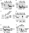
Fig. 2. PK PDP of p50 dimers and monomers. (A) PK PDP of p50 dimers. p50 dimers translated in vitro were treated with PK (10 μg/ml) at 4°C for the times indicated. This treatment resulted in two fragments of ∼37 (p37) and 32 kDa (p32). Both fragments could be immunoprecipitated by anti-NLS antidodies (lanes 6–10) but not by antibodies to the N-terminal T7 gp10 epitope tag (lanes 1–5). (B) PK cleavage occurs within the N-terminal sequences of the p50 dimer, not at the joint of p50 and the epitope tag. p50 dimers either containing (lanes 1–4) or lacking (lanes 5–8) the T7 gp10 epitope tag generated identical PK PDPs, as measured by immunoprecipitation of anti-NLS antibodies. (C) p50 monomers and p50 dimers exhibit different PK PDPs. mRNAs encoding wild-type p50 and p50 dimerization mutants (Δ251–270 and Δ302–310) were translated in vitro and treated with PK (10 μg/ml) at 4°C for 10 min. Aliquots (20 μl) of the reaction mixtures were analyzed directly by SDS–PAGE. Wild-type p50 generated p37 and p32 (lane 1), while both dimerization mutants generated two smaller fragments of ∼27 (p27) and 24 kDa (p24) (lanes 2 and 3). (D) The p27 and p24 PK-resistant fragments generated from the dimerization mutants of p50 are not immunoprecipitated by anti-NLS antibodies. Wild-type p50 and the p50 dimerization mutants were treated with PK and immunoprecipitated with anti-NLS antibodies. Neither p27 nor p24 reacted with anti-NLS antibodies (lanes 5–12) while the p37 and p32 proteins derived were immunoprecipitated (lanes 1–4). (E) Sequences located downstream of the p50 NLS are not included in the dimeric PK-resistant folding core. Both wild-type p50 and a p50 mutant containing only the 364 residues of p50 extending to the NLS were subjected to PK treatment. p50 (364 residues) generated a PDP (lanes 5–8) identical to that obtained with wild-type p50 (lanes 1–4). For an unknown reason, untreated p50 (364 residues) migrated as a doublet (lane 5). (F) Heterodimeric p50 generates a PK PDP indistinguishable from that of p50 homodimers. mRNAs encoding 780 residues of the N-terminal portion of p105 (p-780) or 497 residues (without stop codon, p-497/XhoI) were translated in vitro and treated with PK (10 μg/ml) at 4°C for 10 min and immunoprecipitated with anti-NLS antibodies. Lanes 1, 3 and 5 show untreated controls, and lanes 2, 4 and 6 show the PK-treated samples.
None
Fig. 3. Sucrose gradient sedimentation of p-780. (A) Analysis of CHX-terminated translation of p-780 by sucrose gradient sedimentation. p-780 was translated in vitro at 25°C for 20–30 min followed by adding CHX. The translation mixture was then sedimented on a 20–45% sucrose gradient (11 ml) (by centrifugation) at 40 000 r.p.m. for 3.75 h. Fractions (1 ml) were then collected from the gradient and divided into two parts. One part was immunoprecipitated directly with anti-NLS antibodies (left panel), and the other part was subjected to PK treatment (10 μg/ml) at 4°C for 30 min before immunoprecipitation (right panel). Due to the ratio of PK versus the substrate, most of the digestions yield only the more stable fragment p32. The fractions containing ribosomes are determined by absorption at 260 nm, and the top and bottom of the gradient are indicated (lanes 6–11). (B) Analysis of the RNase-treated p-780 translation mixture by sucrose gradient sedimentation. p-780 was translated as described in (A) and then treated with RNase (5 μg/ml) at 37°C for 15 min before sucrose gradient sedimentation. The resulting fractions were analyzed as in (A).
None
Fig. 4. Heterodimeric nature of the NFKB1 gene products associated with the ribosomes. mRNA encoding T7 gp10 epitope-tagged p105 N-terminal fragments of 646 or 780 residues (p-646/EcoRI and p-780/EcoRI, respectively) lacking stop codons were translated in vitro at 30°C for 20 min. The translation mixtures were then divided into two parts, one was treated with CHX (final concentration of 0.25 mg/ml) and the other was treated with puromycin (final concentration of 1 mM). Both samples were incubated at 30°C for an additional 20 min. After diluting with 100 μl of sucrose gradient buffer and centrifuging at 14 000 g for 10 min to remove insoluble material, the samples were loaded on to 350 μl of 25% sucrose and centrifuged at 100 000 r.p.m. for 40 min in a TLA.100.2 rotor. The pellets were washed twice with 0.5 ml of sucrose gradient buffer and suspended in 0.5 ml of the same buffer. Both pellets and supernatants were then immunoprecipitated with anti-T7 gp10 antibodies and analyzed by SDS–PAGE. Lanes 1, 2, 5 and 6 correspond to translation mixtures treated with CHX; and lanes 3, 4, 7 and 8 represent samples treated with puromycin. The supernatant (S) and pellet (P) fractions are also indicated. Asterisks appearing on the right side of bands indicate peptidyl-tRNA species, while those appearing on the left side of bands indicate possible ubiquitylation conjugates.
None
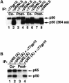
Fig. 5. NF-κB p50 dimerizes on the same polysome. (A) p50 dimerizes on the same polysome. mRNAs encoding T7 gp10-tagged p50 (433 residues) and untagged p50 (364 residues) were either cotranslated (Co-, lanes 1 and 2) or separately translated and posttranslationally mixed (Post-, lanes 3 and 4). The translation mixtures were divided into two parts; one part was immunoprecipitated with anti-T7 gp10 antibodies (lanes 1 and 3), and the other part was immunoprecipitated with anti-NLS (lanes 2 and 4). Similarly, mRNA encoding T7 gp10-tagged p50 (364 residues) and untagged p50 (433 residues) were either cotranslated (lanes 5 and 6) or translated separately and then combined posttranslationally (lanes 7 and 8), followed by immunoprecipitation with anti-T7 gp10 antibodies (lanes 5 and 7) or anti-NLS antibodies (lanes 6 and 8). (B) p50 and p65 form heterodimers when cotranslated. mRNAs encoding T7 gp10-tagged p50 and untagged p65 were cotranslated (Co-, lanes 1 and 2) or translated separately and then combined (Post-, lanes 3 and 4). The translation mixtures were divided into two parts; one part was immunoprecipitated with antibodies specific for the N-terminus of p65 [αp65 (A), lanes 1 and 3], and the other part was immunoprecipitated with antibodies to p65 and the T7 gp10 epitope tag present on p50 (lanes 2 and 4).
None
Fig. 6. In vivo evidence of cotranslational dimerization of p50 homodimers. (A) Diagram of the bicistronic construct that expresses two different epitope-tagged versions of p50 (also different in size). (B) Immunoprecipitation of p50 species expressed in CHO-CD14 cells by the bicistronic construct. The bicistronic expression vector was transfected into CHO-CD14 cells and labeled with [35S]methionine/cysteine for 1 h. The cell lysates were prepared and divided into three parts for immunoprecipitation. Lane 1, immunoprecipitation with anti-HA; lane 2, immunoprecipitation with anti-gp10; lane 3, immunoprecipitation with anti-HA and anti-gp10; lane 4, pIRES vector that expresses only the HA-tagged full-length p50; lane 5, pIRES vector that expresses only the gp10-tagged shorter form of p50; lane 6, pIRES vector immunoprecipitated with anti-HA and anti-gp10. Since the overall expression of the IRES-dependent gp10-p50 is less than that of HA-p50, twice the amount of lysates was used for immunoprecipitation in lanes 2 and 3.
None
Fig. 7. Mutations affecting dimerization significantly reduce p50 production. (A) Mutations in sd2 affecting dimerization significantly reduce p50 production. The gp10-tagged wild-type p105 (WT) and the dimerization mutants (deletion and composite point mutation) were transfected into CHO-CD14 cells and labeled with [35S]methionine/cysteine for 1 h and immunoprecipitated with anti-gp10 antibodies. Lane 1, wild-type p105; lanes 2 and 3, dimerization mutants [p105 (Δ251–270) and p105 (Δ302–310), respectively]; lane 4, p105 containing two point mutations, Y267D and L269D; lane 5, vector control. (B) Deletions that do not affect dimerization do not affect p50 production. p105 mutants containing a deletion of a 33 amino acid region [residues 166–198, p105 (ΔNO)] in sd1, and deletion of the NLS (residues 358–366) located at the end of sd2 [p105 (ΔNLS), see Figure 1A] were expressed in CHO-CD14 cells, immunoprecipitated with antibodies to their gp10 tag and analyzed for p50 production as described in (A) (lanes 4 and 5). Lane 1, wild-type p105, lanes 2 and 3, dimerization mutants. (C) Pulse–chase radiolabeling studies of the p105 and p50 dimerization mutants. The wild-type p50 and p105 as well as p50 (Δ302–310) and p105 (Δ302–310) mutants were transfected into CHO-CD14 cells, pulse radiolabeled with [35S]methionine/cysteine for 1 h and chased for the time periods indicated. Lanes 1–5, wild-type p50; lanes 6–10, p50 (Δ302–310); lanes 11–15, wild-type p105 and lanes 16–20, p105 (Δ302–310).
None
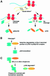
Fig. 8. Models for p50 homodimer formation (see text for more detailed description). (A) Formation of p50–p105 heterodimer. (B) Posttranslational processing model. (C) Chaperone-assisted dimerization model.

Similar articles

Cited by

References

    1. Baeuerle P.A. and Baltimore,D. (1988) IκB: a specific inhibitor of the NF-κB transcription factor. Science, 242, 540–546. - PubMed
    1. Baeuerle P.A. and Baltimore,D. (1989) A 65-kD subunit of active NF-κB is required for inhibition of NF–κB by IκB. Genes Dev., 3, 1689–1698. - PubMed
    1. Baumeister W., Walz,J., Zühl,F. and Seemüller,E. (1998) The proteasome: paradigm of a self-compartmentalizing protease. Cell, 92, 367–380. - PubMed
    1. Belich M.P., Salmerón,A., Johnson,L.H. and Ley,S.C. (1999) TPL-2 kinase regulates the proteolysis of the NF-κB-inhibitory protein NF-κB 1 p105. Nature, 397, 363–368. - PubMed
    1. Betts J.C. and Nabel,G.J. (1996) Differential regulation of NF-κB2 (p100) processing and control by amino-terminal sequences. Mol. Cell. Biol., 16, 6363–6371. - PMC - PubMed

Publication types