. 2000 Sep;74(18):8513-23.
doi: 10.1128/jvi.74.18.8513-8523.2000.

Selective inhibition of splicing at the avian sarcoma virus src 3' splice site by direct-repeat posttranscriptional cis elements

Affiliations

Selective inhibition of splicing at the avian sarcoma virus src 3' splice site by direct-repeat posttranscriptional cis elements

W Guo et al. J Virol. 2000 Sep.

Abstract

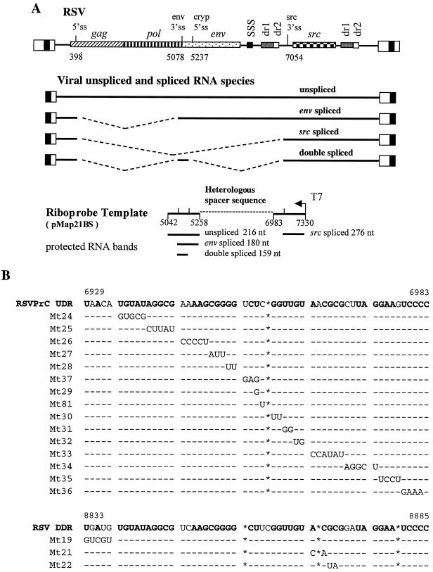
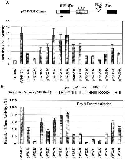
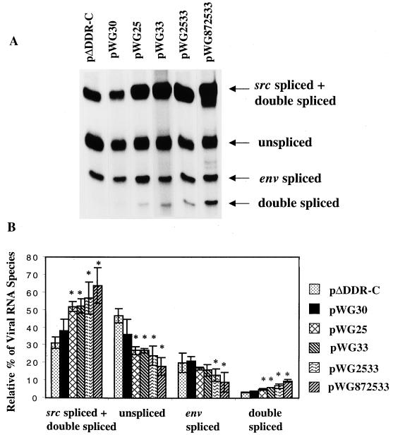
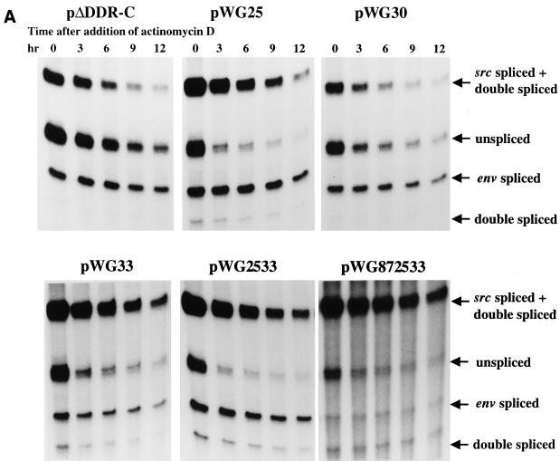
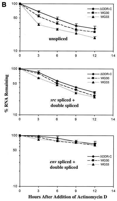
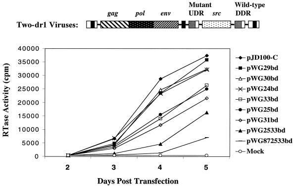
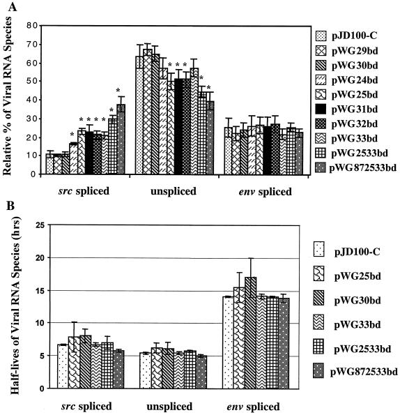
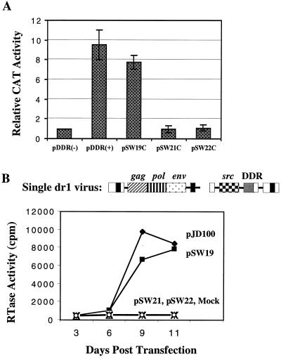
The direct-repeat elements (dr1) of avian sarcoma virus (ASV) and leukosis virus have the properties of constitutive transport elements (CTEs), which facilitate cytoplasmic accumulation of unspliced RNA. It is thought that these elements represent binding sites for cellular factors. Previous studies have indicated that in the context of the avian sarcoma virus genome, precise deletion of both ASV dr1 elements results in a very low level of virus replication. This is characterized by a decreased cytoplasmic accumulation of unspliced RNA and a selective increase in spliced src mRNA. Deletion of either the upstream or downstream dr1 results in a delayed-replication phenotype. To determine if the same regions of the dr1 mediate inhibition of src splicing and unspliced RNA transport, point mutations in the upstream and downstream elements were studied. In the context of viral genomes with single dr1 elements, the effects of the mutations on virus replication and increases in src splicing closely paralleled the effects of the mutations on CTE activity. For mutants strongly affecting CTE activity and splicing, unspliced RNA but not spliced RNA turned over in the nucleus more rapidly than wild-type RNA. In the context of wild-type virus containing two dr1 elements, mutations of either element that strongly affect CTE activity caused a marked delay of virus replication and a selective increase in src splicing. However, the turnover of the mutant unspliced RNA as well as the spliced mRNA species did not differ significantly from that of the wild type. These results suggest the dr1 elements in ASV act to selectively inhibit src splicing and that both elements contribute to the fitness of the wild-type virus. However, a single dr1 element is sufficient to stabilize unspliced RNA.


Figures

FIG. 1
FIG. 1
Schematic representations of the pJD100-C infectious RSV proviral construct, riboprobe template, and sequence alignment. (A) Diagrams of the wild-type RSV proviral DNA, showing sites of interest marked by nucleotide numbers, viral unspliced and spliced RNA species, and antisense riboprobe template used to analyze viral RNA by RNase protection assays. pMap21BS spans the env 3′ splice site (nt 5042 to 5258) and the src 3′ splice site (nt 6983 to 7330), with a heterologous spacer sequence between the RSV sequences. The sizes of the fragments protected by each RNA species are indicated below the riboprobe map. 5′ss, 5′ splice donor; env 3′ss, env 3′ splice acceptor; cryp 5′ss, cryptic 5′ splice site; src 3′ss, src 3′ splice acceptor. (B) Sequence comparison of the PrC RSV UDR and the DDR. Mutations of the UDR and DDR used in this study are indicated. Asterisks indicate lack of corresponding bases. Dashes represent the identity with the wild-type sequence. Nucleotides in boldface type indicate identity between the UDR and DDR sequences.
FIG. 2
FIG. 2
Effects of UDR mutations on CTE activity correlate with effects on virus production. (A) UDR (nt 6864 to 7037) was cloned into the intron region of pCMV138 in both the sense and antisense orientation [pUDR-C(+) and UDR(−), respectively]. UDR mutations shown in Fig. 1B were also cloned into pCMV138. Values shown are based on at least four independent transfections. Transfected cells were harvested at 48 h posttransfection, corresponding to peak CAT levels. (B) RTase activities of the indicated UDR single-dr1 virus mutants on day 9 posttransfection compared to ΔDDR-C. At this time, cells were 50 to 100% infected as determined by morphological transformation, and the values represented peak levels of RTase. The values shown are the average of three independent experiments. Standard deviations are indicated by error bars.
FIG. 3
FIG. 3
Selective increase in src mRNA in cells transfected with single dr1 virus clones containing UDR point mutants. (A) Representative RNase protection assays of total cellular RNA (10 μg) for viral RNA species. The locations of the protected bands are indicated. (B) Percentages of different viral RNA species in infected cells were determined by measurements of radioactivity as discussed in Materials and Methods. Values are the average of three independent experiments. Asterisks indicate values significantly different (P < 0.05) from the parental single dr1 virus (ΔDDR-C). Standard deviations are indicated by error bars.
FIG. 4
FIG. 4
Rapid initial turnover rate of unspliced RNA of single-dr1 UDR mutant viruses. (A) RNase protection assays of total cellular RNA harvested at various time points after addition of dactinomycin (1 μg/ml). (B) The amounts of radioactivity remaining at the indicated time points after addition of drug were measured and the amounts of viral RNA remaining versus time after the drug addition were determined. The values shown are averages of three independent experiments. Standard deviations are indicated by error bars.
FIG. 4
FIG. 4
Rapid initial turnover rate of unspliced RNA of single-dr1 UDR mutant viruses. (A) RNase protection assays of total cellular RNA harvested at various time points after addition of dactinomycin (1 μg/ml). (B) The amounts of radioactivity remaining at the indicated time points after addition of drug were measured and the amounts of viral RNA remaining versus time after the drug addition were determined. The values shown are averages of three independent experiments. Standard deviations are indicated by error bars.
FIG. 5
FIG. 5
The unstable fraction of UDR mutant unspliced RNA is located in the nucleus. ΔDDR-C- and WG33-infected cells were treated as in Fig. 4 with dactinomycin, and fractionation of the nucleus and cytoplasm was carried out at the indicated time points after the drug addition. Shown are the protected bands for the unspliced RNA species in the nuclear and cytoplasmic fractions. Percent of remaining RNA are given for two independent experiments (Exp1 and Exp2).
FIG. 6
FIG. 6
UDR mutations in the context of two-dr1 virus constructs cause a delayed virus replication phenotype. All of the proviral constructs used contain a wild-type DDR with a mutated UDR. After transfection of CEF, culture media were harvested at indicated time points and analyzed for RTase activity. At least three independent experiments were carried out for each mutant. Representative data are shown.
FIG. 7
FIG. 7
UDR mutations in two-dr1 virus constructs cause a selective increase in src mRNA splicing but do not significantly affect RNA stability. (A) Relative molar ratios of viral RNA species in cells infected with mutant viruses based on RNase protection assay of total cellular RNA harvested on day 5 posttransfection. The values shown are the average of data from at least three independent experiments. Asterisks indicate values significantly different (P < 0.05) from those for wild-type pJD100-C. (B) Half-lives of RNA species of mutant viruses were determined as described in the legend to Fig. 4. Values shown are averages of three independent experiments. Standard deviations are indicated by error bars.
FIG. 8
FIG. 8
Effects of DDR mutations on CTE activity correlate with effects on viral production of single-dr1 DDR viruses. (A) DDR mutations were cloned into pCMV138 as described in the legend to Fig. 2. Locations of mutations are shown in Fig. 1B. The values shown are the average of six independent experiments. (B) RTase activity of indicated single-dr1 viruses with DDR mutations at different times after transfection. At least three independent experiments were carried out for each mutant. Representative data are shown.
FIG. 9
FIG. 9
DDR mutations in two-dr1 virus constructs resulted in a delayed virus replication phenotype as described in the legend to Fig. 6 (A) and selective increases in src mRNA splicing as described in Fig. 7 (B). The results are based on three experiments, and in panel B, values significantly different from those for the wild type (P < 0.05) are labeled with asterisks. Standard deviations are indicated by error bars.

Similar articles

Cited by

References

    1. Amendt B A, Simpson S B, Stoltzfus C M. Inhibition of RNA splicing at the Rous sarcoma virus src 3′ splice site is mediated by an interaction between a negative cis element and a chicken embryo fibroblast nuclear factor. J Virol. 1995;69:5068–5076. - PMC - PubMed
    1. Aschoff J M, Foster D, Coffin J M. Point mutations in the avian sarcoma/leukosis virus 3′ untranslated region result in a packaging defect. J Virol. 1999;73:7421–7429. - PMC - PubMed
    1. Belshen M, Park G S, Bilodeau P, Stoltzfus C M, Carpenter S. Binding of equine infectious anemia virus Rev to an exon splicing silencer mediates alternative splicing and nuclear export of viral mRNAs. Mol Cell Biol. 2000;20:3550–3557. - PMC - PubMed
    1. Berberich S L, Macias M, Zhang L, Turek L P, Stoltzfus C M. Comparison of Rous sarcoma virus RNA processing in chicken and mouse fibroblasts: evidence for double-spliced RNA in nonpermissive mouse cells. J Virol. 1990;64:4313–4320. - PMC - PubMed
    1. Berberich S L, Stoltzfus C M. Analysis of spliced and unspliced Rous sarcoma virus RNAs early and late after infection of chicken embryo fibroblasts: effect of cell culture conditions. Virology. 1991;182:135–144. - PubMed

Publication types

LinkOut - more resources