. 2000 Sep;20(17):6287-99.
doi: 10.1128/MCB.20.17.6287-6299.2000.

The RNA-binding protein TIA-1 is a novel mammalian splicing regulator acting through intron sequences adjacent to a 5' splice site

Affiliations

The RNA-binding protein TIA-1 is a novel mammalian splicing regulator acting through intron sequences adjacent to a 5' splice site

F Del Gatto-Konczak et al. Mol Cell Biol. 2000 Sep.

Abstract

Splicing of the K-SAM alternative exon of the fibroblast growth factor receptor 2 gene is heavily dependent on the U-rich sequence IAS1 lying immediately downstream from its 5' splice site. We show that IAS1 can activate the use of several heterologous 5' splice sites in vitro. Addition of the RNA-binding protein TIA-1 to splicing extracts preferentially enhances the use of 5' splice sites linked to IAS1. TIA-1 can provoke a switch to use of such sites on pre-mRNAs with competing 5' splice sites, only one of which is adjacent to IAS1. Using a combination of UV cross-linking and specific immunoprecipitation steps, we show that TIA-1 binds to IAS1 in cell extracts. This binding is stronger if IAS1 is adjacent to a 5' splice site and is U1 snRNP dependent. Overexpression of TIA-1 in cultured cells activates K-SAM exon splicing in an IAS1-dependent manner. If IAS1 is replaced with a bacteriophage MS2 operator, splicing of the K-SAM exon can no longer be activated by TIA-1. Splicing can, however, be activated by a TIA-1-MS2 coat protein fusion, provided that the operator is close to the 5' splice site. Our results identify TIA-1 as a novel splicing regulator, which acts by binding to intron sequences immediately downstream from a 5' splice site in a U1 snRNP-dependent fashion. TIA-1 is distantly related to the yeast U1 snRNP protein Nam8p, and the functional similarities between the two proteins are discussed.


Figures

FIG. 1
FIG. 1
IAS1 activates splicing of a heterologous tropomyosin exon. (A) Schematic representations of pre-mRNAs used for in vitro splicing. In Tropo 6A-7 pre-mRNA, exons 6A and 7 are separated by a 284-nucleotide intron including the S4 activating sequence. In 6A-P3AS-7 and IAS1 down, S4 has been replaced with a purine-rich sequence and with IAS1, respectively. In 6A-Δ4-7, the S4 sequence is deleted. In IAS1 up and RAN, IAS1 and the random RAN sequence, respectively, have been inserted immediately downstream of the 5′ss. The last nucleotides of exon 6A are boxed, and the IAS1 and RAN sequences are shown in boxes. (B) In vitro splicing assays using pre-mRNAs shown in panel A in HeLa cell nuclear extract. mRNAs obtained by splicing exons 6A and 7 together are identified, as well as excised introns. The space of migration between the pre-mRNAs and mRNA has been reduced. The amounts of excised introns (I) and remaining pre-mRNA (P) were quantified using a Fuji phosphorimager, and the percentage of splicing was determined as I/(I + P) × 100%. The mean of three determinations is given below the lanes. nt, nucleotides.
FIG. 2
FIG. 2
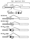
Schematic representations of pre-mRNAs with competing 5′ss used for in vitro splicing. Part of the E1A pre-mRNA is shown, with the natural competing D2 and D1 5′ss. In the other pre-mRNAs, the D1 5′ss has been replaced with a pair of competing 5′ss as shown, and the major splicing reactions observed are indicated.
FIG. 3
FIG. 3
Effect of IAS1 and TIA-1 on competing D2 5′ss in vitro. In vitro splicing assays were performed using pre-mRNAs shown in Fig. 2. For spliced mRNAs, the donor (D) and acceptor (A) (the E1A 3′ss) splice sites used are identified. (A) Splicing was carried out in HeLa cell nuclear extract (NE). Note that a cryptic splicing reaction occurs with the D2/D2-wt pre-mRNA using a cryptic 5′ss located 88 nucleotides upstream of the distal D2 site. The cryptic intron is visible on the photo, but the corresponding mRNA has not been retained. (B) Splicing was in cytoplasmic S100 extract (9 μl) with 0.5 μg of SR proteins added (S100+SR). Lanes 1 to 3, D2/D2-IAS1 pre-mRNA spliced in extract alone (lane 1) or in extract with 600 ng of TIA-1 (lane 2) or 600 ng of hnRNP C1 (lane 3). Lanes 4 to 6, D2/D2-wt pre-mRNA spliced in extract alone (lane 4) or with 600 ng of TIA-1 (lane 5) or 600 ng of hnRNP C1 (lane 6) added.
FIG. 4
FIG. 4
Effects of IAS1 and TIA-1 on competing D1 and DSAM 5′ss in vitro. In vitro splicing assays were performed using pre-mRNAs shown in Fig. 2. For spliced mRNAs, the donor (D) and acceptor (A) (the E1A 3′ss) splice sites used are identified. (A) Splicing was carried out in cytoplasmic S100 extract (9 μl) with 0.5 μg of SR proteins added (S100+SR). Lane 1, D1/DSAM-IAS1 pre-mRNA starting material. Lanes 2 to 5, D1/DSAM-IAS1 pre-mRNA spliced in extract alone (lane 2), in extract with 300 or 600 ng of TIA-1 added (lanes 3 and 4, respectively), or in extract with 600 ng of hnRNP C1 added (lane 5). Lanes 6 to 8, D1/DSAM-RAN pre-mRNA spliced in extract alone (lane 6), extract with 600 ng of TIA-1 added (lane 7), or extract with 600 ng of hnRNP C1 added (lane 8). (B) Splicing was in a 6:4 mixture of nuclear extract and S100 extract (NE/S100). Lane 1, D1/DSAM-IAS1 pre-mRNA starting material. Lanes 2 to 6, D1/DSAM-IAS1 pre-mRNA spliced in extract alone (lane 2); in extract with 200, 400, or 600 ng of TIA-1 added (lanes 3 to 5, respectively); or in extract with 400 ng of hnRNP C1 added (lane 6). Lanes 7 to 9, D1/DSAM-RAN pre-mRNA spliced in extract alone (lane 7) or in extract with 400 ng of TIA-1 (lane 8) or 400 ng of hnRNP C1 (lane 9) added. The radioactivities present in the mRNAs were determined by a phosphorimager, corrected for their content in C residues, and used to calculate the ratio of use of D1 versus that of DSAM.
FIG. 5
FIG. 5
Interaction between TIA-1 and IAS1. (A and B) RNA probes as shown were incubated with various extracts either alone (−) or with 150 ng of added recombinant TIA-1 (+ TIA-1) or hnRNP C1 (+ C1) as indicated before UV cross-linking and SDS-PAGE analysis. NE, HeLa nuclear extract; S100, HeLa S100 extract. WCE, WCE from 293-EBNA cells. WCE/TIA-1, WCE from cells transfected with pTIA-1. After UV cross-linking and RNase treatment, equivalent aliquots were resolved directly on an SDS-polyacrylamide gel. The positions of prestained protein standards (NOVEX) are indicated. Note that the apparent molecular mass of the adduct proteins is usually 3 to 5 kDa higher than that of the corresponding protein. (C and D) RNA probes were incubated with various extracts as in panels A and B. After UV cross-linking and RNase treatment, samples were divided into three parts. One was analyzed directly (total); the others were analyzed after immunoprecipitation with antibodies against either TIA-1 (αTIA-1) or hnRNP C1 (αC1). Analysis was performed by SDS-PAGE. The aliquots loaded on the gel for the samples analyzed directly (total) were one-third of the amount used for those analyzed after immunoprecipitation.
FIG. 6
FIG. 6
U1 snRNP is involved in TIA-1 binding to IAS1. WCE from 293-EBNA cells (WCE) or from transfected 293-EBNA cells (WCE/TIA-1) were mock preincubated (−) or preincubated with oligonucleotides complementary to the 5′ end of U1 snRNA (U1) or complementary to the T7 promoter (T7) before cross-linking to the 5′ss-IAS1 probe. For lanes 1 to 3 and 6 to 8, each assay mixture was analyzed directly. In addition, immunoprecipitation with anti-TIA-1 antibodies was performed on the mock-preincubated samples (lanes 4 and 9) and the samples preincubated with the oligonucleotide complementary to the 5′ end of U1 snRNA (lanes 5 and 10). The aliquots loaded on the gels for the samples analyzed directly represent one-third of the amount used for those analyzed after immunopurification.
FIG. 7
FIG. 7
Schematic representations of FGFR-2 minigenes. The parent minigene RK3 is shown, with the Rous sarcoma virus long terminal repeat promoter (RSV), the alternative exons K-SAM and BEK, the upstream and downstream constitutive exons C1 and C2, and the bovine growth hormone polyadenylation sequence (BGH). Locations of primers P3 and P4 used for RT-PCR are marked. The U-rich intron-activating sequence IAS1 is identified. RK20 is similar to RK3, except for the replacement of IAS1 with a random sequence. In RK97, the BEK exon's polypyrimidine sequence and 3′ss have been deleted. RK98 was derived from RK97 by replacing IAS1 with the random sequence described in the text. RK-MS2 is derived from RK3 by replacing IAS1 and some downstream sequences with bacteriophage MS2 coat binding sites (MS2). In addition, the BEK exon is deleted. RK99 is similar to RK20, except that the BEK exon is deleted and bacteriophage MS2 coat binding sites (MS2) have been placed well downstream of the K-SAM exon's 5′ss.
FIG. 8
FIG. 8
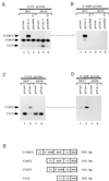
TIA-1 activation of the K-SAM exon requires IAS1. Cells were cotransfected with minigenes and expression vectors for bacteriophage MS2 coat protein, TIA-1, or hnRNP C1 as shown. RT-PCR was carried out on transfected cell RNA using primers P3 and P4 shown in Fig. 7, and products were subjected to Southern analysis. Hybridization was performed first to a probe corresponding to the K-SAM exon (B and D), and then the same blot was dehybridized and rehybridized to a probe made up of exons C1 and C2 (A and C). RT-PCR products are identified using names corresponding to structures shown in panel E.
FIG. 9
FIG. 9
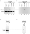
TIA-1 activates splicing if recruited close to the 5′ss. Cells were cotransfected with minigenes and the empty expression vector pCI-neo or expression vectors for bacteriophage MS2 coat protein or TIA-1 or the following fusions with coat protein: TIA-1–coat fusion (TIA-coat), hnRNP C1-coat fusion (C1-coat), and hnRNP A1-coat fusion (A1-coat). RT-PCR was carried out on transfected cell RNA using primers P3 and P4 shown in Fig. 7, and products were subjected to Southern analysis. Hybridization was performed first to a probe corresponding to the K-SAM exon (B and D), followed by dehybridization and rehybridization to a probe made up of exons C1 and C2 (A and C). RT-PCR products are identified using names corresponding to structures shown in Fig. 8E.
FIG. 10
FIG. 10
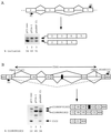
Effects of TIA-1 and hnRNP C1 on splicing in vivo. (A) The preprotachykinin minigene was cotransfected into SVK14 cells with pCI-neo (lane 1), pTIA-1 (lane 2), or phnRNP C1 (lane 3). RT-PCR was carried out on transfected cell RNA using primers P1 and P2, and products were subjected to Southern analysis with hybridization to a probe made up of exons 2 to 5. (B) A hybrid FGFR-2–CD44 minigene was cotransfected into 293-EBNA cells with pCI-neo (lane 1), pTIA-1 (lane 2), or phnRNP C1 (lane 3). RT-PCR was carried out on transfected cell RNA using primers P3 and P4 as marked, and products were subjected to Southern analysis with hybridization to a probe made up of exons C1 and C2 of the FGFR-2 gene. The 6.9-kb ClaI-SmaI fragment of the human CD44 gene used is identified by arrows and contains alternative exons v8, v9, and v10, as well as an additional alternative exon (50) represented by a black box. Note that exon v9 has two alternative 5′ss (50). On the minigene map, the three major splicing events seen in 293-EBNA cells transfected with the minigene and pCI-neo are illustrated. The corresponding RT-PCR products are illustrated below the map. RSV, Rous sarcoma virus long terminal repeat; BGH, bovine growth hormone polyadenylation signal. Radioactivities present in bands were determined by phosphorimager and used to calculate splicing percentages.

Similar articles

Cited by

References

    1. Balvay L, Libri D, Gallego M, Fiszman M Y. Intronic sequence with both negative and positive effects on the regulation of alternative transcripts of the chicken beta tropomyosin transcripts. Nucleic Acids Res. 1992;20:3987–3992. - PMC - PubMed
    1. Beck A, Miller I J, Anderson P, Streuli M. RNA-binding protein TIAR is essential for primordial germ cell development. Proc Natl Acad Sci USA. 1998;95:2331–2336. - PMC - PubMed
    1. Beck A R, Medley Q G, O'Brien S, Anderson P, Streuli M. Structure, tissue distribution and genomic organization of the murine RRM-type RNA binding proteins TIA-1 and TIAR. Nucleic Acids Res. 1996;24:3829–3835. - PMC - PubMed
    1. Black D L. Finding splice sites within a wilderness of RNA. RNA. 1995;1:763–771. - PMC - PubMed
    1. Bourgeois C F, Popielarz M, Hildwein G, Stevenin J. Identification of a bidirectional splicing enhancer: differential involvement of SR proteins in 5′ or 3′ splice site activation. Mol Cell Biol. 1999;19:7347–7356. - PMC - PubMed

Publication types

MeSH terms

Substances