. 2000 Aug;74(16):7451-61.
doi: 10.1128/jvi.74.16.7451-7461.2000.

The murine gammaherpesvirus 68 v-cyclin is a critical regulator of reactivation from latency

Affiliations

The murine gammaherpesvirus 68 v-cyclin is a critical regulator of reactivation from latency

L F van Dyk et al. J Virol. 2000 Aug.

Abstract

Gamma-2 herpesviruses encode a homolog of mammalian D-type cyclins. The v-cyclin encoded by murine gammaherpesvirus 68 (gammaHV68) induces cell cycle progression and is an oncogene (L. F. van Dyk, J. L. Hess, J. D. Katz, M. Jacoby, S. H. Speck, and H. W. Virgin IV, J. Virol. 73:5110-5122, 1999). However, the role of the pro-proliferative v-cyclins in gamma-2 herpesvirus pathogenesis is not known. Here we report the generation and characterization of a gammaHV68 v-cyclin mutant (v-cyclin.LacZ) that is unable to express a functional v-cyclin protein. Notably, although the gammaHV68 v-cyclin is expressed from an early-late lytic transcript, v-cyclin. LacZ replicated normally in fibroblasts in vitro and during acute infection in the spleen, liver, and lungs in vivo. Moreover, v-cyclin.LacZ exhibited wild-type (wt) virulence in mice with severe combined immunodeficiency. In addition, in a model of gammaHV68-induced chronic disease in mice lacking the gamma interferon receptor (IFNgammaR(-/-)), v-cyclin.LacZ virus was similar to wt gammaHV68 in terms of the incidence of mortality and vasculitis. Further analysis revealed that the frequencies of splenocytes and peritoneal cells harboring the latent gammaHV68 genome in normal and B-cell-deficient mice infected with wt gammaHV68 or v-cyclin.LacZ were very similar. However, v-cyclin.LacZ was significantly compromised in its capacity to reactivate from latency. This phenotype was conclusively mapped to the v-cyclin gene by (i) generating a marker rescue virus (v-cyclin.MR) from the v-cyclin.LacZ mutant, which restored the frequency of cells in which virus reactivated from latency to the levels observed with wt gammaHV68; and (ii) generating a second v-cyclin mutant virus containing a translation stop codon within the v-cyclin gene (v-cyclin.stop), which was compromised in reactivation from latency. These studies demonstrate that despite expression as a lytic cycle gene, the pro-proliferative gammaHV68 v-cyclin is not required for gammaHV68 replication either in vitro or during acute infection in vivo but rather is a critical determinant of reactivation from latency.


Figures
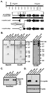
FIG. 1
FIG. 1
Genomic structures of v-cyclin.LacZ and v-cyclin.stop mutants. (A) Shown is a schematic representation of the region of the γHV68 genome encoding the v-cyclin gene (gene 72). Mutations in the v-cyclin gene, as well as the marker rescue virus, were generated as described in Materials and Methods. Genome coordinates of the surrounding ORFs in wt γHV68 region are as follows: 100-bp repeat region, bp 98981 to 101170; ORF72, bp 103181 to 102426; M11 (v-bcl-2), bp 103418 to 103930; ORF73, bp 104868 to 103927; and ORF74 (v-GPCR), bp 105057 to 106067. Genome coordinates are based on the γHV68 WUMS sequence (67). The cyclin region probe is depicted as a bar below the wt γHV68 schematic and spans bp 101654 to 105377. Also shown, at the left end of the viral genome, are the locations of the NotI and NsiI sites used to diagnose for the presence of deletions in this region of the genome (see panel C, below). (B) Southern analysis of wt γHV68, v-cyclin.LacZ, v-cyclin.stop, and v-cyclin.MR viruses. Purified genomic viral DNA was digested with BamHI or EcoRI, electrophoresed, blotted, and hybridized with the cyclin region probe. 32P-labeled molecular size markers were included on each Southern blot, and the locations of positions of size standards are indicated. The Southern blot shown in the right-hand panel demonstrates the presence of the predicted 0.5-kb diagnostic EcoRI band in the v-cyclin.LacZ mutant, which was run off the blot shown in the left-hand panel. (C) To assess the integrity of the left end of the viral genome, purified viral DNA was digested with NotI and NsiI, electrophoresed, blotted, and probed with a fragment of the viral genome containing gene 6 (bp 11100 to 14026). The locations of the NotI and NsiI sites at the left end of the genome are depicted in panel A. (D) Immunoblot analysis of v-cyclin protein expression in NIH 3T12 cells. NIH 3T12 fibroblasts were lytically infected with wt γHV68, v-cyclin.LacZ, v-cyclin.stop, or v-cyclin.MR or were mock infected, and cell lysates collected at 24 h postinfection were probed with a polyclonal rabbit antiserum to the v-cyclin (upper panel). The blot was reprobed with a monoclonal antibody against β-actin (Sigma catalog no. AC74) (lower panel).
FIG. 2
FIG. 2
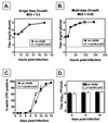
γHV68-cyclin.LacZ grows similarly to wt γHV68. NIH 3T12 cells were infected with 5.0 (A) or 0.05 (B) PFU of wt γHV68 (closed squares) or v-cyclin.LacZ (open circles) per cell, samples were harvested at the times indicated, and virus was quantitated by plaque assay. Data are representative of two independent experiments. The dotted line indicates the sensitivity of the plaque assay (50 PFU). The inoculum was removed 1 h postinfection and replaced with fresh medium to measure single-step growth (A) or was left in place to measure multistep growth (B). (C) Viral growth in MEFs. MEF monolayers in 96-well plates were infected with 1 PFU (MOI, ca. 0.0001) of wt γHV68 or v-cyclin.LacZ. The CPE in 24 wells per virus was recorded over the course of 15 days postinfection. Results are the means of values for four independent dilution series of wt γHV68 and v-cyclin.LacZ (the standard errors of the means [SEM] are indicated by error bars). (D) Six wells of MEFs infected with 1 PFU of wt γHV68 or v-cyclin.LacZ were harvested at days 6, 8, and 14 for determination of viral titer by plaque assay. Results are the means of values for six wells each of wt γHV68 and v-cyclin.LacZ at each time point (SEMs are indicated by error bars).
FIG. 3
FIG. 3
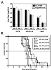
In vivo growth and virulence. (A) Viral titers in spleens, livers, and lungs of mice 4 and 9 days after i.p. infection. C57BL/6 mice were infected with 106 PFU of wt γHV68 (closed bars) or v-cyclin.LacZ (open bars). Data from two independent experiments with seven to nine mice total per time point were pooled (the standard errors of the means are indicated by error bars). (B) Virulence of wt γHV68 and v-cyclin.LacZ in SCID mice. C.B-17 SCID mice were infected by i.p. injection with 106, 103, or 101 PFU of wt γHV68 (closed symbols) or v-cyclin.LacZ (open symbols), and mortality was recorded over the course of the experiment. Data from two independent experiments were pooled, and the total number of mice at each dose is indicated. mut, mutant; mock, mock-infected mouse data.
FIG. 4
FIG. 4
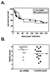
Analysis of chronic infection of the vascular system in IFNγR−/− mice. (A) Survival of wt γHV68- or v-cyclin.LacZ-infected IFNγR−/− mice. IFNγR−/− mice were infected with 2 × 10⁷ PFU of wt γHV68 (open squares) or v-cyclin.LacZ (closed squares), and mortality was recorded over the course of the experiment. Data from three independent experiments were compiled (wt γHV68, n = 37 mice; v-cyclin.LacZ, n = 38 mice). (B) Severity of arteritic lesions at the base of the aorta in wt γHV68- or v-cyclin.LacZ-infected IFNγR−/− mice. The histologic scoring criteria are described in detail elsewhere (Dal Canto et al., submitted). In this scale, 0 represents no lesion and 5 represents the most severe lesion. Comparison of wt γHV68 (open squares)- and v-cyclin.LacZ (closed squares)-induced lesions is depicted by individual points and means (horizontal lines). ∗∗, v-cyclin.LacZ-induced lesions were slightly less severe than wt γHV68-induced lesions (P = 0.0326.)
FIG. 5
FIG. 5
The v-cyclin.LacZ mutant establishes latency but fails to reactivate efficiently from C57Bl/6 mice. C57Bl/6 mice were infected i.p. with 106 PFU of v-cyclin.LacZ (triangles), wt γHV68 (squares), or v-cyclin.MR (circles) virus, and cells were harvested 42 days postinfection for quantitation of the frequency of viral genome positive cells (A and B) and the frequency of cells reactivating virus (C and D). (A and B) Latently infected splenocytes or PECs were analyzed for the frequency of viral-genome-positive cells by limiting-dilution nested PCR. Ten PCRs were performed per cell dilution for each experiment, and PCR controls were run within each experiment. The numbers of individual experiments are indicated, and the standard errors of the means are shown (error bars). (C and D) Limiting-dilution ex vivo reactivation analyses using the same populations of splenocytes and PECs as for the experiments shown in panels A and B. Intact (live) cells were plated on MEF indicator monolayers to determine the frequency of ex vivo reactivation (closed symbols). In parallel, samples of each cell population were mechanically disrupted to measure preformed infectious virus (open symbols), which was demonstrated to be absent. Curve fit lines were derived by nonlinear-regression analysis, and symbols represent means and SEMs (error bars) of data from individual experiments as indicated. The dashed line is at 63%, the value which was used to calculate the frequency of genome-positive cells or the frequency of reactivating cells as indicated by a Poisson distribution. Data represent independent experiments as indicated, with each experiment containing cells pooled from four to seven mice. ∗∗∗, frequencies of reactivation for v-cyclin.LacZ and v-cyclin.MR were statistically different (PECs, P = 0.003; splenocytes, P = 0.043).
FIG. 6
FIG. 6
The v-cyclin.LacZ mutant establishes latency but fails to reactivate efficiently in cells from B-cell-deficient mice. B-cell-deficient mice were infected with 106 PFU of v-cyclin.LacZ (triangles), wt γHV68 (squares), or v-cyclin.MR (circles), and cells were harvested 42 days postinfection for quantitation of the frequency of viral-genome-positive cells (A and B) and the frequency of cells reactivating virus (C and D). (A and B) Latently infected splenocytes and PECs were analyzed for the frequency of viral-genome-positive cells by limiting-dilution nested PCR, as described in the legend to Fig. 5 and in Materials and Methods. (C and D) Cells were harvested 42 days postinfection for limiting-dilution ex vivo reactivation analysis on MEF indicator monolayers, as described in the legend to Fig. 5 and in Materials and Methods. The presence of preformed infectious virus was assessed by limiting-dilution analysis of disrupted cells, with a control being included in each experiment (open symbols). Curve fit lines were derived by nonlinear-regression analysis (standard errors of the means are shown [error bars]). The dashed line is at 63%, the value which was used to calculate the frequency of genome-positive cells (A and B) or the frequency of reactivating cells (C and D) as indicated by a Poisson distribution. Data represent independent experiments as indicated, with each experiment utilizing cells pooled from four to seven mice. ∗∗∗, differences in the frequencies of reactivation for v-cyclin.LacZ and wt γHV68 were statistically significant (PECs, P = 0.0005; splenocytes, P = 0.0162).
FIG. 7
FIG. 7
The v-cyclin.stop mutant exhibits the same reactivation phenotype as the v-cyclin.LacZ mutant. C57Bl/6 mice were infected with 106 PFU of v-cyclin.LacZ (triangles), wt γHV68 (squares), or v-cyclin.stop (circles) virus, and cells were harvested 28 days postinfection for quantitation of the frequency of virus reactivation in latently infected PECs, as described in the legend to Fig. 5 and in Materials and Methods. The data shown were pooled from two independent experiments with a total of 10 mice per virus. The diminished frequencies of reactivation of both the v-cyclin.LacZ (P = 0.0029) and v-cyclin.stop (P = 0.0023) compared to wt γHV68 were statistically significant.

Similar articles

Cited by

References

    1. Albrecht J-C, Fleckenstein B. New member of the multigene family of complement control proteins in herpesvirus saimiri. J Virol. 1992;66:3937–3940. - PMC - PubMed
    1. Albrecht J-C, Nicholas J, Biller D, Cameron K R, Biesinger B, Newman C, Wittmann S, Craxton M A, Coleman H, Fleckenstein B, Honess R W. Primary structure of the herpesvirus saimiri genome. J Virol. 1992;66:5047–5058. - PMC - PubMed
    1. Arvanitakis L, Yaseen N, Sharma S. Latent membrane protein-1 induces cyclin D2 expression, pRb hyperphosphorylation, and loss of TGF-beta-1-mediated growth inhibition on EBV-positive B cells. J Immunol. 1995;155:1047–1056. - PubMed
    1. Austin P J, Flemington E, Yandava C N, Strominger J L, Speck S H. Complex transcription of the Epstein-Barr virus BamHI fragment H rightward open reading frame 1 (BHRF1) in latently and lytically infected B lymphocytes. Proc Natl Acad Sci USA. 1988;85:3678–3682. - PMC - PubMed
    1. Bosma M, Schuler W, Bosma G. The scid mouse mutant. Curr Top Microbiol Immunol. 1988;137:197–202. - PubMed

Publication types

MeSH terms

LinkOut - more resources