. 2000 Jul 1;14(13):1678-90.

Control of her1 expression during zebrafish somitogenesis by a delta-dependent oscillator and an independent wave-front activity

Affiliations

Control of her1 expression during zebrafish somitogenesis by a delta-dependent oscillator and an independent wave-front activity

S A Holley et al. Genes Dev. .

Abstract

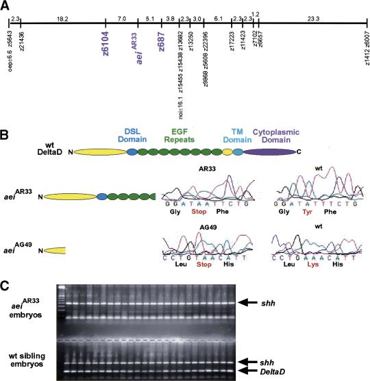
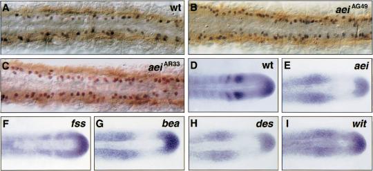
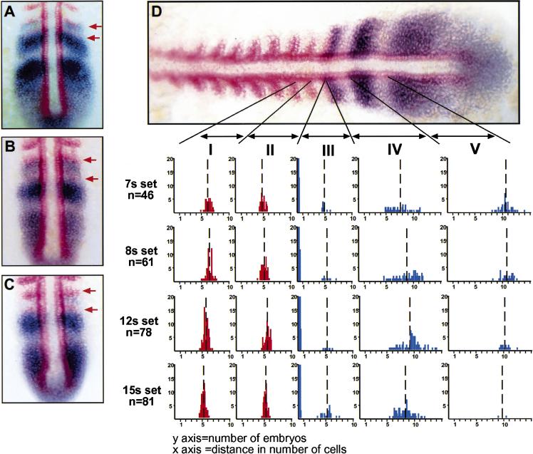
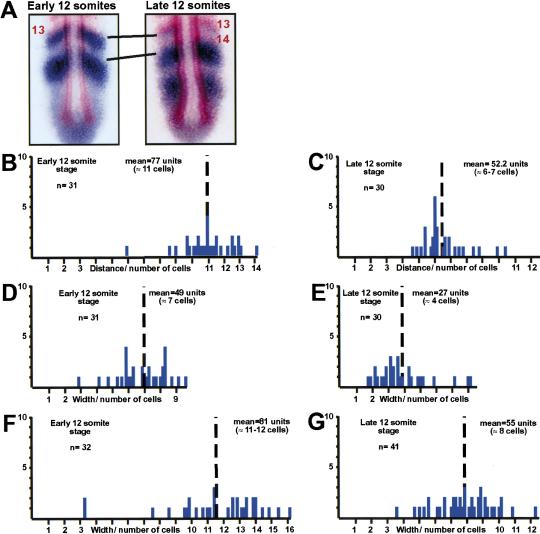
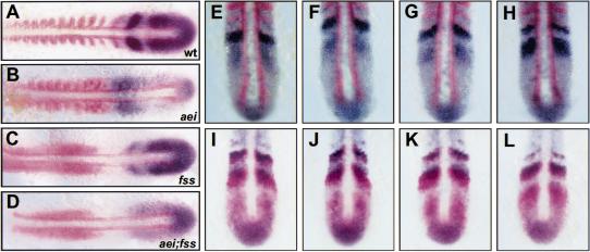
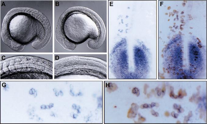
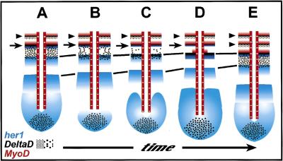
Somitogenesis has been linked both to a molecular clock that controls the oscillation of gene expression in the presomitic mesoderm (PSM) and to Notch pathway signaling. The oscillator, or clock, is thought to create a prepattern of stripes of gene expression that regulates the activity of the Notch pathway that subsequently directs somite border formation. Here, we report that the zebrafish gene after eight (aei) that is required for both somitogenesis and neurogenesis encodes the Notch ligand DeltaD. Additional analysis revealed that stripes of her1 expression oscillate within the PSM and that aei/DeltaD signaling is required for this oscillation. aei/DeltaD expression does not oscillate, indicating that the activity of the Notch pathway upstream of her1 may function within the oscillator itself. Moreover, we found that her1 stripes are expressed in the anlage of consecutive somites, indicating that its expression pattern is not pair-rule. Analysis of her1 expression in aei/DeltaD, fused somites (fss), and aei;fss embryos uncovered a wave-front activity that is capable of continually inducing her1 expression de novo in the anterior PSM in the absence of the oscillation of her1. The wave-front activity, in reference to the clock and wave-front model, is defined as such because it interacts with the oscillator-derived pattern in the anterior PSM and is required for somite morphogenesis. This wave-front activity is blocked in embryos mutant for fss but not aei/DeltaD. Thus, our analysis indicates that the smooth sequence of formation, refinement, and fading of her1 stripes in the PSM is governed by two separate activities.


Figures

Figure 1
Figure 1
aei is DeltaD. (A) The genetic map of linkage group 13 is shown with aei and the markers to which it was linked (purple). Numbers indicate genetic distance in cM. (B) Schematics are shown of a wild-type DeltaD protein and of the two predicted protein products produced in aeiAR33 and aeiAG49. Sequencing trace profiles of the region altered in each mutant allele are presented along with the corresponding wild-type sequences. (C) The products of mapping PCR reactions performed on 24 aeiAR33 embryos and 24 wild-type embryos are displayed. Although all embryos produce the shh control product, only the sibling embryos produce the DeltaD product.
Figure 2
Figure 2
aei/DeltaD is required for both neurogenesis and patterning of aei/DeltaD expression in the PSM. (A–C) Examination of Islet-1-expressing neurons (black/blue) in the trunk of aei embryos revealed a neuronal hyperplasia. Using myosin (brown) as a reference for somite position, Islet-1-expressing neurons anterior to the posterior border of somite 5 were counted in wild-type (A) and aei embryos (B,C). Wild-type embryos (n = 20) averaged 33 Islet-1-expressing neurons in this region, whereas aeiAG49 (n = 19) and aeiAR33 (n = 20) averaged 58 and 61 such neurons, respectively. All embryos are at about the 13-somite stage. Photos show dorsal views of the trunk region. (D–I) Expression of aei/DeltaD is mispatterned in each of the fss-type mutants. Note, in fss embryos (F), 10%–15% show some evidence of aei/DeltaD stripe formation. Most embryos, however, exhibit expression patterns as seen in aei/DeltaD and the other fss-type mutants. All embryos are at about the 15-somite stage. Photos show dorsal views of the tailbud and posterior trunk. In all panels, anterior is to the left and posterior to the right.
Figure 3
Figure 3
her1 is expressed in the anlage of consecutive somites. Embryos (7-, 8-, 12-, and 15-somite stage) were double-stained for her1 (blue) and MyoD (red). Some embryos from each data set were found to have her1 stripes immediately posterior to consecutive MyoD stripes: (A) 7 somites; (B) 12 somites; (C) 15 somites. The last two MyoD stripes are indicated by red arrows. (D) A summary of all of the measurement data is shown. The distance between the posterior of the third to last and penultimate MyoD stripes (interval I), between the posterior of the penultimate and the last MyoD stripes (interval II), between the posterior of the last MyoD stripe and the anterior of the first her1 stripe (interval III), between the anterior of the first and the anterior of the second her1 stripe (interval IV), and between the anterior of the second and the anterior of the third her1 stripe (interval V) were made. Vertical broken lines represent mean values. Note that in most cases, including the embryo shown in D, the last MyoD stripe and the first her1 stripe are directly juxtaposed giving a value of 0 for that measurement. This is indicated by the large number of data points at 0 in interval III. Adobe Photoshop 5.0 was used to analyze the expression domains of the digitally photographed in situ images. Distances between expression domains were measured in pixels, and the number of pixels per cell (seven) was calibrated using the in situ as well as DAPI stained embryos.
Figure 4
Figure 4
The stripes of her1 expression progress anteriorly during somitogenesis. (A) Early 12-somite embryos and late 12-somite embryos were stained for her1 (blue) and MyoD (red). Measurements were made between the anterior of the her1 stripe immediately posterior to the thirteenth MyoD stripe and the anterior of the next, posterior her1 stripe. In many of the late 12-somite embryos (as pictured), the fourteenth MyoD stripe already had formed, but the her1 stripe immediately posterior to the thirteenth MyoD stripe had not faded. (B–G) Plots of individual measurements are shown with the y-axis representing the number of embryos and the x-axis representing distance from anterior (left) to posterior (right). The distance between the two her1 stripes decreases with time (cf. B to C). Likewise, the width of the two her1 stripes along the anterior–posterior axis decreases with time at the same rate (cf. D to E and F to G). Mean values are indicated by the broken vertical line. Means were compared by two-sample t-test. The difference between the means in B and C is 29 pixels with a 95% C.I. from 19.5 to 30.7 pixels. The difference between the means in D and E is 21.6 pixels with a 95% C.I. from 15.9 to 27.4 pixels. The difference between the means in F and G is 25.7 pixels with a 95% C.I. from 17.8 to 33.7 pixels. Thus, the differences between the means in each comparison is statistically significant.
Figure 5
Figure 5
aei/DeltaD signaling is required for oscillation of her1 expression. (A–D) Anterior is left and posterior is right. Expression of her1 (blue) and MyoD (red) in wild-type (A), aei (B), fss (C), and aei;fss (D) embryos. (E–H) A hypothetical time course illustrating aei/DeltaD (blue) stripe formation relative to MyoD expression (red) is depicted. Anterior is top and posterior is bottom. In E, one aei/DeltaD stripe is seen immediately posterior to the most posterior MyoD stripe. Posterior to this aei/DeltaD stripe is a broad, weaker domain of DeltaD expression. This weaker domain of expression appears to refine such that a region of nonexpression begins to form immediately posterior to the strong aei/DeltaD stripe (F,G). As this intervening region not expressing aei/DeltaD becomes more refined, the posterior stripe of expression increases in intensity (H). (I–L) A hypothetical time course comparing aei/DeltaD expression (blue) with her1 expression (red) is depicted. aei/DeltaD expression goes through the same process of refinement as shown in E–H, whereas the her1 stripes proceed anteriorly. Although in J and K the anterior limit of the posterior her1 and aei/DeltaD stripes are not aligned as the anterior limits of the more anterior stripes are, ultimately, these more posterior stripes do align (L). Measurements of the distance between the anterior border of the two aei/DeltaD stripes suggest that the posterior aei/DeltaD stripe may progress anteriorly over one to two cell diameters.
Figure 6
Figure 6
fss activity is required cell autonomously to propagate a molecular and morphogenic wave-front activity. (A–D) Approximately 17-somite stage embryos are shown. C and D are higher magnification views of the embryos in A and B, respectively. aei/DeltaD embryos (A,C) have a qualitatively different phenotype than fss embryos (B,D). The “unsegmented” posterior somitic mesoderm in aei/DeltaD embryos is convoluted with irregular somite borders (A,C), whereas no border morphogenesis is observed in fss embryos (B,D). (E–H) her1 expression (blue) in a fss embryo that has received wild-type cells (brown) via transplantation is shown. G and H are higher magnification views of E and F, respectively, with anterior to the left. Embryos were first stained for her1 expression, dissected, mounted, and photographed. Then, the dissected tailbuds were stained for the biotin–dextran-labeled donor cells. All her1-expressing cells in the anterior domain are wild-type donor cells (cf. G to H). This result was observed in a total of 61 embryos from five independent experiments.
Figure 7
Figure 7
A model for the concomitant changes in MyoD, her1, and aei/DeltaD gene expression during zebrafish somite formation. Anterior is top and posterior is bottom. (AE) A time series during one cycle of somite formation is represented. Somite furrows are indicated by arrowheads. MyoD is expressed in the posterior of each somite, whereas aei/DeltaD is weakly expressed in the anterior of each somite. The anterior-most stripe of her1 overlaps with a strong stripe of aei/DeltaD (arrows). These two stripes refine and fade together (A–D) leaving only a faint stripe of aei/DeltaD expression (E). Around this time, the next, posterior stripe of MyoD emerges (D), and somite furrow formation occurs (second arrowhead in E). During this time period, the more posterior stripes of her1 expression progress anteriorly (A–E) (equivalent stripes are indicated by connecting lines). Meanwhile, the weak domain of aei/DeltaD expression immediately posterior to the strong stripe of expression (arrow) in the anterior PSM (A) begins to refine (B,C). Ultimately, this process results in a strong stripe of expression that aligns anteriorly with a stripe of her1 expression (D). Posterior to these overlapping stripes, a broad, weak domain of expression of aei/DeltaD forms (E) as was originally present in A. In the more posterior PSM and tailbud, an additional stripe of her1 expression emerges (B–E). The expression of aei/DeltaD in the posterior tailbud remains relatively constant (A–E).

Similar articles

Cited by

References

    1. Amacher SL, Kimmel CB. Promoting notochord fate and repressing muscle development in zebrafish. Development. 1998;125:1397–1406. - PubMed
    1. Aoyama H, Asamoto K. Determination of somite cells: Independence of cell differentiation and morphogenesis. Development. 1988;104:15–28. - PubMed
    1. Artavanis-Tsakonas S, Rand MD, Lake RJ. Notch signaling: Cell fate control and signal integration in development. Science. 1999;284:770–776. - PubMed
    1. Aulehla A, Johnson RL. Dynamic expression of lunatic fringe suggests a link between notch signaling and an automomous cellular oscillator driving somite segmentation. Dev Biol. 1999;207:49–61. - PubMed
    1. Bierkamp C, Campos-Ortega JA. A zebrafish homologue of the Drosophila neurogenic gene Notch and its pattern of transcription during early embryogenesis. Mech Dev. 1993;43:87–100. - PubMed

Publication types

MeSH terms

Substances

LinkOut - more resources