. 2000 Jul;20(13):4910-21.
doi: 10.1128/MCB.20.13.4910-4921.2000.

Orphan receptor DAX-1 is a shuttling RNA binding protein associated with polyribosomes via mRNA

Affiliations

Orphan receptor DAX-1 is a shuttling RNA binding protein associated with polyribosomes via mRNA

E Lalli et al. Mol Cell Biol. 2000 Jul.

Abstract

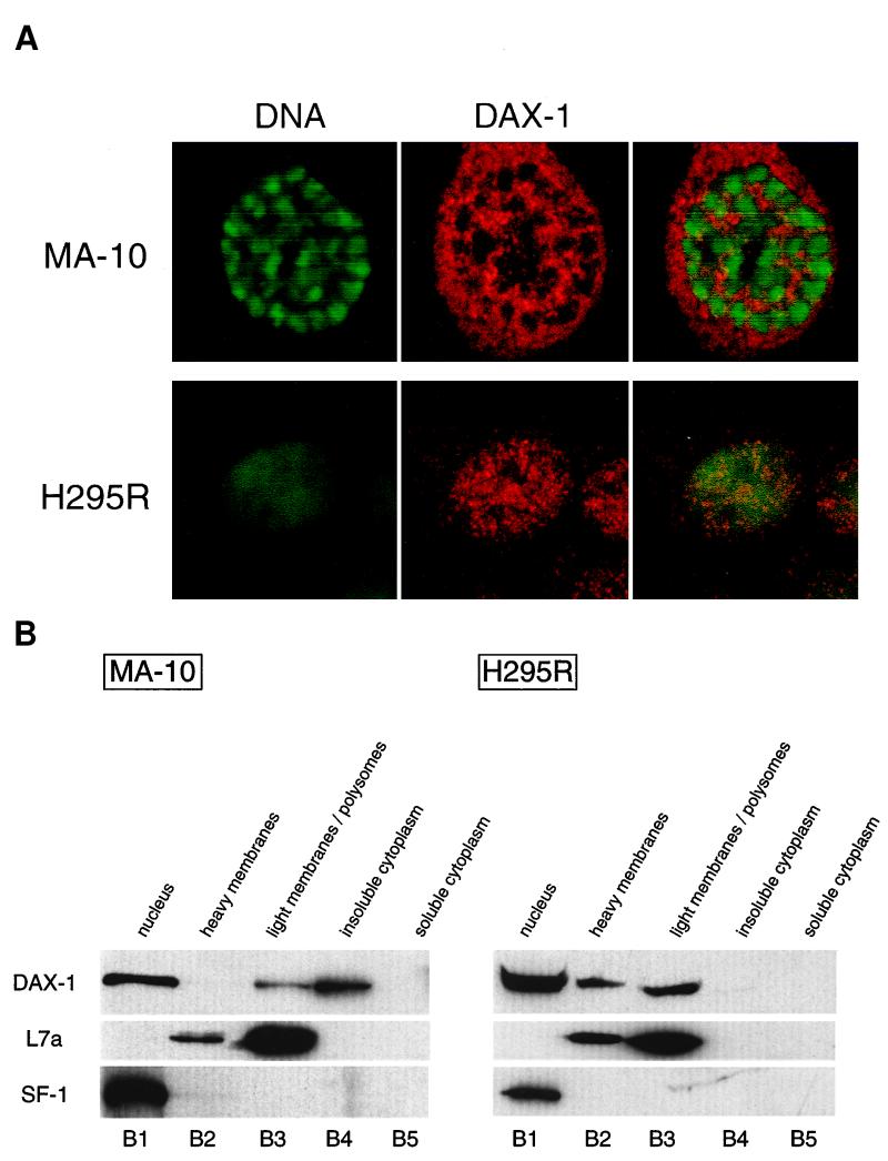
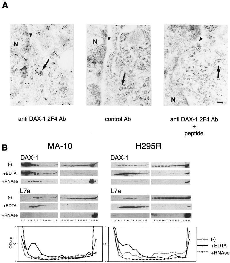
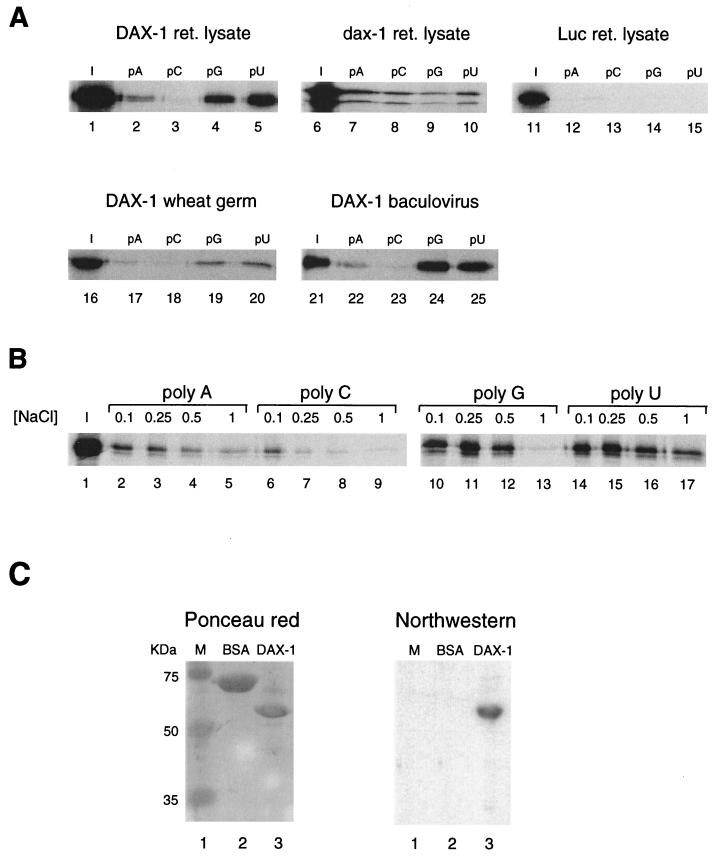
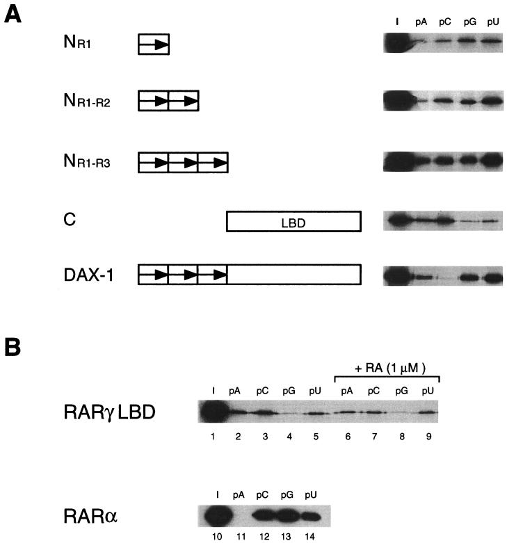
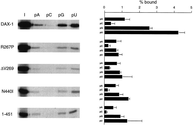
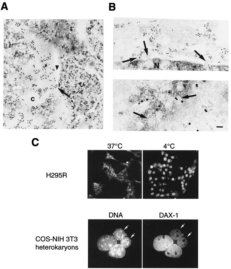
The DAX-1 (NR0B1) gene encodes an unusual member of the nuclear hormone receptor superfamily which acts as a transcriptional repressor. Mutations in the human DAX-1 gene cause X-linked adrenal hypoplasia congenita (AHC) associated with hypogonadotropic hypogonadism (HHG). We have studied the intracellular localization of the DAX-1 protein in human adrenal cortex and mouse Leydig tumor cells and found it to be both nuclear and cytoplasmic. A significant proportion of DAX-1 is associated with polyribosomes and is found complexed with polyadenylated RNA. DAX-1 directly binds to RNA, two domains within the protein being responsible for cooperative binding activity and specificity. Mutations in DAX-1 found in AHC-HHG patients significantly impair RNA binding. These findings reveal that DAX-1 plays multiple regulatory roles at the transcriptional and posttranscriptional levels.


Figures

FIG. 1
FIG. 1
Subcellular localization of DAX-1. (A) Confocal microscopy analysis of DAX-1 distribution in steroidogenic mouse MA-10 Leydig cells (top) and human adrenocortical H295R cells (bottom). DNA is shown in green, and DAX-1 is in red. (B) Fractionation of H295R and MA-10 cell extracts on a discontinuous sucrose gradient (10). Proteins from the nuclear (B1), heavy membrane (B2), light membrane-polysome (B3), insoluble cytoplasm (B4), and soluble cytoplasm (B5) fractions were run on an SDS–10% PAGE gel and transferred to nitrocellulose. The membrane was sequentially probed with the anti-DAX-1 (top), anti-L7a ribosomal protein (center), and anti-SF-1 (bottom) antibodies.
FIG. 2
FIG. 2
DAX-1 is associated with polyribosomes. (A) DAX-1 immunogold labeling in MA-10 cells. Gold particles corresponding to DAX-1 (left) are found in the nucleus localized at the periphery of chromatin, in the interchromatin space, and in the cytoplasm, mostly colocalized with polyribosomes, which appear as electron-dense clusters. No significant staining was observed when the anti-DAX-1 2F4 antibody was replaced with a control antibody (Ab) at the same dilution (center) or when the anti-DAX-1 antibody was preadsorbed with the specific peptide (right). N, nucleus. Perinuclear cisternae are indicated with an arrowhead, and polyribosomes are marked with an arrow. Bar, 0.1 μm. (B) Fractionation of MA-10 (left) and H295R (right) cell extracts on a 15 to 45% continuous sucrose gradient. Twenty-four fractions were collected from the top and analyzed by Western blot using the anti-DAX-1 2F4 antibody and the anti-L7a ribosomal protein antiserum. Distribution of DAX-1 and L7a in the gradients is also shown for extracts treated with EDTA and RNase, as described in Materials and Methods. Optical density profiles of the gradient fractions at 260 nm are shown for each treatment in the graphs at the bottom of the figure. A large proportion of the cytoplasmic DAX-1 protein is found in fractions containing polyribosomes, as shown by staining with the anti-L7a antibody. Polyribosome dissociation by both EDTA and RNase treatment modifies DAX-1 localization in the gradient fractions.
FIG. 3
FIG. 3
DAX-1 associates with poly(A)+ RNA. (A) Extracts from UV-irradiated H295R cells were subjected to oligo(dT) chromatography under native conditions. Aliquots from total cell extract (lane 1), flowthrough (lane 2), high-salt (lane 3), and low-salt (lane 4) washes and eluate (lane 5) fractions were subjected to SDS-PAGE and analyzed by Western blot using the anti-DAX-1 2F4 antibody (top) and the anti-LDH antibody (center). In vitro-translated DAX-1 from a rabbit reticulocyte lysate was assayed for direct association with the chromatographic column under the same purification conditions (bottom). Previous RNase treatment of cell extracts (1.2 mg of RNase A and 30 U of RNase T1 per ml for 15 min at 37°C) abolishes DAX-1 fractionation in the eluate (lanes 6 and 7). (B) Extracts from MA-10 cells were fractioned on a 15 to 45% continuous sucrose gradient. Fractions 1 to 3, 4 to 10, and 11 to 24, corresponding to the top of the gradient, isolated ribosomal subunits, and polyribosomal particles with a density higher than the 80S monosomes, respectively (see Fig. 2B), were pooled and subjected to oligo(dT) chromatography under native conditions. On the left side is an ethidium bromide-stained agarose gel showing that poly(A)+ RNA is selectively eluted from gradient fractions 11 to 24. Lane M, DNA molecular size markers. On the right side, oligo(dT) column eluates from each pool (lanes 2 to 4) were analyzed for DAX-1 content by Western blot. Lane L, DAX-1 protein present in 5% of the volume of the cell extract loaded on the sucrose gradient.
FIG. 4
FIG. 4
DAX-1 binds to RNA. (A) RNA homopolymer binding assay. Binding to agarose beads coupled to poly(A), poly(C), poly(G), and poly(U) is shown for human (lanes 2 to 5) and mouse (lanes 7 to 10) DAX-1 and for luciferase (Luc) (lanes 12 to 15) translated in a rabbit reticulocyte (ret.) lysate. Binding to RNA homopolymers is also shown for DAX-1 translated in a wheat germ system (lanes 17 to 20) and expressed in insect cells (lanes 22 to 25). A 1:10 ratio of the input protein is shown in each case (lanes 1, 6, 11, 16, and 21). The assay was performed in a buffer containing 0.25 M NaCl. 35S-labeled proteins were detected by fluorography, and DAX-1 expressed in a baculovirus system was detected by Western blot. (B) Salt sensitivity of the binding of DAX-1 to poly(A) (lanes 2 to 5), poly(C) (lanes 6 to 9), poly(G) (lanes 10 to 13), and poly(U) (lanes 14 to 17) was tested in buffers containing NaCl concentrations of 0.1, 0.25, 0.5, and 1 M. Lane I, 1:10 of the input protein (lane 1). (C) Northwestern binding assay using a 32P-labeled 240-nucleotide riboprobe transcribed from PvuII-linearized pBluescript and proteins immobilized on a nitrocellulose membrane. The Ponceau red-stained membrane is also shown on the left. Lanes show molecular size markers (lane 1), BSA (10 μg, lane 2), and DAX-1 (10 μg, lane 3).
FIG. 5
FIG. 5
DAX-1 domains involved in RNA binding. (A) Full-length DAX-1 and truncated proteins corresponding to aa 1 to 69 (NR1), 1 to 135 (NR1-R2), 1 to 204 (NR1-R3), and 205 to 470 (C, corresponding to the DAX-1 putative LBD) were translated in the rabbit reticulocyte system and tested in the RNA homopolymer binding assay. Lane I, 1:10 of the input protein. The assay was performed in a buffer containing 0.25 M NaCl. (B) RARγ LBD (aa 178 to 423) was translated in the rabbit reticulocyte system, and binding to RNA homopolymers was tested in the presence (lanes 6 to 9) and in the absence (lanes 2 to 5) of 1 μM all-trans-retinoic acid. Lane I, 1:10 of the input protein (lane 1). Bacterially expressed RARα was used in the RNA homopolymer binding assay and detected by Western blot, as described (12) (lanes 11 to 14). Lane I, 1:10 of the input protein (lane 10). The assay was performed in a buffer containing 0.25 M NaCl.
FIG. 6
FIG. 6
DAX-1 binding to RNA is impaired by mutations found in AHC-HHG patients. Wild-type DAX-1 and the R267P, ΔV269, N440I, and 1-451 mutants were expressed in the rabbit reticulocyte lysate and tested in the RNA homopolymer binding assay. Proteins eluted from beads were run on an SDS-PAGE gel and subjected to fluorography. The amount of radioactivity retained on beads was measured by phosphoimaging. Lane I, 1:10 of the input protein. The assay was performed in a buffer containing 0.25 M NaCl. Data are expressed as a percentage of the input protein retained on beads and represent the averages of three independent experiments + standard error of the mean.
FIG. 7
FIG. 7
DAX-1 associates with nuclear RNP and pore structures and is exported from and reimported to the nucleus. (A) DAX-1 immunogold labeling in MA-10 cells processed with the EDTA-regressive staining technique to selectively visualize RNP structures. In the nucleus, gold particles are found in the nucleoplasm associated with perichromatin fibrils and in the interchromatin space. No particles are found in correspondence with the bleached chromatin. Cytoplasmic labeling is found associated with ribosome-rich areas (open arrow; see also Fig. 2A). An image of a cluster of gold particles associated with a nuclear pore structure is indicated with an arrow. c, chromatin; i, interchromatin space. A perinuclear cisterna is indicated with an arrowhead. (B) Gold particles associated with nuclear pore structures. Top: side view. Bottom: tangential view. Nuclear pore structures are indicated with an arrow. Gold particles are larger in the bottom panel because 1-nm immunogold particles were used, followed by silver enhancement. N, nucleus. Bar, 0.1 μm. (C) Top: anti-DAX-1 immunofluorescence in H295R cells cultured at 37°C (left) or kept at 4°C for 4 h (right). Bottom: COS monkey cells transfected with a DAX-1 expression vector were fused to NIH 3T3 mouse cells using the method described in Materials and Methods. Four hours after fusion, cells were fixed with paraformaldehyde, and DAX-1 distribution was detected by immunofluorescence (right). DNA was stained with Hoechst 33342 (left). NIH 3T3 cells (arrows) are easily recognized by their heterochromatin clumps brightly stained by the Hoechst dye. DAX-1 transfer into the NIH 3T3 cell nucleus is readily detectable (arrows). No DAX-1 staining was detected in the nucleus of NIH 3T3 cells mock-fused to COS cells (not shown).

Similar articles

Cited by

References

    1. Bae D S, Schaefer M L, Partan B W, Muglia L. Characterization of the mouse DAX-1 gene reveals evolutionary conservation of a unique amino-terminal motif and widespread expression in mouse tissue. Endocrinology. 1996;137:3921–3927. - PubMed
    1. Bardoni B, Zanaria E, Guioli S, Floridia G, Worley K C, Tonini G, Ferrante E, Chiumello G, McCabe E R B, Fraccaro M, Zuffardi O, Camerino G. A dosage sensitive locus at chromosome Xp21 is involved in male to female sex reversal. Nat Genet. 1994;7:497–501. - PubMed
    1. Bernhard W. A new staining procedure for electron microscopical cytology. J Ultrastruct Res. 1969;27:250–265. - PubMed
    1. Bertrandy S, Burlet P, Clermont O, Huber C, Fondrat C, Thierry-Mieg D, Munnich A, Lefebvre S. The RNA-binding properties of SMN: deletion analysis of the zebrafish orthologue defines domains conserved in evolution. Hum Mol Genet. 1999;8:775–782. - PubMed
    1. Caricasole A, Duarte A, Larsson S H, Hastie N D, Little M, Holmes G, Todorov I, Ward A. RNA binding by the Wilms tumor suppressor zinc finger proteins. Proc Natl Acad Sci USA. 1996;93:7562–7566. - PMC - PubMed

Publication types

MeSH terms