. 2000 Jun 5;191(11):1881-94.
doi: 10.1084/jem.191.11.1881.

Receptor revision of immunoglobulin heavy chain variable region genes in normal human B lymphocytes

Affiliations

Receptor revision of immunoglobulin heavy chain variable region genes in normal human B lymphocytes

P C Wilson et al. J Exp Med. .

Abstract

Contrary to the general precepts of the clonal selection theory, several recent studies have provided evidence for the secondary rearrangement of immunoglobulin (Ig) genes in peripheral lymphoid tissues. These analyses typically used transgenic mouse models and have only detected secondary recombination of Ig light chain genes. Although Ig heavy chain variable region (V(H)) genes encode a substantial element of antibody combining site specificity, there is scant evidence for V(H) gene rearrangement in the periphery, leaving the physiological importance of peripheral recombination questionable. The extensive somatic mutations and clonality of the IgD(+)Strictly-IgM(-)CD38(+) human tonsillar B cell subpopulation have now allowed detection of the first clear examples of receptor revision of human V(H) genes. The revised VDJ genes contain "hybrid" V(H) gene segments consisting of portions from two separate germline V(H) genes, a phenomenon previously only detected due to the pressures of a transgenic system.


Figures

Figure 1
Figure 1
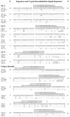
7 related recombinant hybrids were isolated from the 65 clones of CP1. A total of 65 clonally related sequences referred to as CP1 were isolated by either random or directed cloning of VH4DJH transcripts from IgD+GC B cells. The proposed germline genes encoding the CP1 founder clonotype consist of VH4-34, D3-10, and JH3 (first line of comparisons). Nucleotides homologous to these germline genes are indicated as dashes. Base exchanges resulting from somatic hypermutation are indicated in boldface type. Asterisks indicate deletional differences between the VH4-34 and VH4-61 germline genes. (A) A comparison of the 7 sister hybrid VH segments of the revised clonotypes with the consensus sequence of the remaining unrevised 57 CP1 clones (second line) and with the VH4-34 (top line) and VH4-61 (bottom line) germline genes. Another single CP1 hybrid that used a different donor VH gene than the seven sister hybrid clones is not shown or included in the consensus. The last nucleotide of homology of the hybrids to VH4-61 is C183 (black arrowhead). Proximal to C183 is a well-conserved cryptic RSS beginning at C201 that we propose is the hybrid junction. A canonical heptamer-nonamer RSS is included for comparison, and the white arrowhead marks the proposed junction. The seven hybrid clones are listed from top to bottom in order of phylogenetic similarity. Note the last three lines of the sequence comparison, demonstrating the undoubted clonal relatedness of the hybrid clonotypes to the rest of the CP1 isolates represented by the consensus. Ig sequence numbering is based on the numbering system of Chothia et al. (reference 43). (B) Phylogeny of CP1. This phylogenetic tree is the most parsimonious tree implied using the “branch and bound” algorithm of this DNAPENNY component of the PHYLIP phylogenetic analysis software package (see Materials and Methods). The tree is rooted on the germline VH4-34/D3-10/JH3 genes with consensus junctional regions (indicated by the boxed “CP1 Germline”), and was produced using only the 3′ end of the VH genes beyond the point of the proposed hybrid junction (white arrowhead at C201 in A) in order to negate effects of the 5′ portion of the hybrid sequences (composed of VH4-61) on this comparative analysis, and allowing an estimation of when the receptor revision event occurred during the development of this clonal pool. The “Consensus” sequence in A is based on the majority sequence of all of the clones in normal print on this tree. The seven hybrid sequences from this figure are indicated in boldface type. Note that the receptor revision event apparently occurred quite early in the phylogeny; however, as discussed in the text, it should be noted that all clones were highly diverged from the germline VH4-34/D3-10/JH3 sequence of the original CP1 founder. A second hybrid involving a different 5′ VH donor from this clonal pool (CP1-HB2, underlined) also occurred early in the phylogeny.
Figure 1
Figure 1
7 related recombinant hybrids were isolated from the 65 clones of CP1. A total of 65 clonally related sequences referred to as CP1 were isolated by either random or directed cloning of VH4DJH transcripts from IgD+GC B cells. The proposed germline genes encoding the CP1 founder clonotype consist of VH4-34, D3-10, and JH3 (first line of comparisons). Nucleotides homologous to these germline genes are indicated as dashes. Base exchanges resulting from somatic hypermutation are indicated in boldface type. Asterisks indicate deletional differences between the VH4-34 and VH4-61 germline genes. (A) A comparison of the 7 sister hybrid VH segments of the revised clonotypes with the consensus sequence of the remaining unrevised 57 CP1 clones (second line) and with the VH4-34 (top line) and VH4-61 (bottom line) germline genes. Another single CP1 hybrid that used a different donor VH gene than the seven sister hybrid clones is not shown or included in the consensus. The last nucleotide of homology of the hybrids to VH4-61 is C183 (black arrowhead). Proximal to C183 is a well-conserved cryptic RSS beginning at C201 that we propose is the hybrid junction. A canonical heptamer-nonamer RSS is included for comparison, and the white arrowhead marks the proposed junction. The seven hybrid clones are listed from top to bottom in order of phylogenetic similarity. Note the last three lines of the sequence comparison, demonstrating the undoubted clonal relatedness of the hybrid clonotypes to the rest of the CP1 isolates represented by the consensus. Ig sequence numbering is based on the numbering system of Chothia et al. (reference 43). (B) Phylogeny of CP1. This phylogenetic tree is the most parsimonious tree implied using the “branch and bound” algorithm of this DNAPENNY component of the PHYLIP phylogenetic analysis software package (see Materials and Methods). The tree is rooted on the germline VH4-34/D3-10/JH3 genes with consensus junctional regions (indicated by the boxed “CP1 Germline”), and was produced using only the 3′ end of the VH genes beyond the point of the proposed hybrid junction (white arrowhead at C201 in A) in order to negate effects of the 5′ portion of the hybrid sequences (composed of VH4-61) on this comparative analysis, and allowing an estimation of when the receptor revision event occurred during the development of this clonal pool. The “Consensus” sequence in A is based on the majority sequence of all of the clones in normal print on this tree. The seven hybrid sequences from this figure are indicated in boldface type. Note that the receptor revision event apparently occurred quite early in the phylogeny; however, as discussed in the text, it should be noted that all clones were highly diverged from the germline VH4-34/D3-10/JH3 sequence of the original CP1 founder. A second hybrid involving a different 5′ VH donor from this clonal pool (CP1-HB2, underlined) also occurred early in the phylogeny.
Figure 2
Figure 2
A pair of related hybrid recombinants with extensively mutated postjunctional regions were isolated from 77 total clones of CP2. All labeling, annotations, and methods are standardized to Fig. 1. “Consensus” in the second line in A of the comparison represents 72 nonhybrids from this clonal pool, which are displayed in normal print in B. Three additional single hybrid VH isolates in this clonal pool are not included in the consensus, but are underlined in the phylogeny in B. Note the eight somatic mutations shared by the two hybrid recombinants and the consensus of the nonhybrids in the postjunctional regions (after C201, white arrowhead) of VH4-34 and JH6. These mutations are highly indicative that somatic hypermutation had begun before the hybrid formation, and additional mutations 5′ to the junction demonstrate continued hypermutation after the hybrid formed. A similar point is evident by the phylogeny of the postjunctional portions of all CP2 clones in B. The hybrids occur in the middle of an extensive branch of the phylogeny, indicating mutational divergence both before and after the recombination event. It is interesting to note that another single hybrid recombinant (CP2-HB2, underlined in B) formed with a different donor gene (VH4-39 donor) occurred immediately after the pair of related hybrid recombinants displayed in bold print (CP2-HB1-1 and CP2-HB1-2).
Figure 2
Figure 2
A pair of related hybrid recombinants with extensively mutated postjunctional regions were isolated from 77 total clones of CP2. All labeling, annotations, and methods are standardized to Fig. 1. “Consensus” in the second line in A of the comparison represents 72 nonhybrids from this clonal pool, which are displayed in normal print in B. Three additional single hybrid VH isolates in this clonal pool are not included in the consensus, but are underlined in the phylogeny in B. Note the eight somatic mutations shared by the two hybrid recombinants and the consensus of the nonhybrids in the postjunctional regions (after C201, white arrowhead) of VH4-34 and JH6. These mutations are highly indicative that somatic hypermutation had begun before the hybrid formation, and additional mutations 5′ to the junction demonstrate continued hypermutation after the hybrid formed. A similar point is evident by the phylogeny of the postjunctional portions of all CP2 clones in B. The hybrids occur in the middle of an extensive branch of the phylogeny, indicating mutational divergence both before and after the recombination event. It is interesting to note that another single hybrid recombinant (CP2-HB2, underlined in B) formed with a different donor gene (VH4-39 donor) occurred immediately after the pair of related hybrid recombinants displayed in bold print (CP2-HB1-1 and CP2-HB1-2).
Figure 3
Figure 3
There are cryptic RSS at each of the VH recombinant hybrid junctions. For each comparison, the germline counterpart of the original or recipient VH gene is the top sequence, the bottom sequence is the germline donor VH gene, and the middle sequence is the hybrid VHDJ clone. Nucleotides similar to the recipient VH gene are shown as dashes. Standard RSS containing both 12- and 23-bp spacer segments are listed at each junction for reference (the short RSS for Set 1 uses a 13-bp rather than a 12-bp spacer, as this spacer length gives a more complete nonamer; the canonical 12-bp spacer is actually 12 ± 1 bp [reference 45]). Three of the cryptic RSS were shared by greater than one hybrid VHDJH clone isolated (listed as Set 1‚ Set 2, and Set 3). The remaining six isolates had unique junctional positions. Note that clone CP1-HB3 has a 24-nucleotide (8-codon) deletion suggestive of exonuclease activity during recombination. Complete Ig VH gene sequences from this study are available from EMBL/GenBank/DDBJ under accession nos. AF262069–AF262209.
Figure 4
Figure 4
VH to VHDJH recombination requires hybrid joint formation. RAG-mediated cleavage occurs at the RSS producing signal ends (triangles) and coding ends (short rectangles). (a) To produce a functional hybrid recombinant, nonstandard V(D)J recombination involving hybrid joint formation with coding to signal end joining must occur (reference 26). Joining of the recipient signal end to the donor coding end produces a functional VH gene consisting of 5′ donor sequence and 3′ recipient and DJH sequence. The end of the original VHDJH gene 5′ to the internal cryptic RSS, and the end of the donor VH gene 3′ to its internal cryptic RSS, plus any intervening DNA, form a circular deletional product by donor signal end to recipient coding end joining. (b) Standard V(D)J recombination involves signal to signal end, and coding to coding end joint formation. If standard recombination were to occur involving a VH to VHDJH recombination, the donor signal end and 3′ donor sequence would be joined to the recipient signal end and 3′ sequence, forming a nonfunctional hybrid consisting of the inverted 3′ end of the donor recombined to the recipient 3′ end DJH. Recipient to donor coding end joining would similarly disrupt the donor VH gene sequence. Note that this inversional recombination would not damage the chromosome and may be a means to deactivate rearrangements allowing rearrangement at the other allele, as suggested by Taki et al. (reference 27). As described in the text, the small arrows in the top panel marked by an asterisk mark the position bound by primers to specifically detect the recombination circle deletion product. Note that only in the deletion circle will these primers drive an efficient reaction.

Comment in

  • Revising B cell receptors.
    Nemazee D, Weigert M. Nemazee D, et al. J Exp Med. 2000 Jun 5;191(11):1813-7. doi: 10.1084/jem.191.11.1813. J Exp Med. 2000. PMID: 10839798 Free PMC article. Review. No abstract available.

Similar articles

Cited by

References

    1. Tonegawa S. Somatic generation of antibody diversity. Nature. 1983;302:575–581. - PubMed
    1. Radic M.Z., Erikson J., Litwin S., Weigert M. B lymphocytes may escape tolerance by revising their antigen receptors. J. Exp. Med. 1993;177:1165–1173. - PMC - PubMed
    1. Gay D., Saunders T., Camper S., Weigert M. Receptor editingan approach by autoreactive B cells to escape tolerance. J. Exp. Med. 1993;177:999–1008. - PMC - PubMed
    1. Tiegs S.L., Russell D.M., Nemazee D. Receptor editing in self-reactive bone marrow B cells. J. Exp. Med. 1993;177:1009–1020. - PMC - PubMed
    1. McMahan C.J., Fink P.J. RAG reexpression and DNA recombination at T cell receptor loci in peripheral CD4+ T cells. Immunity. 1998;9:637–647. - PubMed

Publication types

MeSH terms

Associated data