Comparative Study
. 2000 Jun 1;19(11):2629-40.
doi: 10.1093/emboj/19.11.2629.

Distribution of acetylated histones resulting from Gal4-VP16 recruitment of SAGA and NuA4 complexes

Affiliations
Comparative Study

Distribution of acetylated histones resulting from Gal4-VP16 recruitment of SAGA and NuA4 complexes

M Vignali et al. EMBO J. .

Abstract

We analyzed the targeting of histone acetyltransferase (HAT) complexes by DNA-binding activators during transcriptional activation and the resulting distribution of acetylated histones. An in vitro competition assay was developed to acetylate and transcribe a nucleosomal array template in the presence of excess non-specific chromatin, which mimics in vivo conditions. Stimulation of transcription from the nucleosomal array template under competitive conditions by the SAGA and NuA4 HAT complexes depended on the presence of the Gal4-VP16 activator, which recognizes sites in the promoter and directly interacts with these HATs. Importantly, the stimulation of transcription by SAGA and NuA4 depended on the presence of Gal4-VP16 during histone acetylation, and Gal4-VP16-bound nucleosomal templates were acetylated preferentially by SAGA and NuA4 relative to the competitor chromatin. While targeting of the SAGA complex led to H3 acetylation of promoter-proximal nucleosomes, targeting of the NuA4 complex led to a broader domain of H4 acetylation of >3 kbp. Thus, either promoter-proximal H3 acetylation by SAGA or broadly distributed acetylation of H4 by NuA4 activated transcription from chromatin templates.


Figures
None
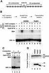
Fig. 1. Targeted histone acetylation by SAGA and NuA4 is required for stimulated transcription in vitro under competitive conditions. (A) Construct used in the transcription experiments, showing the position of the Gal4 DNA-binding sites and the 5S rDNA repeats. The chromatin template was generated as described in Materials and methods. The filled arrow signals the initiation site and the direction of transcription, while the open arrowhead shows the position and orientation of the oligonucleotide used for RNA analysis by primer extension. (B) Transcription assay examining the influence of targeted histone acetylation on transcription. The G5E4-5S nucleosomal array was transcribed following HAT reactions including (+) or omitting (–) Gal4-VP16, the G5E4-5S array and Superose 6-purified HAT complexes as indicated. All HAT reactions contained acetyl-CoA, a 50-fold molar excess of purified chromatin relative to the G5E4-5S array and an HIV-1 plasmid as an internal control for recovery. As indicated in the top diagram, acetyl-CoA was removed from the reaction after the acetylation step and before the transcription step. In lanes 3–10, spin column eluates lacking either Gal4-VP16 (lanes 4 and 8), the G5E4-5S array (lanes 5 and 9) or HAT complexes (lanes 6 and 10) were supplemented with the omitted component, so that transcription was performed under constant conditions. (C) The G5E4-5S nucleosomal array was transcribed following HAT reactions in the presence (+) or absence (–) of competitor chromatin and the NuA3 complex as indicated. All lanes contained Gal4-VP16. As in (B), the HIV-1 plasmid was included in the reaction as a recovery control. (D) The Superose 6 fractions of partially purified SAGA (lane 2), NuA3 (lane 3) and NuA4 (lane 4) were tested for their ability to acetylate nucleosomal histones. The samples were separated by SDS–PAGE, and the gel was stained with Coomassie Blue to determine the position of the core histones (top panel) and treated for fluorography to reveal the acetylation pattern (lower panel).
None
Fig. 2. Gal4-VP16 targets the HAT activity of SAGA and NuA4, but not that of NuA3, to the G5E4-5S array in the presence of competitor chromatin. (A) Diagram of the experimental protocol. (B) Ethidium bromide-stained agarose gel showing the migration profiles of the chromatin used as competitor (lane 1) and the reconstituted G5E4-5S array (lane 3) under the electrophoretic conditions used in (C). Lane 2 is HindIII-digested λ DNA. (C) The reconstituted array was incubated in the absence (lanes 1, 2, 5, 6, 9 and 10) or presence (3, 4, 7, 8, 11 and 12) of Gal4-VP16, and competitor chromatin was added to even-numbered lanes as indicated. Next, the reactions were incubated with SAGA, NuA3 or NuA4 in the presence of [3H]acetyl-CoA, and Gal4-VP16 was competed off by incubation with an oligonucleotide corresponding to the consensus Gal4-binding site. The samples were then separated by agarose gel electrophoresis. Finally, the gels were treated for fluorography and exposed. The arrowhead indicates the position of the 5S array (also compare with lane 3 of B).
None
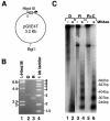
Fig. 3. The HAT activity of SAGA and NuA4, but not that of NuA3, is recruited to the pG5E4T nucleosomal array by Gal4-VP16. (A) Diagram showing the location of the Gal4-binding sites (open rectangles) and the adenovirus 2 E4 minimal promoter (arrow) in the pG5E4T plasmid, as well as selected restriction enzyme sites. (B) Agarose electrophoresis of the in vitro reconstituted pG5E4T template (R) and of non-reconstituted DNA (D). Lane 1 corresponds to HindIII-digested λ DNA and lane 4 is the 1 kb ladder from NEB. (C) The in vitro reconstitution of pG5E4T generates a regular nucleosomal ladder upon MNase digestion. Naked DNA (D) and reconstituted array +/– competitor chromatin (R and R+C, respectively) were either mock digested (–) or digested with appropriate amounts of MNase (+). DNA was extracted from these reactions, separated by electrophoresis using 32P-labeled pBR322/MspI as the molecular weight marker, and subjected to Southern blotting using hexanucleotide-labeled pG5E4T DNA as a probe. (D) Fluorography of agarose gels showing the targeting of the different HAT activities to the reconstituted pG5E4T array. The experiment was performed as described in Figure 2C. (E) The recruitment of SAGA and NuA4 to the pG5E4T template depends on the ability of Gal4-VP16 to bind DNA. The experiment was performed as in (D), but all reactions contain Gal4-VP16 and competitor chromatin, as indicated. A large molar excess of an oligonucleotide corresponding to the consensus Gal4-binding site (Gal4 oligo) was added either after the HAT reaction (lanes 1 and 3, also labeled –) or together with Gal4-VP16 (lanes 2 and 4, labeled +).
None
Fig. 4. In vitro ChIP assays confirm the targeting of the HAT activity of SAGA and NuA4 to the pG5E4T array in the presence of competitor chromatin. (A) The reconstituted array was incubated in the absence (rows A, B, E and F) or presence (rows C, D, G and H) of Gal4-VP16 as in Figure 3. Competitor chromatin was added as indicated (columns 2, 4 and 6). After acetylation by SAGA (columns 1 and 2), NuA3 (3 and 4) or NuA4 (5 and 6), the substrates were immunoprecipitated with antibodies directed against acetylated H3 (SAGA and NuA3) or H4 (NuA4). DNA was extracted from the unbound (rows A–D) and bound (rows E–H) fractions and applied to a Zeta-Probe membrane by slot blot. The membrane was hybridized with hexanucleotide-labeled pG5E4T DNA. (B–D) Graphic representation of the data shown in (A). The membranes were exposed to a PhosphorImager and quantitated. The graphs show the average and standard deviation of 3–4 repeats of each experiment with the SAGA (B), NuA3 (C) or NuA4 (D) complexes. (E) Fold stimulation by each HAT under the different conditions. The y-axis corresponds to the ratio of the material immunoprecipitated in the presence of each HAT complex (% IP with HAT) divided by the amount precipitated in the absence of the complex (% IP without HAT).
None
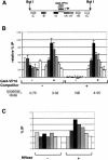
Fig. 5. Gal4-VP16 directs the HAT activity of the SAGA complex to promoter-proximal nucleosomes. The experimental set-up for these ‘scanning ChIPS’ is similar to that used in Figure 4: the template was incubated in the absence (–) or presence (+) of Gal4-VP16, and competitor chromatin was added where indicated. Next, the reactions were incubated with SAGA and acetyl-CoA (or mock acetylated in the absence of HAT complex), MNase digested and immunoprecipitated with anti-acetylated H3 antibody. DNA was extracted from the bound and unbound fractions and slot-blotted. The membranes were hybridized successively with a series of short probes (between 250 and 300 bp) that scan the length of the template, generated by PCR and labeled by primer extension from random hexanucleotides (Boehringer Mannheim). (A) Diagram showing the localization of the different probes when the plasmid is digested with BglI. (B) Average values and standard deviation of normalized data from three repeats of the experiment. The background signal (–HAT) was subtracted from the values obtained in the presence of SAGA for each condition. The numbers under the graph show the ratio of proximal (average of +A and –A) versus distal (average of +C and –C) signal for each condition tested. ND, not determined. (C) The reconstituted array was pre-incubated with Gal4-VP16 and competitor chromatin was added. Then, the template was acetylated by SAGA in the presence of acetyl-CoA, reactions were digested with MNase (+) or mock-digested (–), and the immunoprecipitation and slot blot were carried out as described above.
None
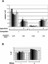
Fig. 6. Scanning ChIPs for the NuA4 complex. Experiments identical to those described in detail in the legend for Figure 5 were carried out for NuA4, except that anti-acetylated H4 antibody was used for the immunoprecipitation step. (A) Average and standard deviation of normalized data from 3–4 repeats of the scanning ChIP performed on BglI-linearized reconstituted template. (B) Control for MNase digestion (see Figure 5C).
None
Fig. 7. The domain of acetylation generated by NuA4 upon Gal4-VP16 targeting is broader than that observed for SAGA. BglI-linearized pG5E4T nucleosomal arrays were pre-incubated with Gal4-VP16 and competitor chromatin was added. Scanning ChIPs were performed as described for previous figures. The data were normalized to the highest peak for comparison purposes. The x-axis shows the distance from the Gal4 sites in base pairs.

Similar articles

Cited by

References

    1. Allard S., Utley,R.T., Savard,J., Clarke,A., Grant,P.A., Brandl,C.J., Pillus,L., Workman,J.L. and Côté,J. (1999) NuA4, an essential transcription adaptor/histone H4 acetyltransferase complex containing Esa1p and the ATM-related cofactor Tra1p. EMBO J., 18, 5108–5119. - PMC - PubMed
    1. Belotserkovskaya R. and Berger,S.L. (1999) Interplay between chromatin modifying and remodeling complexes in transcriptional regulation. Crit. Rev. Eukaryot. Gene Express., 9, 221–230. - PubMed
    1. Berger S.L., Piña,B., Silverman,N., Marcus,G.A., Agapite,J., Regier,J.L., Triezenberg,S.J. and Guarente,L. (1992) Genetic isolation of ADA2: a potential transcriptional adaptor required for function of certain acidic activation domains. Cell, 70, 251–265. - PubMed
    1. Braunstein M., Sobel,R.E., Allis,C.D., Turner,B.M. and Broach,J.R. (1996) Efficient transcriptional silencing in Saccharomyces cerevisiae requires a heterochromatin histone acetylation pattern. Mol. Cell. Biol., 16, 4349–4356. - PMC - PubMed
    1. Brown C.E., Lechner,T., Howe,L. and Workman,J.L. (2000) The many HATs of transcription coactivators. Trends Biochem. Sci., 25, 15–19. - PubMed

Publication types

MeSH terms

LinkOut - more resources