. 2000 Jun;20(12):4359-70.
doi: 10.1128/MCB.20.12.4359-4370.2000.

Mechanism for specificity by HMG-1 in enhanceosome assembly

Affiliations

Mechanism for specificity by HMG-1 in enhanceosome assembly

K B Ellwood et al. Mol Cell Biol. 2000 Jun.

Abstract

Assembly of enhanceosomes requires architectural proteins to facilitate the DNA conformational changes accompanying cooperative binding of activators to a regulatory sequence. The architectural protein HMG-1 has been proposed to bind DNA in a sequence-independent manner, yet, paradoxically, it facilitates specific DNA binding reactions in vitro. To investigate the mechanism of specificity we explored the effect of HMG-1 on binding of the Epstein-Barr virus activator ZEBRA to a natural responsive promoter in vitro. DNase I footprinting, mutagenesis, and electrophoretic mobility shift assay reveal that HMG-1 binds cooperatively with ZEBRA to a specific DNA sequence between two adjacent ZEBRA recognition sites. This binding requires a strict alignment between two adjacent ZEBRA sites and both HMG boxes of HMG-1. Our study provides the first demonstration of sequence-dependent binding by a nonspecific HMG-box protein. We hypothesize how a ubiquitous, nonspecific architectural protein can function in a specific context through the use of rudimentary sequence recognition coupled with cooperativity. The observation that an abundant architectural protein can bind DNA cooperatively and specifically has implications towards understanding HMG-1's role in mediating DNA transactions in a variety of enzymological systems.


Figures

FIG. 1
FIG. 1
HMG-1 binds to a specific site in the BHLF-1 proximal promoter. (A) Schematic representation of the BHLF-1 promoter. The ZEBRA sites are numbered Z-1 through Z-4, where Z-1 represents the site most proximal to the start of transcription. A bracket represents the 97-bp region (HLZ3,4) subcloned for use in Fig. 2 to 6. (B) HMG-1 induces cooperative binding of ZEBRA and generates a novel DNase I footprint from positions −122 to −134. A 32P-labeled promoter fragment from positions +38 to −174, bearing Z-1 through Z-4, was incubated with the proteins indicated and subjected to DNase I footprinting analysis. An autoradiograph of the polyacrylamide-urea sequencing gel surrounding the Z-3 and Z-4 footprints is shown. Lane 1, cleavage pattern of naked DNA; lanes 2 to 6, twofold decreasing titration of ZEBRA from 10 to 0.6 ng of protein; lanes 7 to 10, the last four points of the same titration of ZEBRA in the presence of 250 ng of HMG-1; lane 11, 250 ng of HMG-1 alone. (C) ZEBRA induces cooperative binding of HMG-1. Lane 1, DNase I cleavage ladder of naked DNA; lane 2, cleavage pattern generated in the presence of a subsaturating concentration of ZEBRA (2.5 ng) alone; lanes 3 to 7, footprints elicited by twofold increasing concentrations of HMG-1 (31 to 500 ng); lanes 8 to 12, the same titration of HMG-1 in the presence of 2.5 ng of ZEBRA. In the volumes used, 2.5 ng of ZEBRA corresponds to a dimer concentration of 3.5 nM.
FIG. 2
FIG. 2
Both ZEBRA binding sites are required for HMG-1-induced cooperative binding of ZEBRA. (A) HMG-1 and ZEBRA bind cooperatively to Z-3 and Z-4. The 32P-labeled 97-bp (Fig. 1A) DNA fragment, bearing Z-3 and Z-4, was incubated with the proteins indicated and subjected to DNase I footprinting analysis. Lane 1, DNase I cleavage pattern of naked DNA; lane 2, protection of Z-3 and Z-4 at saturating concentrations of ZEBRA (100 ng); lanes 3 to 5, footprints generated by twofold decreasing steps of ZEBRA (2.5 to 0.6 ng); lanes 6 to 8, footprints generated by ZEBRA (2.5 to 0.6 ng) in the presence of 250 ng of recombinant HMG-1. Note the HMG-1 footprint between the ZEBRA sites in lanes 6 to 8. (B) Mutation of individual ZEBRA binding sites abolishes cooperative binding of ZEBRA and HMG-1. The cooperative binding effect of HMG-1 on ZEBRA to the wild-type (WT) 97-bp promoter fragment is shown in lanes 1 to 4. In the context of the ΔZ-3 and ΔZ-4 promoter mutants, HMG-1 was no longer able to facilitate cooperative binding of ZEBRA (2.5 ng), as shown in lanes 5 to 8, where Z-3 has been mutated (ΔZ-3), or in lanes 9 to 12, where Z-4 has been mutated (ΔZ-4). Note in lanes 6 and 10 that ZEBRA binds to the remaining site in the mutant promoters when saturating concentrations of protein (100 ng) were used. (C) The Z-DBD is sufficient to mediate cooperative binding. The Z-DBD (Δ161) was not added (lane 1) or was added in twofold decreasing steps ranging from 24 to 3 ng either alone (lanes 2 to 5) or in the presence of 250 ng of HMG-1 (lanes 6 to 9). A 3-ng quantity of the Z-DBD corresponds to a dimer concentration of approximately 12 nM.
FIG. 2
FIG. 2
Both ZEBRA binding sites are required for HMG-1-induced cooperative binding of ZEBRA. (A) HMG-1 and ZEBRA bind cooperatively to Z-3 and Z-4. The 32P-labeled 97-bp (Fig. 1A) DNA fragment, bearing Z-3 and Z-4, was incubated with the proteins indicated and subjected to DNase I footprinting analysis. Lane 1, DNase I cleavage pattern of naked DNA; lane 2, protection of Z-3 and Z-4 at saturating concentrations of ZEBRA (100 ng); lanes 3 to 5, footprints generated by twofold decreasing steps of ZEBRA (2.5 to 0.6 ng); lanes 6 to 8, footprints generated by ZEBRA (2.5 to 0.6 ng) in the presence of 250 ng of recombinant HMG-1. Note the HMG-1 footprint between the ZEBRA sites in lanes 6 to 8. (B) Mutation of individual ZEBRA binding sites abolishes cooperative binding of ZEBRA and HMG-1. The cooperative binding effect of HMG-1 on ZEBRA to the wild-type (WT) 97-bp promoter fragment is shown in lanes 1 to 4. In the context of the ΔZ-3 and ΔZ-4 promoter mutants, HMG-1 was no longer able to facilitate cooperative binding of ZEBRA (2.5 ng), as shown in lanes 5 to 8, where Z-3 has been mutated (ΔZ-3), or in lanes 9 to 12, where Z-4 has been mutated (ΔZ-4). Note in lanes 6 and 10 that ZEBRA binds to the remaining site in the mutant promoters when saturating concentrations of protein (100 ng) were used. (C) The Z-DBD is sufficient to mediate cooperative binding. The Z-DBD (Δ161) was not added (lane 1) or was added in twofold decreasing steps ranging from 24 to 3 ng either alone (lanes 2 to 5) or in the presence of 250 ng of HMG-1 (lanes 6 to 9). A 3-ng quantity of the Z-DBD corresponds to a dimer concentration of approximately 12 nM.
FIG. 3
FIG. 3
Both HMG boxes of HMG-1 are required to mediate cooperative binding of ZEBRA. (A) The relative DNA-bending activities of the deletion derivatives in ligase-mediated circularization assays are given. +++, wild-type levels of bending; ++, a threefold increase in protein concentration is required to reach wild-type bending efficiency; +, an approximately 10-fold increase in protein concentration is required to reach wild-type bending efficiency. (B) DNase I footprinting reveals that both HMG boxes (A and B) are required for cooperative binding of ZEBRA. Lane 1, cleavage ladder generated by DNase I on naked DNA; lane 2, protections of Z-3 and Z-4 in the presence of saturating concentrations of ZEBRA (100 ng); lane 3, protections observed with subsaturating concentrations of ZEBRA (2.5 ng); lanes 4 to 11, protections induced upon incubation of 250 ng of each HMG-1 deletion derivatives in the presence of 2.5 ng of ZEBRA.
FIG. 4
FIG. 4
HMG-1 binding to DNA requires a specific sequence. (A) Mutations to sequences between Z-3 and Z-4 are deleterious to HMG-1 binding and reduce its ability to stimulate cooperative binding of ZEBRA. The effect of HMG-1 was measured on the unaltered promoter (WT) and two mutants, termed HMGΔ1 and HMGΔ2, encompassing the HMG-1 DNase I footprint. No protein (lanes 1, 5, and 9) or saturating amount of ZEBRA (100 ng) (lanes 2, 6, and 10), a subsaturating amount of ZEBRA (2.5 ng) (lanes 3, 7, and 11), or 2.5 ng of ZEBRA and 250 ng of HMG-1 (lanes 4, 8, 12) were incubated with the DNA fragments indicated (see text for details about the promoter mutants) and subjected to cleavage by DNase I. HMGΔ1 and HMGΔ2 failed to support either HMG-1 binding or cooperative binding of ZEBRA (lanes 8 and 12). (B) HMGΔ2 failed to support HMG-1-stimulated transcription. Quantities (50 ng each) of reporter templates bearing the wild-type HLZ3,4-promoter fragment upstream of E4-Lux or the HMGΔ2 mutant were cotransfected via lipofection into BHK21 cells either in the presence or absence of 500 ng of pBXG0-ZEBRA or 1,000 ng of pBXG0-HMG-1 or both together. Luciferase activity was determined (relative luciferase units), and the relative activation of each reporter in the presence or absence of a specific effector was calculated by subtracting out the basal level of activity. The results shown are the averages of three experiments. (C) Refining the HMG-1 target sequence. DNase I footprinting was performed on the listed promoter mutants (see text for details) and the fold cooperativity was calculated based on titrations of ZEBRA (10 to 0.6 ng) in the presence and absence of 250 ng of HMG-1. The underlined regions represent the positions of the mutations in Δ3, Δ4, and Δ5. Δ6 and Δ7 represent insertion mutants of +5 and +10 bp. (D) Increased DNA flexibility does not bypass the requirement for HMG-1 in promoting cooperative binding of ZEBRA. Heteroduplex DNA was generated by mixing equal volumes of the wild-type 32P-end-labeled HLZ3,4 promoter with either the HMGΔ1 or HMGΔ2 mutant promoters. The mixtures were then heated at 93°C for 3 min and allowed to cool slowly to room temperature. Samples were then fractionated on a mutation detection enhancement gel, which separates the parental homoduplexes from the heteroduplexes (49). The heteroduplex DNAs were purified and validated by digestion with 1 U of mung bean nuclease for 10 min, followed by polyacrylamide-urea gel analysis. Arrows point to the positions of cleavage at the heteroduplex region. These probes were employed in DNase I footprinting assays. The heteroduplex joints did not stimulate ZEBRA binding versus the parental probes and supported only a modest effect when HMG-1 was added. The fold cooperativity in the presence of HMG-1 was calculated and is summarized.
FIG. 4
FIG. 4
HMG-1 binding to DNA requires a specific sequence. (A) Mutations to sequences between Z-3 and Z-4 are deleterious to HMG-1 binding and reduce its ability to stimulate cooperative binding of ZEBRA. The effect of HMG-1 was measured on the unaltered promoter (WT) and two mutants, termed HMGΔ1 and HMGΔ2, encompassing the HMG-1 DNase I footprint. No protein (lanes 1, 5, and 9) or saturating amount of ZEBRA (100 ng) (lanes 2, 6, and 10), a subsaturating amount of ZEBRA (2.5 ng) (lanes 3, 7, and 11), or 2.5 ng of ZEBRA and 250 ng of HMG-1 (lanes 4, 8, 12) were incubated with the DNA fragments indicated (see text for details about the promoter mutants) and subjected to cleavage by DNase I. HMGΔ1 and HMGΔ2 failed to support either HMG-1 binding or cooperative binding of ZEBRA (lanes 8 and 12). (B) HMGΔ2 failed to support HMG-1-stimulated transcription. Quantities (50 ng each) of reporter templates bearing the wild-type HLZ3,4-promoter fragment upstream of E4-Lux or the HMGΔ2 mutant were cotransfected via lipofection into BHK21 cells either in the presence or absence of 500 ng of pBXG0-ZEBRA or 1,000 ng of pBXG0-HMG-1 or both together. Luciferase activity was determined (relative luciferase units), and the relative activation of each reporter in the presence or absence of a specific effector was calculated by subtracting out the basal level of activity. The results shown are the averages of three experiments. (C) Refining the HMG-1 target sequence. DNase I footprinting was performed on the listed promoter mutants (see text for details) and the fold cooperativity was calculated based on titrations of ZEBRA (10 to 0.6 ng) in the presence and absence of 250 ng of HMG-1. The underlined regions represent the positions of the mutations in Δ3, Δ4, and Δ5. Δ6 and Δ7 represent insertion mutants of +5 and +10 bp. (D) Increased DNA flexibility does not bypass the requirement for HMG-1 in promoting cooperative binding of ZEBRA. Heteroduplex DNA was generated by mixing equal volumes of the wild-type 32P-end-labeled HLZ3,4 promoter with either the HMGΔ1 or HMGΔ2 mutant promoters. The mixtures were then heated at 93°C for 3 min and allowed to cool slowly to room temperature. Samples were then fractionated on a mutation detection enhancement gel, which separates the parental homoduplexes from the heteroduplexes (49). The heteroduplex DNAs were purified and validated by digestion with 1 U of mung bean nuclease for 10 min, followed by polyacrylamide-urea gel analysis. Arrows point to the positions of cleavage at the heteroduplex region. These probes were employed in DNase I footprinting assays. The heteroduplex joints did not stimulate ZEBRA binding versus the parental probes and supported only a modest effect when HMG-1 was added. The fold cooperativity in the presence of HMG-1 was calculated and is summarized.
FIG. 5
FIG. 5
HMG-1 forms a ternary complex with ZEBRA and the HLZ3,4 promoter fragment. EMSAs on the wild-type (WT) HLZ3,4 and mutant HMGΔ2 promoters were performed as described in Materials and Methods. Panels A and B represent lanes from the same gel except panel B is a darker exposure of selected lanes. Lane 1, the migration of free probe through a native 6% polyacrylamide gel; lanes 2 and 3, complexes formed on the wild-type HLZ3,4 promoter with saturating (25 ng) and subsaturating amounts (0.4 ng) of Δ161, respectively; lanes 4, 9, and 13, 12.5 ng of HMG-1 or FLAG-HMG-1 incubated with subsaturating Δ161 (0.4 ng); lanes 5 and 14, binding of HMG-1 and FLAG-HMG-1 alone (12.5 ng) to the wild-type HLZ3,4 probe; lane 10, HMG-1 (12.5 ng) binding to the mutant HMGΔ2 probe; lanes 16 and 18, FLAG antibody added to complexes incorporating FLAG-HMG-1 with either the WT (lane 16) or the HMGΔ2 (lane 18) promoter. Note that the HMGΔ2 DNA template failed to induce cooperative binding of ZEBRA and the HMG-1-supershifted complexes seen with the wild-type template (compare lanes 3 and 4 and 8 and 9). A schematic diagram of the various complexes is shown to the left and is explained in Results.
FIG. 6
FIG. 6
Mapping the HMG-1 binding site by hydroxyl radical footprinting. The figure compares HMG-1-mediated cooperative binding of ZEBRA on the wild-type (WT) HLZ3,4 probe by DNase I and hydroxyl (OH) radical footprinting or on the HMGΔ2 mutant by hydroxyl radical. Lane 3 of the DNase I footprint shows the protections to Z-3 and Z-4 generated by Δ161 using saturating concentrations of protein (800 ng). The protections generated by twofold-decreasing steps of Δ161 ranging from 200 to 50 ng are shown in lanes 4 to 6, while lanes 7 to 9 illustrate the cooperative effect of 450 ng of HMG-1. Lane 10 represents the DNase I footprint of 450 ng of HMG-1 alone. The hydroxyl radical footprints on HLZ3,4 are also shown. Lanes 1 and 2 show the cleavage ladder of naked DNA generated in 2.5- and 5-min reactions. Lanes 3 to 9 show 5-min cleavage reactions performed on twofold-decreasing concentrations (200 to 50 ng) of Δ161 in the absence (lanes 3 to 5) or presence (lanes 6 to 8) of 450 ng of HMG-1. Lane 9 represents the hydroxyl radical protections generated by 450 ng of HMG-1 alone. The HMG-1 footprints between Z-3 and Z-4 are numbered 1 and 2. Identical reactions performed on the HMGΔ2 promoter mutant are shown. The individual bands generated by hydroxyl radical cleavage were boxed and quantitated using ImageQuant software. The values obtained from three different footprints were normalized to the most intense lane in each experiment and the percent saturation was calculated; strongest protections were mapped in Fig. 7. Note, higher concentrations of both HMG-1 and Δ161 were used in the hydroxyl radical footprinting experiments (see text).
FIG. 7
FIG. 7
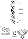
Modeling the interaction of HMG-1 with its site. (A) A helical projection of the promoter sequences from Z-3 to Z-4 of BHLF-1. The 7-bp ZEBRA binding sites are indicated by brackets. Hydroxyl radical protections are projected onto the helical backbone. Open circles, base pairs that were partially (50 to 70%) saturated; closed circles, complete (80 to 100%) protections in the presence of ZEBRA and HMG-1. (B) Model depicting how HMG-1 induces cooperative binding of ZEBRA to the Z-3 and Z-4 pair of binding sites. In the presence of both proteins, ZEBRA stabilizes HMG-1 interaction with DNA between Z-3 and Z-4 and HMG-1 induces cooperative binding of ZEBRA.

Similar articles

Cited by

References

    1. Aidinis V, Bonaldi T, Beltrame M, Santagata S, Bianchi M E, Spanopoulou E. The RAG1 homeodomain recruits HMG1 and HMG2 to facilitate recombination signal sequence binding and to enhance the intrinsic DNA-bending activity of RAG1-RAG2. Mol Cell Biol. 1999;19:6532–6542. - PMC - PubMed
    1. Allain F H, Yen Y M, Masse J E, Schultze P, Dieckmann T, Johnson R C, Feigon J. Solution structure of the HMG protein NHP6A and its interaction with DNA reveals the structural determinants for non-sequence-specific binding. EMBO J. 1999;18:2563–2579. - PMC - PubMed
    1. Boonyaratanakornkit V, Melvin V, Prendergast P, Altmann M, Ronfani L, Bianchi M E, Taraseviciene L, Nordeen S K, Allegretto E A, Edwards D P. High-mobility group chromatin proteins 1 and 2 functionally interact with steroid hormone receptors to enhance their DNA binding in vitro and transcriptional activity in mammalian cells. Mol Cell Biol. 1998;18:4471–4487. - PMC - PubMed
    1. Bustin M. Regulation of DNA-dependent activities by the functional motifs of the high-mobility-group chromosomal proteins. Mol Cell Biol. 1999;19:5237–5246. - PMC - PubMed
    1. Bustin M, Reeves R. High-mobility-group chromosomal proteins: architectural components that facilitate chromatin function. Prog Nucleic Acid Res Mol Biol. 1996;54:35–100. - PubMed

Publication types