Human cytomegalovirus replicates abortively in polymorphonuclear leukocytes after transfer from infected endothelial cells via transient microfusion events
- PMID: 10823870
- PMCID: PMC112050
- DOI: 10.1128/jvi.74.12.5629-5638.2000
Human cytomegalovirus replicates abortively in polymorphonuclear leukocytes after transfer from infected endothelial cells via transient microfusion events
Abstract
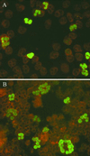
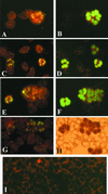
Using a recently developed model for in vitro generation of pp65-positive polymorphonuclear leukocytes (PMNLs), we demonstrated that PMNLs from immunocompetent subjects may harbor both infectious human cytomegalovirus (HCMV) and viral products (pp65, p72, DNA, and immediate-early [IE] and pp67 late mRNAs) as early as 60 min after coculture with human umbilical vein endothelial cells (HUVEC) or human embryonic lung fibroblasts (HELF) infected with a clinical HCMV isolate (VR6110) or other wild-type strains. The number of PMNLs positive for each viral parameter increased with coculture time. Using HELF infected with laboratory-adapted HCMV strains, only very small amounts of viral DNA and IE and late mRNAs were detected in PMNLs. A cellular mRNA, the vascular cell adhesion molecule-1 mRNA, which is abundantly present in both infected and uninfected HUVEC, was detected in much larger amounts in PMNLs cocultured with VR6110-infected cells than in controls. Coculture of PMNLs with VR6110-infected permissive cells in the presence or absence of RNA, protein, and viral DNA synthesis inhibitors showed that only IE genes were transcribed in PMNLs during coculture. Synthesis of IE transcripts in PMNLs was also supported by the finding that only the copy number of IE mRNA (and not the DNA or the pp67 mRNA) per infected PMNL increased markedly with time, and the pp67 to IE mRNA copy number ratio changed from greater than 10 in infected HUVEC to less than 1 in cocultured PMNLs. Fluorescent probe transfer experiments and electron microscopy studies indicated that transfer of infectious virus and viral products from infected cells to PMNLs is likely to be mediated by microfusion events induced by wild-type strains only. In addition, HCMV pp65 and p72 were both shown to localize in the nucleus of the same PMNLs by double immunostaining. Two different mechanisms may explain the virus presence in PMNLs: (i) one major mechanism consists of transitory microfusion events (induced by wild-type strains only) of HUVEC or HELF and PMNLs with transfer of viable virus and biologically active viral material to PMNLs; and (ii) one minor mechanism, i.e., endocytosis, occurs with both wild-type and laboratory strains and leads to the acquisition of very small amounts of viral nucleic acids. In conclusion, HCMV replicates abortively in PMNLs, and wild-type strains and their products (as well as cellular metabolites and fluorescent dyes) are transferred to PMNLs, thus providing evidence for a potential mechanism of HCMV dissemination in vivo.
Figures


Similar articles
-
In vitro selection of human cytomegalovirus variants unable to transfer virus and virus products from infected cells to polymorphonuclear leukocytes and to grow in endothelial cells.J Gen Virol. 2001 Jun;82(Pt 6):1429-1438. doi: 10.1099/0022-1317-82-6-1429. J Gen Virol. 2001. PMID: 11369888
-
Human cytomegalovirus and human umbilical vein endothelial cells: restriction of primary isolation to blood samples and susceptibilities of clinical isolates from other sources to adaptation.J Clin Microbiol. 2002 Jan;40(1):233-8. doi: 10.1128/JCM.40.1.233-238.2002. J Clin Microbiol. 2002. PMID: 11773121 Free PMC article.
-
Lack of transmission to polymorphonuclear leukocytes and human umbilical vein endothelial cells as a marker of attenuation of human cytomegalovirus.J Med Virol. 2002 Mar;66(3):335-9. doi: 10.1002/jmv.2150. J Med Virol. 2002. PMID: 11793385
-
Relationship of human cytomegalovirus-infected endothelial cells and circulating leukocytes in the pathogenesis of disseminated human cytomegalovirus infection: A narrative review.Rev Med Virol. 2024 Jan;34(1):e2496. doi: 10.1002/rmv.2496. Rev Med Virol. 2024. PMID: 38282408 Review.
-
The pentameric complex of human Cytomegalovirus: cell tropism, virus dissemination, immune response and vaccine development.J Gen Virol. 2017 Sep;98(9):2215-2234. doi: 10.1099/jgv.0.000882. Epub 2017 Aug 15. J Gen Virol. 2017. PMID: 28809151 Review.
Cited by
-
Virion Glycoprotein-Mediated Immune Evasion by Human Cytomegalovirus: a Sticky Virus Makes a Slick Getaway.Microbiol Mol Biol Rev. 2016 Jun 15;80(3):663-77. doi: 10.1128/MMBR.00018-16. Print 2016 Sep. Microbiol Mol Biol Rev. 2016. PMID: 27307580 Free PMC article. Review.
-
Isolation of human monoclonal antibodies that potently neutralize human cytomegalovirus infection by targeting different epitopes on the gH/gL/UL128-131A complex.J Virol. 2010 Jan;84(2):1005-13. doi: 10.1128/JVI.01809-09. Epub 2009 Nov 4. J Virol. 2010. PMID: 19889756 Free PMC article.
-
Human cytomegalovirus virion protein complex required for epithelial and endothelial cell tropism.Proc Natl Acad Sci U S A. 2005 Dec 13;102(50):18153-8. doi: 10.1073/pnas.0509201102. Epub 2005 Nov 30. Proc Natl Acad Sci U S A. 2005. PMID: 16319222 Free PMC article.
-
Specialization for Cell-Free or Cell-to-Cell Spread of BAC-Cloned Human Cytomegalovirus Strains Is Determined by Factors beyond the UL128-131 and RL13 Loci.J Virol. 2020 Jun 16;94(13):e00034-20. doi: 10.1128/JVI.00034-20. Print 2020 Jun 16. J Virol. 2020. PMID: 32321807 Free PMC article.
-
Cytomegalovirus Latency and Reactivation: An Intricate Interplay With the Host Immune Response.Front Cell Infect Microbiol. 2020 Mar 31;10:130. doi: 10.3389/fcimb.2020.00130. eCollection 2020. Front Cell Infect Microbiol. 2020. PMID: 32296651 Free PMC article. Review.
References
-
- Bitsch A, Kirchner H, Dupke R, Bein G. Cytomegalovirus transcripts in peripheral blood leukocytes of actively infected transplant patients detected by reverse-transcription-polymerase chain reaction. J Infect Dis. 1993;167:740–743. - PubMed
-
- Blok M J, Goossens V J, Vanherle S J V, Top B, Tacken N, Middeldorp J M, Christiaans M H L, van Hooff J P, Bruggeman C A. Diagnostic value of monitoring human cytomegalovirus late pp67 mRNA expression in renal-allograpt recipients by nucleic acid-sequence based amplification. J Clin Microbiol. 1998;36:1341–1346. - PMC - PubMed
-
- Boivin G, Handfield J, Toma E, Lalonde R, Bergeron M G. Expression of the late cytomegalovirus (CMV) pp150 transcript in leukocytes of AIDS patients is associated with a high viral DNA load in leukocytes and presence of CMV DNA in plasma. J Infect Dis. 1999;179:1101–1107. - PubMed
-
- Brown J M, Kaneshima H, Mocarski E S. Dramatic interstrain differences in the replication of human cytomegalovirus in SCID-hu mice. J Infect Dis. 1995;171:1599–1603. - PubMed
-
- Butini L, De Fougerolles A R, Vaccarezza M, Graziosi C, Cohen D I, Montroni M, Springer T A, Pantaleo G, Fauci A S. Intracellular adhesion molecules (ICAM)-1, ICAM-2, and ICAM-3 function as counter-receptor for lymphocyte function-associated molecule 1 in human immunodeficiency virus-mediated syncytia formation. Eur J Immunol. 1994;24:2191–2195. - PubMed
Publication types
MeSH terms
Substances
LinkOut - more resources
Full Text Sources
Other Literature Sources

