. 2000 May 15;149(4):863-74.
doi: 10.1083/jcb.149.4.863.

Microtubule interactions with the cell cortex causing nuclear movements in Saccharomyces cerevisiae

Affiliations

Microtubule interactions with the cell cortex causing nuclear movements in Saccharomyces cerevisiae

N R Adames et al. J Cell Biol. .

Abstract

During mitosis in budding yeast the nucleus first moves to the mother-bud neck and then into the neck. Both movements depend on interactions of cytoplasmic microtubules with the cortex. We investigated the mechanism of these movements in living cells using video analysis of GFP-labeled microtubules in wild-type cells and in EB1 and Arp1 mutants, which are defective in the first and second steps, respectively. We found that nuclear movement to the neck is largely mediated by the capture of microtubule ends at one cortical region at the incipient bud site or bud tip, followed by microtubule depolymerization. Efficient microtubule interactions with the capture site require that microtubules be sufficiently long and dynamic to probe the cortex. In contrast, spindle movement into the neck is mediated by microtubule sliding along the bud cortex, which requires dynein and dynactin. Free microtubules can also slide along the cortex of both bud and mother. Capture/shrinkage of microtubule ends also contributes to nuclear movement into the neck and can serve as a backup mechanism to move the nucleus into the neck when microtubule sliding is impaired. Conversely, microtubule sliding can move the nucleus into the neck even when capture/shrinkage is impaired.


Figures
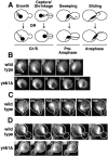
Figure 1
Figure 1
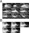
Nuclear movement caused by microtubule growth, capture/shrinkage, sweeping and sliding. (A) This schematic illustrates the different microtubule behaviors and associated nuclear movements described in the paper. (B) Nuclear movements to the incipient bud site accompanied by microtubule capture/shrinkage in G1. (C) Nuclear movements in a budded cell accompanied first by microtubule growth and later by capture/shrinkage. (D) Microtubule sweeping and spindle movement during G2/M before anaphase. The end of the spindle closer to the neck pivots as the microtubule sweeps in the direction of the mother-bud axis. Spindles in the yeb1Δ strain do not make the large movements seen in the wild-type strain. The spindle shown is at a large angle relative to the mother-bud axis and seen end-on. Budded cells are outlined for clarity. Time (s) is indicated. See supplemental videos 1–5 at http://www. jcb.org/cgi/content/full/149/4/863/DC1. Bar, 2 μm.
Figure 2
Figure 2
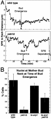
Nuclear movement to the neck. (A) Representative plots of the distance of the SPB from the bud neck vs. time for a wild-type cell and a yeb1Δ cell. (B) The percentage of cells in which the nucleus was positioned at the bud neck when the bud formed. Nuclei were considered to be at the bud neck if they were within the third of the mother cell nearest the neck (∼1.5–2 μm from the neck). Numbers of cells in each group: wild-type, 54; yeb1Δ, 61; ts-arp1, 122; ts-arp1 yeb1Δ, 33. Error bars represent standard error of proportion (SEP).
Figure 3
Figure 3
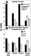
Spindle position and orientation before anaphase. (A) Position of the short spindle relative to the neck in pre-anaphase cells. Numbers of cells: wild-type, 112; yeb1Δ, 189; ts-arp1, 134; ts-arp1 yeb1Δ, 190. Error bars represent SEP. (B) Angle of the short spindle from the mother-bud axis in cells with spindles positioned at the neck. Numbers of cells: wild-type, 88; yeb1Δ, 51; ts-arp1, 102; ts-arp1 yeb1Δ, 34. Error bars represent SEP.
Figure 4
Figure 4
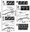
Microtubule sliding and capture/shrinkage during anaphase spindle movements. Microtubules made lateral associations with the bud cortex before and during spindle movements into the bud neck. For A, C, and D, the position of the distal end of a cytoplasmic microtubule (open triangles) and its SPB (filled squares) are plotted vs. time (s). Zero distance is defined as the position of the SPB at t = 0. The length of the microtubule (open circles) is also plotted. The insets show three frames from movies, with time (s) indicated. (A) In a wild-type cell, a cytoplasmic microtubule slides along the bud cortex as the spindle moves into the neck. (B) Bending of a spindle in a wild-type cell (arrow). (C) In a wild-type cell with the spindle in the neck, a cytoplasmic microtubule slides along the mother cortex as the spindle moves back toward the mother. (D) A yeb1Δ cell showing microtubule sliding along the bud cortex and spindle movement into the neck. (E) Capture/shrinkage events in wild-type cells with the spindle in the neck. (Top) Capture/shrinkage in the mother and (bottom) in the bud. Time (s) is indicated. See supplemental videos 6–11 at http://www.jcb.org/cgi/content/full/149/4/863/DC1. Bar, 2 μm.
Figure 6
Figure 6
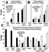
Movement and orientation of mid-anaphase spindles in relation to pre-anaphase spindle position and orientation. Time-lapse movies were analyzed for movement of spindles into the neck and orientation of spindles along the mother-bud axis. Each bar is the proportion of cells in the indicated pre-anaphase category showing delayed spindle movement into the neck. (A) The percentage of mid-anaphase spindles that failed to move into the neck is plotted in relation to their pre-anaphase position. Mid-anaphase was defined as half of the mean time from start of spindle elongation to spindle breakdown in wild-type cells. In wild-type and ts-arp1 cells, nearly all spindles were at the bud neck before anaphase. Numbers of cells: wild-type, 112; yeb1Δ next to neck, 107; yeb1Δ intermediate distance, 54; yeb1Δ far from neck, 28; ts-arp1, 128; ts-arp1 yeb1Δ next to neck, 45; ts-arp1 yeb1Δ intermediate distance, 86; ts-arp1 yeb1Δ far from neck, 59. (B) The percentage of mid-anaphase spindles that failed to move into the neck, in relation to their pre-anaphase orientation. Results are grouped by pre-anaphase spindle orientation in increments of 30°. Only spindles positioned at the bud neck were included. Numbers of cells: wild-type 0–30°, 54; wild-type 30–60°, 32; wild-type 60–90°, 2; yeb1Δ 0–30°, 22; yeb1Δ 30–60°, 20; yeb1Δ 60–90°, 9; ts-arp1 0–30°, 67; ts-arp1 30–60°, 34; ts-arp1 60–90°, 1; ts-arp1 yeb1Δ 0–30°, 11; ts-arp1 yeb1Δ 30–60°, 18; ts-arp1 yeb1Δ 60–90°, 5. (C) Orientation of mid-anaphase spindles. Results are grouped by the range of angles of the mid-anaphase spindles from the mother-bud axis in increments of 30°, in addition to the pre-anaphase spindle position, as in A. Percentages are of mid-anaphase spindles with the indicated orientation and are derived from all spindles having the indicated pre-anaphase position. Numbers of cells are as in A. Error bars are SEP.
Figure 7
Figure 7
Microtubule sweeping and capture/shrinkage during anaphase spindle movements in ts-arp1 cells. (A) In this cell, microtubules do not make lateral associations with the bud cortex and do not slide but do sweep. The spindle does not move into the neck. Later a microtubule grows very long and buckles as the spindle is pushed out of the neck. (B) In this cell, a capture/shrinkage event in the bud occurs as the spindle moves into the neck. Here, the spindle movement into the neck was not caused solely by spindle elongation since both ends of the spindle moved toward the bud. The starting positions of the SPBs are indicated with arrows. Time (s) is indicated. See supplemental videos 14 and 15 at http://www.jcb.org/cgi/content/full/149/4/863/DC1. Bar, 2 μm.
Figure 8
Figure 8
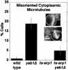
Misorientation of cytoplasmic microtubules during anaphase. In some cells, the cytoplasmic microtubules from both SPBs were temporarily directed toward the rear of the mother, as shown in the inset. The percentage of cells in which this occurred is plotted. Numbers of cells: wild-type, 112; yeb1Δ, 189; ts-arp1, 128; ts-arp1 yeb1Δ, 190. Error bars are SEP. Bar, 2 μm.

Similar articles

Cited by

References

    1. Amberg D.C., Zahner J.E., Mulholland J.W., Pringle J.R., Botstein D. Aip3p/Bud6p, a yeast actin-interacting protein that is involved in morphogenesis and the selection of bipolar budding sites. Mol. Biol. Cell. 1997;8:729–753. - PMC - PubMed
    1. Carminati J.L., Stearns T. Microtubules orient the mitotic spindle in yeast through dynein-dependent interactions with the cell cortex. J. Cell Biol. 1997;138:629–641. - PMC - PubMed
    1. Clark S.W., Meyer D.I. ACT3 - a putative centractin homologue in S. cerevisiae is required for proper orientation of the mitotic spindle. J. Cell Biol. 1994;127:129–138. - PMC - PubMed
    1. Cottingham F.R., Hoyt M.A. Mitotic spindle positioning in Saccharomyces cerevisiae is accomplished by antagonistically acting microtubule motor proteins. J. Cell Biol. 1997;138:1041–1053. - PMC - PubMed
    1. DeZwaan T.M., Ellingson E., Pellman D., Roof D.M. Kinesin-related Kip3 of Saccharomyces cerevisiae is required for a distinct step in nuclear migration. J. Cell Biol. 1997;138:1023–1040. - PMC - PubMed

Publication types

MeSH terms