. 2000 May 15;149(4):811-24.
doi: 10.1083/jcb.149.4.811.

The condensin complex governs chromosome condensation and mitotic transmission of rDNA

Affiliations

The condensin complex governs chromosome condensation and mitotic transmission of rDNA

L Freeman et al. J Cell Biol. .

Abstract

We have characterized five genes encoding condensin components in Saccharomyces cerevisiae. All genes are essential for cell viability and encode proteins that form a complex in vivo. We characterized new mutant alleles of the genes encoding the core subunits of this complex, smc2-8 and smc4-1. Both SMC2 and SMC4 are essential for chromosome transmission in anaphase. Mutations in these genes cause defects in establishing condensation of unique (chromosome VIII arm) and repetitive (rDNA) regions of the genome but do not impair sister chromatid cohesion. In vivo localization of Smc4p fused to green fluorescent protein showed that, unexpectedly, in S. cerevisiae the condensin complex concentrates in the rDNA region at the G2/M phase of the cell cycle. rDNA segregation in mitosis is delayed and/or stalled in smc2 and smc4 mutants, compared with separation of pericentromeric and distal arm regions. Mitotic transmission of chromosome III carrying the rDNA translocation is impaired in smc2 and smc4 mutants. Thus, the condensin complex in S. cerevisiae has a specialized function in mitotic segregation of the rDNA locus. Chromatin immunoprecipitation (ChIP) analysis revealed that condensin is physically associated with rDNA in vivo. Thus, the rDNA array is the first identified set of DNA sequences specifically bound by condensin in vivo. The biological role of higher-order chromosome structure in S. cerevisiae is discussed.


Figures
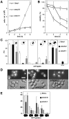
Figure 2
Figure 2
Characterization of smc2-8 and smc4-1 mutants. (A) Growth curves of smc mutants (strains 1bAS330 and 1bAS344) at 37°C, compared with the isogenic strain YPH499 (Smc+). (B) Viability of Smc+ and Smc strains (YPH499, 1bAS330, and 1bAS344) at 37°C determined by plating assay. (C) Distribution of cell types within arrested populations of smc mutants (strains listed in A) after a 3-h exposure to restrictive temperature (37°C). (D) Cell morphology (phase-contrast) and nuclear DNA position (4,6-diamidino-2-phenylindole staining) in smc mutant strains after a 3-h exposure to 37°C. Bar, 5 μm. (E) Sister chromatid separation in wild-type cells and smc mutants with a GFP tag at the ura3 locus after 3 h at 37°C. Smc +: YPH499bV; smc2-8: 1bAS330bV; smc4-1: 1aAS342bV. The classes of cells are as follows (only cells with buds were counted): single dot in any cell body; two dots in the same cell body; one dot in each cell body with the distance between two dots <2/3 of maximal cell axis; one dot in each cell body with the distance between two dots >2/3 of maximal axial measurement; more than two dots per mother and daughter cell body.
Figure 3
Figure 3
Establishment of rDNA condensation is impaired in smc mutants. (A) rDNA FISH. Red, propidium iodide staining corresponding to nuclear DNA; green, rDNA FISH signal. α-Factor–arrested Smc+ cells are shown for comparison. Smc +: YPH499b; smc2-8: 1bAS330b; smc4-1: 1aAS342b. Bar, 5 μm. (B) Quantitative analysis of condensation of the rDNA locus normalized to the Smc+ value. Arrest conditions, quantification approach, and correction to minimize contribution of cell-to-cell differences in spreading are described in Materials and Methods. (C) Chromosome VIII multiprobe FISH painting. Strains are as in A. A minimal circle was used as an approximation of the area occupied by fluorescent signals (see Materials and Methods). Bar, 5 μm. (D) Quantitative analysis of condensation of chromosome VIII normalized to the Smc+ value.
Figure 1
Figure 1
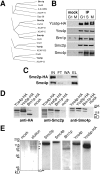
Characterization of the condensin protein complex. (A) CLUSTALW alignment of the condensin subunits from different organisms. Arabidopsis thaliana and human sequences are from GenBank. (B) Immunoprecipitation of condensin throughout the cell cycle. Strains YPH499bp (mock) and YP499bp5 were arrested with α-factor (G1, 100% of cells arrested), hydroxyurea (S, 84% arrest), and nocadazole (M, 96% arrest). 12CA5 immunoprecipitates from the corresponding protein extracts are shown for YPH499bp5. Smc2p, Smc4p, Brn1p, and Ycs4p are detected with the corresponding rabbit polyclonal antibodies. The precipitates were probed with antibodies against chromatin proteins, including Hmo1, Smc1p, Mcd1p, and Top2p, but no positive signal was detected (data not shown). (C) Smc2p and Smc4p form an obligatory complex. Asynchronous cells of YPH499bp2 were analyzed by immunoprecipitation. Saturating amount of anti-HA beads were added, allowing quantitative depletion of Smc2p-HA. IN, input; FT, flow-through; WA, wash; and EL, elution. (D) Stoichiometry of non-SMC condensin subunits. Extracts from YPH499bp6, BY4733bp4, and YPH499bp5, grown to equal optical density were analyzed by anti-HA immunoprecipitation. Immunoprecipitates were probed with anti-HA, anti-Smc2p, and anti-Smc4p antibodies. (E) Immunoaffinity purification of condensin complex. 10 g of the YPH499bp5 cell was subjected to the modified immunoprecipitation protocol, followed by the peptide elution with AS4 (GYPYDVPDYAG) (0.5 mg/ml) in place of SDS. 10 g of YPH499bp cells was processed identically for the mock purification. The silver-stained gel (4–12% PAGE) shows stoichiometry of condensin subunits. Asterisks indicate positions of the condensin subunit bands on the gel. Proteins were identified by Western blot with the corresponding antibodies. Smc2p (predicted molecular mass 134 kD) and Ycs4p (133 kD) comigrate.
Figure 4
Figure 4
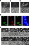
Condensin localization. (A) Condensin localization visualized by Smc4p-GFP in AS335/pLF640 diploid strain grown asynchronously. Arrowhead points to two nucleolar chromatin signals in one nucleus. (B) AS335 without pLF640: negative control for autofluorescence. (C) Strain NOY891/pLF640 lacking rDNA tandem repeats: subnuclear clustering of Smc4p-GFP signal is replaced by diffuse nuclear signal. Arrowhead points to diffuse GFP signal along the putative position of the spindle. (D) Colocalization of Net1p and condensin. Strain WY53 (Shou et al. 1999) was transformed with pLF640, allowing simultaneous detection of Smc4p-GFP (green) and Net1p-MYC (red, anti-MYC antibody, Cy3-conjugated secondary antibody). Arrowhead in the overlap panel points to the nucleolar region. Cells were released from nocadazole block for 30 min, fixed for 5 min with formaldehyde, and processed for immunofluorescence. DNA was visualized with 4,6-diamidino-2-phenylindole. (E) Time course analysis of YPH499b/pLF640 expressing Smc4p-GFP after release from G1 arrest (α-factor) at 23°C in selective media. GFP and phase-contrast images are shown. Arrowheads indicate subnuclear signal corresponding to nucleolar chromatin. Bars, 5 μm.
Figure 5
Figure 5
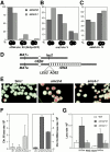
Segregation of the rDNA locus is impaired in condensin mutants. (A) rDNA segregation after a 3-h shift to 37°C in the strains 3-1bAS330b/pAS622 (smc2-8) and 6-1bAS344b/pAS622 (smc4-1). rDNA was visualized by GFP fused to Sir2p. Only budded cells were scored. Numbers indicate percentage of the given cell type among budded cells. (B) Segregation of pericentromeric ura3 locus after a 3-h shift to 37°C in the strains 1bAS330bV (smc2-8) and 1aAS342bV (smc4-1). The chromosomal tag at ura3 was visualized by GFP fused to LacI. Only budded cells were scored. (C) Segregation of the centromere-distal (850 kb from CEN4) ade8 locus after a 3-h shift to 37°C in the strains 1bAS330bIV (smc2-8) and 1aAS342bIV (smc4-1). The chromosomal tag at ade8 was visualized by GFP fused to LacI. Only large-budded cells were scored. (D) Schematic of two chromosome III homologues in strains AS389, AS388, and AS386 used for experiments in E and F. (E) Loss of the rDNA-carrying chromosome III in Smc+, smc2-8, and smc4-1 diploid strain at 35°C. Red sectors correspond to chromosome loss events. The sectoring in Smc+ (diploid strain AS389) is due to ADE2 loss as a result of mitotic recombination. (F) Comparison of the ADE2-marked native chromosome III loss in strains AS260–1 (wt), AS359–1 (smc2-8), and AS362–1 (smc4-1) to the loss of the rDNA-carrying chromosome III in AS389 (wt), AS388 (smc2-8), and AS386 (smc4-1) diploid strains after a transient 6-h shift to 37°C. (G) Loss rates of a 200-kb YAC in strains 4419 (wt), 2aAS415 (smc2-8), and 1aAS416 (smc4-1), compared with the loss rates of a 900-kb YAC in strains 6228 (wt), 5bAS413 (smc2-8), and 1aAS414 (smc4-1) after a 6-h shift to 37°C.
Figure 6
Figure 6
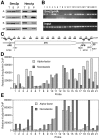
Condensin binding to chromatin at the rDNA locus. (A) A significant portion of Smc2p is chromatin-bound throughout the cell cycle. Chromatin binding of Smc2p and Hmo1p (a reference chromatin protein) throughout the cell cycle determined by chromatin solubilization assay (Liang and Stillman 1997). Strain YPH499bp2 was grown asynchronously or arrested in G1, S, or G2/M phase. Lysates were centrifuged to yield a chromatin-unbound fraction (supernatant 2 according to Liang and Stillman 1997) and a chromatin-bound fraction (pellet 3). Unfractionated lysate (T), supernatant (S), and pellet (P) were probed with antibodies to Smc2p and Hmo1p (Lu et al. 1996). (B) Agarose gel electrophoresis of ChIP analysis of the rDNA repeat in the asynchronously growing YPH499bp2 strain with PCR probes shown in C. (D) Quantification of the Smc2p-HA ChIP in strain YPH499bp2 arrested with α-factor and nocadazole. The PCR signals were quantified using ImageQuant (Molecular Dynamics). All volume scans were normalized to the signal from probe 12 that was only marginally above background (Relative ChIP) and to the corresponding PCR on the input DNA. (E) Quantification of the Ycs5p-HA ChIP in strain YPH499bp5 arrested with α-factor and nocadazole. Analysis was as in Fig. 5 D.

Similar articles

Cited by

References

    1. Bhat M.A., Philp A.V., Glover D.M., Bellen H.J. Chromatid segregation at anaphase requires the barren product, a novel chromosome-associated protein that interacts with Topoisomerase II. Cell. 1996;87:1103–1114. - PubMed
    1. Britton R.A., Lin D.C., Grossman A.D. Characterization of a prokaryotic SMC protein involved in chromosome partitioning. Genes Dev. 1998;12:1254–1259. - PMC - PubMed
    1. Bryk M., Banerjee M., Murphy M., Knudsen K.E., Garfinkel D.J., Curcio M.J. Transcriptional silencing of Ty1 elements in the RDN1 locus of yeast. Genes Dev. 1997;11:255–269. - PubMed
    1. Chuang P., Albertson D., Meyer B. DPY-27a chromosome condensation protein homolog that regulates C. elegans dosage compensation through association with the X chromosome. Cell. 1994;79:459–474. - PubMed
    1. Chuang P.T., Lieb J.D., Meyer B.J. Sex-specific assembly of a dosage compensation complex on the nematode X chromosome. Science. 1996;274:1736–1739. - PubMed

Publication types

MeSH terms