. 2000 May 15;19(10):2193-203.
doi: 10.1093/emboj/19.10.2193.

mu1A-adaptin-deficient mice: lethality, loss of AP-1 binding and rerouting of mannose 6-phosphate receptors

Affiliations

mu1A-adaptin-deficient mice: lethality, loss of AP-1 binding and rerouting of mannose 6-phosphate receptors

C Meyer et al. EMBO J. .

Abstract

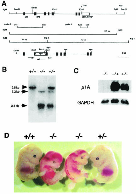
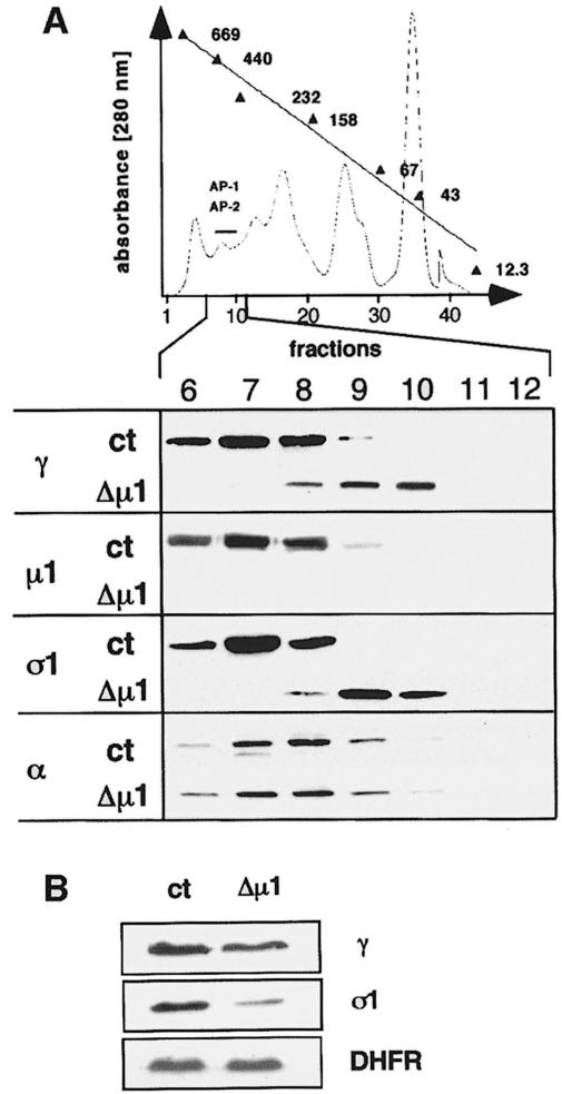
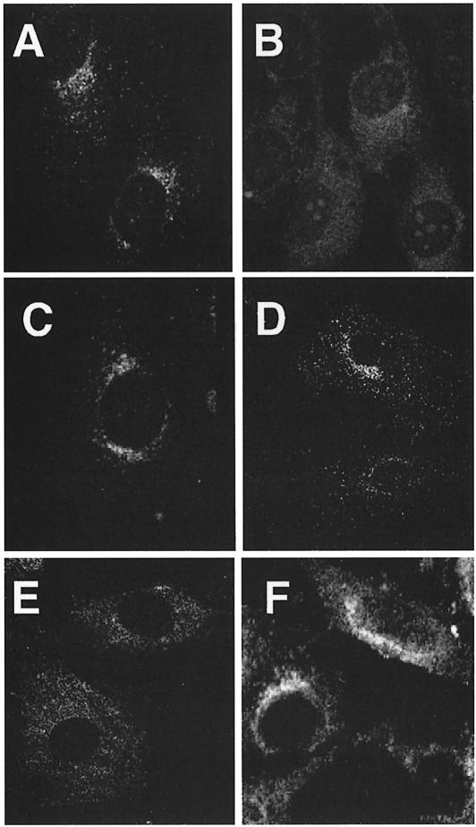
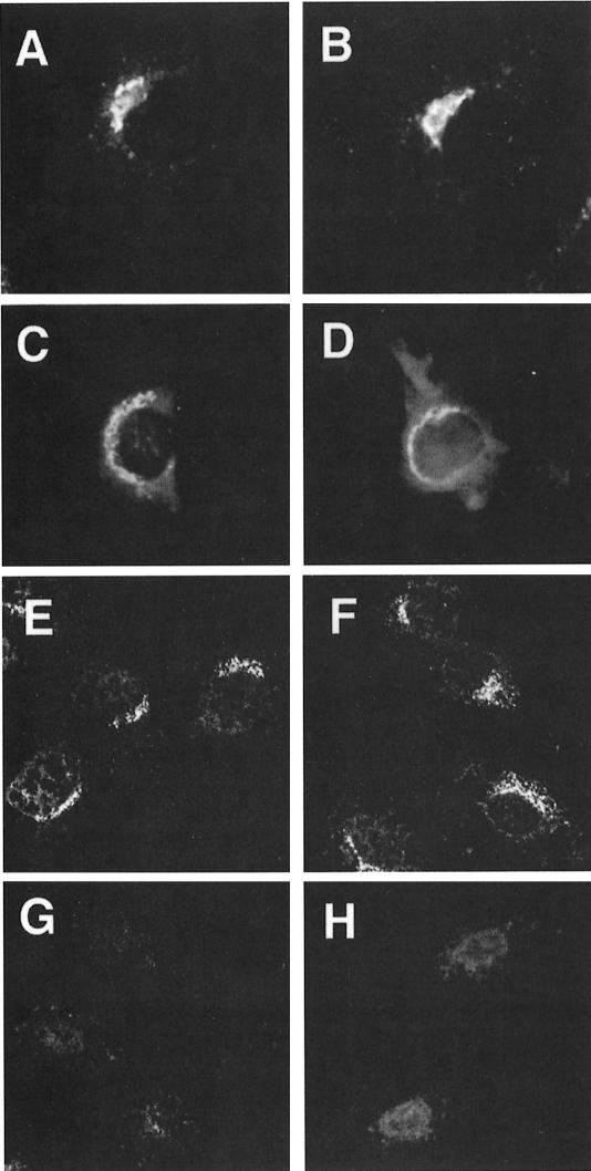
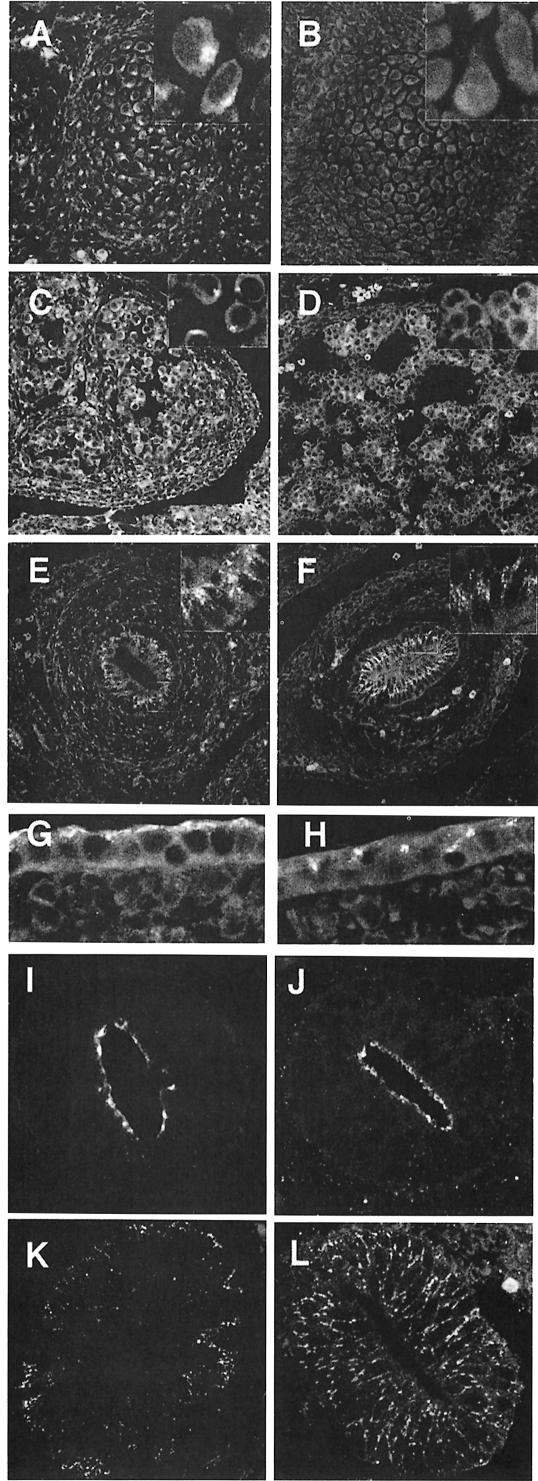
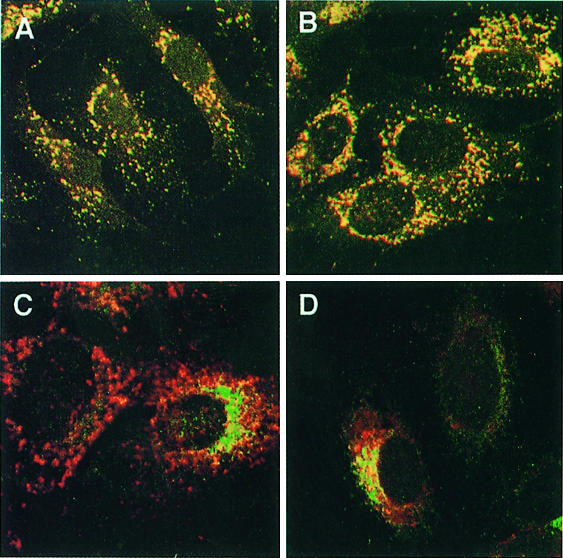
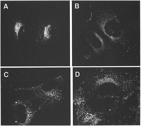
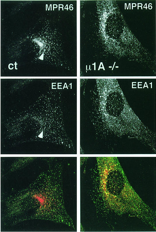
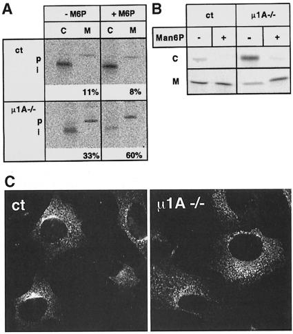
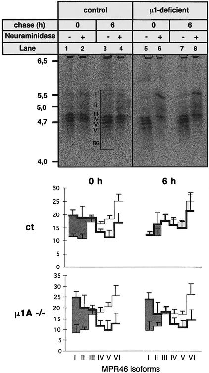
The heterotetrameric AP-1 complex is involved in the formation of clathrin-coated vesicles at the trans-Golgi network (TGN) and interacts with sorting signals in the cytoplasmic tails of cargo molecules. Targeted disruption of the mouse mu1A-adaptin gene causes embryonic lethality at day 13.5. In cells deficient in micro1A-adaptin the remaining AP-1 adaptins do not bind to the TGN. Polarized epithelial cells are the only cells of micro1A-adaptin-deficient embryos that show gamma-adaptin binding to membranes, indicating the formation of an epithelial specific AP-1B complex and demonstrating the absence of additional mu1A homologs. Mannose 6-phosphate receptors are cargo molecules that exit the TGN via AP-1-clathrin-coated vesicles. The steady-state distribution of the mannose 6-phosphate receptors MPR46 and MPR300 in mu1A-deficient cells is shifted to endosomes at the expense of the TGN. MPR46 fails to recycle back from the endosome to the TGN, indicating that AP-1 is required for retrograde endosome to TGN transport of the receptor.


Figures

None
Fig. 1. Targeted disruption of the µ1A-adaptin gene. (A) Genomic 9.5 kb EcoRI µ1A locus (top) and 5.6 kb EcoRI–KpnI targeting construct (bottom). Exons are indicated by black bars. Arrows indicate µ1A and neoR ORF. Lines mark chromosomal DNA fragments generated by BglII digest in control and mutant cells used to demonstrate homologous recombination and the DNA fragments used as internal (probe 1) and external (probe 2) probes for hybridization of BglII-digested chromosomal DNA. (B) Southern blot analysis of BglII-digested chromosomal DNA isolated from amnion epithelia of day 12.5 p.c. embryos hybridized with probe 2. (C) Northern blot of total embryonic fibroblast RNA hybridized first with 32P-labeled mouse µ1A cDNA and then with GAPDH cDNA. (D) Embryos from a litter isolated at day 13.5 of embryonic development.
None
Fig. 2. AP-1 adaptins in µ1A-deficient cells. (A) AP-1 complex assembly. Shown is a gel-filtration profile of a µ1A-deficient cytosol prepared from embryonic fibroblasts. Fractions containing adaptor complexes AP-1 and AP-2 are indicated by a bar. Numbers are kilodaltons of marker proteins used for calibration (see Materials and methods). Western blot analysis of fractions 6–12 of control (ct) and µ1A-deficient cytosols. AP-2 distribution detected with an anti-α-adaptin antibody served as a control for the adaptor fractionation by gel filtration. (B) γ- and σ1-adaptin in cell extracts of control (ct) and µ1A-deficient cells analyzed by Western blotting. Dihydrofolate reductase (DHFR) was used as control. γ-adaptin is reduced to 70% and σ1-adaptin to 30% of control.
None
Fig. 3. Localization of AP-1 and clathrin in embryonic fibroblasts. AP-1 was detected with anti-γ-adaptin antibodies (A, B and C) and clathrin with anti-chc antibodies (D, E and F). (A and D) Control; (B and E) µ1A-deficient cells; (C and F) µ1A-deficient cells with ectopic expression of µ1A.
None
Fig. 4. Binding of AP-1 to the TGN. (AD) Staining of the Golgi with BODIPY FL C5-ceramide in control (A) and µ1A-deficient cells (B) and by the TGN marker TGN38 (epifluorescence) by indirect immunofluorescence analysis in control (C) and µ1A-deficient fibroblasts (D). (EH) Binding of AP-1 to the TGN of control (E and H) and µ1A-deficient cells (F and G) permeabilized with digitonin. Bound AP-1 was detected with an anti-γ-adaptin antibody (confocal images). Permeabilized cells were incubated with GTPγS-supplemented cytosol from control (E, F and G) or µ1A-deficient (H) cells. In (G) brefeldin A was present during the binding.
None
Fig. 5. Labeling of cryosections of the embryos for γ-adaptin staining. Shown are vertebral bodies of control (A) and µ1A-deficient (B) embryos, liver of control (C) and µ1A-deficient (D) embryos, the gut of control (E) and µ1A-deficient (F) embryos, and epidermis of ct (G) and µ1A-deficient (H) embryos. Polarization of gut epithelial cells demonstrated by staining for tight junctions with anti-ZO-1 in ct (I) and µ1A-deficient cells (J) and for the LDL receptor by fluorescent LDL in ct (K) and µ1A-deficient cells (L).
None
Fig. 6. LAMP-1 sorting in µ1A-deficient cells. (A and B) Co-localization at steady state of LAMP-1 and the lysosomal enzyme β-glucuronidase in control (A) and µ1A-deficient cells (B). (C and D) Anti-LAMP-1 antibody endocytosis over 2 h. AP-3-deficient mouse mocha cells and µ1A-deficient cells were co-cultured and γ-adaptin labeling was used to distinguish between AP-3- and µ1A-deficient cells. (C) Steady-state labeling of LAMP-1 (red) and γ-adaptin (green). (D) Steady-state labeling of γ-adaptin and labeling of LAMP-1 after anti-LAMP-1 antibody endocytosis.
None
Fig. 7. Distribution of MPR46 in control (A) and µ1A-deficient (B) cells and of MPR300 in control (C) and µ1A-deficient (D) cells.
None
Fig. 8. Co-localization of MPR46 (red) and EEA1 (green) by indirect immunofluorescence (confocal images). The arrow marks vesicle stained for both proteins in control cells.
None
Fig. 9. Sorting and endocytosis of lysosomal enzymes in µ1A-deficient cells. (A) Sorting of newly synthesized cathepsin D in control (ct) and µ1A-deficient (µ1A–/–) cells in the absence and presence of 5 mM Man6P and restoration of cathepsin D sorting by ectopic expression of µ1A. Cells were metabolically labeled for 1 h and chased for 4 h. Cathepsin D was immunoprecipitated from the cells (C) and the medium (M). The precursor (p) and proteolytically processed intermediate forms (i) of cathepsin D are indicated. Numbers indicate the percentage of cathepsin D secreted into the medium as quantified by phosphoimager analysis. (B) Endocytosis of metabolically labeled [35S]ASA by MPR300 from the medium by control and µ1A-deficient fibroblasts at 37°C during a 4 h incubation in the absence (–) or presence (+) of 5 mM Man6P in the medium. (C) Endocytosis of anti-MPR46 antibodies from the medium by control (ct) and µ1A-deficient (µ1A–/–) cells.
None
Fig. 10. Desialylation and resialylation of MPR46. Cells were metabolically labeled with [35S]methionine for 16 h. After a 1 h chase cells were incubated for 1 h at 37°C in the absence (–) or presence (+) of neuraminidase. Neuraminidase-containing medium was replaced with medium containing neuraminidase inhibitor. Cells were either harvested or further incubated for 6 h. MPR46 was immunoprecipitated and separated by IEF. Upper panel: representative IEF gel. Areas in lane 3: I–VI represent non-sialylated (I) and sialylated isoforms (II–VI); BG is the area subtracted for background signal. Lower panel: quantification of MPR46 isoforms of three independent experiments by phosphoimager analysis. Thick lines, MPR46 isoforms in neuraminidase-treated cells; thin lines, MPR46 isoforms in untreated cells. The net desialylation is represented by the shaded area.

Similar articles

Cited by

References

    1. Alconada A., Bauer,U. and Hoflack,B. (1996) A tyrosine-based motif and a casein kinase II phosphorylation site regulate the intracellular trafficking of the varicella–zoster virus glycoprotein I, a protein localized in the trans-Golgi network. EMBO J., 15, 6096–6110. - PMC - PubMed
    1. Allan B. and Balch,W. (1999) Protein sorting by directed maturation of Golgi compartments. Science, 285, 63–66. - PubMed
    1. Bonifacino J.S. and Dell’Angelica,E.C. (1999) Molecular bases for the recognition of tyrosine-based sorting signals. J. Cell Biol., 145, 923–926. - PMC - PubMed
    1. Braulke T., Gartung,C., Hasilik,A. and von Figura,K. (1987) Is movement of mannose 6-phosphate-specific receptor triggered by binding of lysosomal enzymes? J. Cell Biol., 104, 1735–1742. - PMC - PubMed
    1. Braun M., Waheed,A. and von Figura,K. (1989) Lysosomal acid phosphatase is transported to lysosomes via the cell surface. EMBO J., 8, 3633–3640. - PMC - PubMed

Publication types

MeSH terms

Substances

LinkOut - more resources