. 2000 Jun;20(11):3942-50.
doi: 10.1128/MCB.20.11.3942-3950.2000.

Mutational analysis of mammalian translation initiation factor 5 (eIF5): role of interaction between the beta subunit of eIF2 and eIF5 in eIF5 function in vitro and in vivo

Affiliations

Mutational analysis of mammalian translation initiation factor 5 (eIF5): role of interaction between the beta subunit of eIF2 and eIF5 in eIF5 function in vitro and in vivo

S Das et al. Mol Cell Biol. 2000 Jun.

Abstract

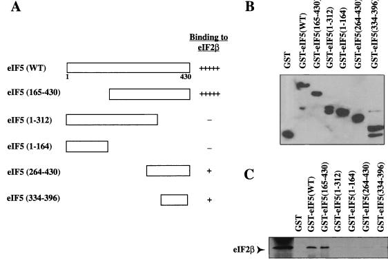
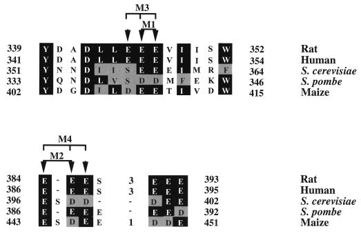
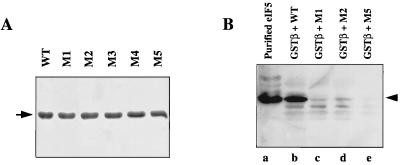
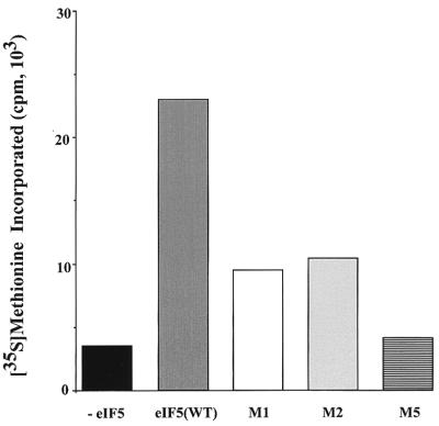
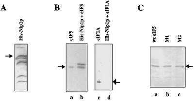
Eukaryotic translation initiation factor 5 (eIF5) interacts with the 40S initiation complex (40S-eIF3-AUG-Met-tRNA(f)-eIF2-GTP) to promote the hydrolysis of ribosome-bound GTP. eIF5 also forms a complex with eIF2 by interacting with the beta subunit of eIF2. In this work, we have used a mutational approach to investigate the importance of eIF5-eIF2beta interaction in eIF5 function. Binding analyses with recombinant rat eIF5 deletion mutants identified the C terminus of eIF5 as the eIF2beta-binding region. Alanine substitution mutagenesis at sites within this region defined several conserved glutamic acid residues in a bipartite motif as critical for eIF5 function. The E346A,E347A and E384A,E385A double-point mutations each caused a severe defect in the binding of eIF5 to eIF2beta but not to eIF3-Nip1p, while a eIF5 hexamutant (E345A,E346A, E347A,E384A,E385A,E386A) showed negligible binding to eIF2beta. These mutants were also severely defective in eIF5-dependent GTP hydrolysis, in 80S initiation complex formation, and in the ability to stimulate translation of mRNAs in an eIF5-dependent yeast cell-free translation system. Furthermore, unlike wild-type rat eIF5, which can functionally substitute for yeast eIF5 in complementing in vivo a genetic disruption of the chromosomal copy of the TIF5 gene, the eIF5 double-point mutants allowed only slow growth of this DeltaTIF5 yeast strain, while the eIF5 hexamutant was unable to support cell growth and viability of this strain. These findings suggest that eIF5-eIF2beta interaction plays an essential role in eIF5 function in eukaryotic cells.


Figures

FIG. 1
FIG. 1
Deletion analysis of eIF5 to determine the eIF2β-binding domain. (A) Schematic representation of deletion mutants of eIF5 expressed as GST fusion proteins in E. coli XL1-Blue cells. The efficiency with which each deletion mutant of eIF5 binds 35S-labeled eIF2β, determined by autoradiography, is shown by + and −. WT, wild type. (B) Expression of GST fusion proteins of eIF5 deletion mutants immobilized on GSH beads was measured by Western blot analysis of 3 μg of protein bound to these beads using anti-GST antibodies. It is not immediately apparent why in case of GST-eIF5(1-312) a polypeptide of the predicted size was not observed. (C) The same eIF5 deletion mutants immobilized on GSH beads (6 μg of bound protein) were incubated at 4°C with 35S-labeled eIF2β synthesized in vitro in bacterial S-30 extracts (Promega) at 4°C for 1 h. The proteins bound to the washed beads were subjected to SDS–15% polyacrylamide gel electrophoresis followed by autoradiography of the dried gel. In the first lane, 35S-labeled eIF2β alone was electrophoresed.
FIG. 2
FIG. 2
Conservation of amino acid residues at the C-terminal bipartite motif of rat eIF5 from different species and locations of alanine substitution mutations within this conserved region. The amino acid sequences of the C-terminal bipartite motif of rat eIF5 (amino acids 339 to 352 and amino acids 384 to 393) were aligned with the corresponding regions in human, S. cerevisiae, Schizosaccharomyces pombe, and maize eIF5 for maximum homology using the program DNASTAR. The sequences of rat, human, and S. cerevisiae eIF5 are from reference ; the sequences of S. pombe and maize eIF5 were obtained from SWISSPROT (accession no. Q09689, and P55876, respectively). The highly conserved amino acid residues between eIF5 of all species are highlighted with dark shading, and the moderately conserved residues are highlighted with light shading. Gaps are represented by broken lines. Residues in rat eIF5 that were targeted for mutagenesis in this study to generate eIF5 mutants M1 to M4 are indicated by arrowheads.
FIG. 3
FIG. 3
Effects of mutations in rat eIF5 on growth of haploid yeast transformants expressing rat eIF5. (A) Haploid yeast strain TMY101 (see Materials and Methods) was transformed separately with different recombinant eIF5 expression plasmids, pTM100-EIF5 expressing either wild-type eIF5 (17) or alanine-mutant eIF5 protein, each containing a single point mutation at E346A, E347A, E384A, or E385A from a GAL1 promoter, and also the vector plasmid pRS315 as indicated. Transformants were initially selected on SGal-Trp-Leu-Ura plates and then replica plated onto both SGal-Trp-Leu-Ura (left) and SGal containing 5-FOA and uracil (SGal-Trp-Leu+Ura + 5-FOA; right). Cells were allowed to grow on these plates for 5 days. (B) Haploid yeast strain TMY101 was transformed separately with mutant eIF5-expressing plasmids pTM100-EIF5(M1), pTM100-EIF5(M2), and pTM100-EIF5(M5) as indicated. Transformants selected on SGal-Trp-Leu-Ura plates (left) were replica plated on SGal-Trp-Leu+Ura + 5-FOA (right). Cells were allowed to grow on the 5-FOA plates for 5 days. (C) Immunoblot analysis of eIF5 in lysates of yeast cells expressing both yeast eIF5 and wild-type (WT) or mutant mammalian eIF5 proteins M1, M2, and M5 from the recombinant plasmids. Yeast cells harboring both the URA3 plasmid pRS316-TIF5 (which expresses yeast eIF5 from its own natural promoter) and the different recombinant LEU expression plasmids expressing either the wild-type or mutant mammalian eIF5 were grown to mid-logarithmic phase in synthetic medium containing 2% galactose as the sole source of carbon. Cell lysates were prepared as described in Materials and Methods and analyzed by Western blotting using rabbit polyclonal anti-rat eIF5 antibodies. Lane a, purified recombinant rat eIF5 as a marker; lanes b to f, extracts from tif5::TRP1 yeast cells harboring yeast eIF5 expression plasmid pRS316-TIF5 and LEU2-based rat eIF5 expression plasmids as follows: lane b, pTM100-EIF5; lane c, pRS315 (vector control); lane d, pTM100-EIF5(M1); lane e, pTM100-EIF5(M2); lane f, pTM100-EIF5(M5).
FIG. 4
FIG. 4
Interaction between eIF5 mutants and the β subunit of eIF2. (A) Recombinant wild-type (WT) and mutant eIF5 proteins M1 to M5 were purified from IPTG-induced XL1-Blue cell lysates as described in Materials and Methods. Purified recombinant eIF5 proteins (3 μg of each) were subjected to SDS-polyacrylamide gel electrophoresis (15% gel) and visualized by Coomassie blue staining. The arrow indicates the position of purified eIF5. (B) Purified recombinant wild-type (lane b) and mutant eIF5 proteins M1 (lane c), M2 (lane d), and M5 (lane e) (9 μg of each) were separately incubated with 12 μg of GST-eIF2β fusion protein immobilized on GSH beads. Following incubation at 4°C with gentle shaking, reaction mixtures were centrifuged, and the beads were washed, suspended in 1× Laemmli buffer, and subjected to Western blot analysis using polyclonal anti-eIF5 antibodies. In lane a, purified rat eIF5 was electrophoresed as a marker and probed with anti-eIF5 antibodies.
FIG. 5
FIG. 5
Analysis of eIF5 mutants for the ability to mediate hydrolysis of GTP bound to the 40S initiation complex and to form the 80S initiation complex. (A) eIF5-mediated GTP hydrolysis. Reaction mixtures (50 μl) contained 20 mM Tris-HCl (pH 7.5), 5 mM MgCl2, 100 mM KCl, and 1 mM dithiothreitol (buffer R), isolated 40S initiation complex (Met-tRNAf–eIF2–[γ-32P]GTP–40S–AUG) containing 2.5 pmol of bound [γ-32P]GTP (38,500 cpm/pmol) isolated as described previously (4), and 20 ng of purified recombinant wild-type (WT) or mutant eIF5 proteins M1 (E346A,E347A), M2 (E384A,E385A), M3 (E345A,E346A,E347A), M4 (E384A,E385A,E386A), and M5 (E345A,E346A,E347A,E384A,E385A,E386A). Following incubation at 25°C, aliquots (8 μl) were removed at each indicated time point and the amount of 32Pi released by the hydrolysis of [γ-32P]GTP was measured by the ammonium phosphomolybdate method as described elsewhere (4). A reaction lacking eIF5 was also included, and the amount of 32Pi released in this control reaction mixture (<0.1 pmol) was subtracted from the results shown. The results shown represent the total amount of 32Pi formed per 50-μl reaction mixture. (B) 80S initiation complex formation. Reaction mixtures containing buffer R, 3 pmol of preformed [35S]Met-tRNAf–eIF2–GTP ternary complex (25,000 cpm/pmol), 0.5 A260 unit of 40S ribosomal subunits, and 0.1 A260 unit of the AUG codon were incubated for 5 min at 37°C to form the 40S initiation complex ([35S]Met-tRNAf–eIF2–GTP–40S–AUG). The chilled reaction mixtures were supplemented with 0.8 A260 unit of 60S ribosomal subunits and purified wild-type or mutant eIF5 at the indicated amount. Following incubation at 37°C for 5 min, the chilled reaction mixtures were sedimented through a 5-ml linear 7.5 to 30% (wt/vol) sucrose gradient in buffer R for 105 min at 48,000 rpm at 4°C in a Beckman 50.1 rotor. Fractions (0.25 ml) collected from the bottom of each tube were counted for 35S radioactivity to quantitate formation of the 80S initiation complex. A control reaction mixture lacking eIF5 formed <0.1 pmol of 80S initiation complex. This value was subtracted from the results shown.
FIG. 6
FIG. 6
Effect of eIF5-Ala mutations on in vitro translation of total yeast RNA. eIF5-depleted cell translation extracts were prepared from TMY201R cells (17), incubated, and analyzed for [35S]methionine incorporation into proteins as described in Materials and Methods. Each reaction mixture (50 μl) contained 15 μCi of [35S]methionine (11 Ci/mmol), 25 μg of total yeast RNA, and where indicated either 100 ng of purified recombinant rat wild-type eIF5 [eIF5(WT)] or 100 ng each of purified mutant eIF5 M1, M2, or M5 protein. Following incubation at 25°C for 40 min, aliquots (10 μl) were withdrawn and analyzed for [35S]methionine incorporation into proteins. A control reaction mixture lacking total yeast RNA and exogenously added eIF5 was also incubated and analyzed. The amount of [35S]methionine incorporated into proteins in this control reaction mixture was subtracted, and the results are shown.
FIG. 7
FIG. 7
Expression of His-Nip1p in bacteria and interaction between His-Nip1p and eIF5. (A) Ni-NTA agarose beads containing bound His-Nip1p were analyzed by Western blot using rabbit anti-yeast Nip1p polyclonal antibodies. The position of intact His-Nip1p is indicated by an arrow. (B) Ni-NTA agarose beads containing 5 μg of His-Nip1p were incubated with 5 μg of either purified recombinant rat eIF5 (lane b) or purified rat eIF1A as a negative control (lane d) for 1 h at 4°C. Proteins bound to the washed beads were analyzed by Western blot using rabbit anti-rat eIF5 (left) and anti-rat eIF1A (right) antibodies. Purified rat eIF5 (lane a) and purified rat eIF1A (lane c) were also run as markers on the same Western blots. (C) Ni-NTA agarose beads containing 5 μg of His-Nip1p were incubated with 5 μg of either purified wild-type (wt) rat eIF5 (lane a) or the mutant proteins M1 (lane b) and M2 (lane c) for 1 h at 4°C. After incubation, the beads were washed and proteins bound to the washed beads were analyzed by Western blot using rabbit anti-rat eIF5 antibodies. The arrow indicates the position of purified rat eIF5. It should be noted that two immunoreactive polypeptides were observed in panels B (lane b) and C. The slower-migrating polypeptide arises from His-Nip1p preparation (data not shown). The intensity of this band varies from preparation to preparation.

Similar articles

Cited by

References

    1. Asano K, Krishnamoorthy T, Phan L, Pavitt G D, Hinnebusch A G. Conserved bipartite motifs in yeast eIF5 and eIF2Bɛ, GTPase-activating and GDP-GTP exchange factors in translation initiation, mediate binding to their common substrate eIF2. EMBO J. 1999;18:1673–1688. - PMC - PubMed
    1. Bandyopadhyay A, Maitra U. Cloning and characterization of the p42 subunit of mammalian translation initiation factor 3 (eIF3): demonstration that eIF3 interacts with eIF5 in mammalian cells. Nucleic Acids Res. 1999;27:1331–1337. - PMC - PubMed
    1. Bourne H R, Sanders D A, McCormick F. The GTPase superfamily: conserved structure and molecular mechanism. Nature. 1991;349:117–127. - PubMed
    1. Chakrabarti A, Maitra U. Function of eukaryotic initiation factor 5 in the formation of an 80S ribosomal polypeptide chain initiation complex. J Biol Chem. 1991;266:14039–14045. - PubMed
    1. Chakravarti D, Maiti T, Maitra U. Isolation and immunochemical characterization of eukaryotic translation initiation factor 5 from Saccharomyces cerevisiae. J Biol Chem. 1993;268:5754–5762. - PubMed

Publication types

MeSH terms