. 2000 May;74(10):4549-61.
doi: 10.1128/jvi.74.10.4549-4561.2000.

Directional transneuronal infection by pseudorabies virus is dependent on an acidic internalization motif in the Us9 cytoplasmic tail

Affiliations

Directional transneuronal infection by pseudorabies virus is dependent on an acidic internalization motif in the Us9 cytoplasmic tail

A D Brideau et al. J Virol. 2000 May.

Abstract

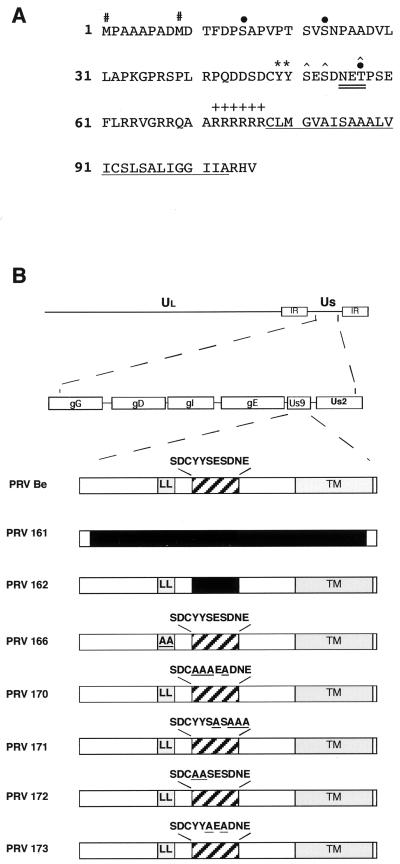
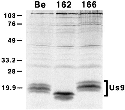
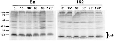
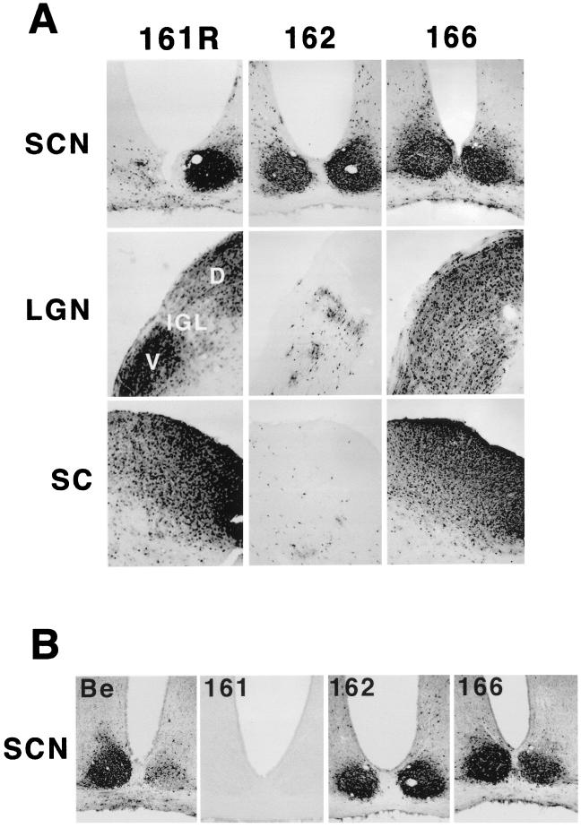
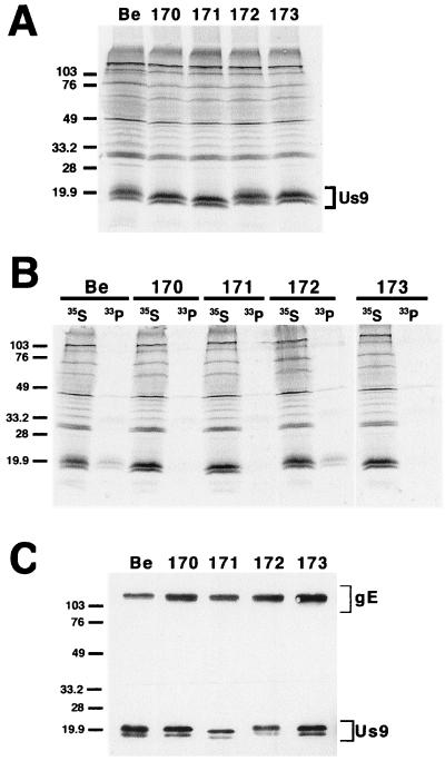
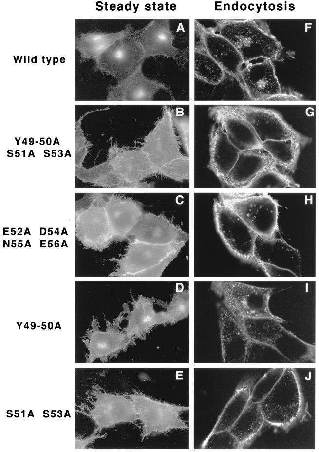
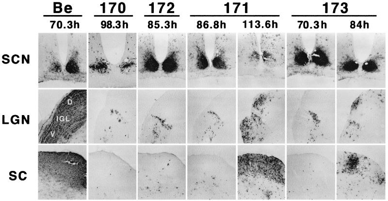
The Us9 gene is conserved among most alphaherpesviruses. In pseudorabies virus (PRV), the Us9 protein is a 98-amino-acid, type II membrane protein found in the virion envelope. It localizes to the trans-Golgi network (TGN) region in infected and transfected cells and is maintained in this compartment by endocytosis from the plasma membrane. Viruses with Us9 deleted have no observable defects in tissue culture yet have reduced virulence and restricted spread to retinorecipient neurons in the rodent brain. In this report, we demonstrate that Us9-promoted transneuronal spread in vivo is dependent on a conserved acidic motif previously shown to be essential for the maintenance of Us9 in the TGN region and recycling from the plasma membrane. Mutant viruses with the acidic motif deleted have an anterograde spread defect indistinguishable from that of Us9 null viruses. Transneuronal spread, however, is not dependent on a dileucine endocytosis motif in the Us9 cytoplasmic tail. Through alanine scanning mutagenesis of the acidic motif, we have identified two conserved tyrosine residues that are essential for Us9-mediated spread as well as two serine residues, comprising putative consensus casein kinase II sites, that modulate the rate of PRV transneuronal spread in vivo.


Figures

FIG. 1
FIG. 1
(A) Amino acid sequence of the Us9 open reading frame product. The two in-frame methionine residues (#) and the potential tyrosine kinase phosphorylation sites (∗) are indicated. The consensus casein kinase I sites [(S/T)X2–3(S/T)X] (31) each are each indicated by a solid circle above the S or T boldfaced here, and the consensus casein kinase II sites [X(S/T)XX(D/E)] are each marked with a caret above the S or T (PROSITE pattern). The potential N-linked glycosylation [NX(S/T)] sequence is double underlined. The putative transmembrane domain is underlined, and the surrounding basic residues are indicated by plus signs. (B) PRV genome and maps of viruses used in this study. The PRV genome is shown on the first line, and the unique short region (Us) is expanded on the second line. The Us9 proteins from the various viruses used in this study are diagrammed below. The dileucine endocytosis motif (LL) and the transmembrane domain (TM) are indicated by shaded boxes. The acidic domain is indicated by a hatched box with the amino acid sequence given above. All amino acid substitutions are underlined, and deletions are indicated by solid boxes. PRV Be is the wild-type strain used in this study. PRV 161 contains a 258-bp deletion in the Us9 open reading frame. PRV 162 contains a 10-amino-acid deletion removing a conserved acid motif in the Us9 cytoplasmic tail. PRV 166 contains a leucine-to-alanine substitution at amino acids 30 and 31 in the Us9 protein. PRV 170 (Y49–50A S51A S53A), PRV 171 (E52A D54A N55A E56A), PRV 172 (Y49–50A), and PRV 173 (S51A S53A) contain alanine substitutions in the conserved acidic domain in the Us9 cytoplasmic tail (see the text for details).
FIG. 2
FIG. 2
Expression of Us9 proteins. PK15 cells were infected with either PRV Be (wild type), PRV 162 (del 46–55), or PRV 166 (L30–31A) at an MOI of 10. The infected monolayers were radiolabeled for 11 h beginning at 5 h postinfection. At 16 h postinfection, cellular lysates were prepared and immunoprecipitated with Us9 polyvalent antiserum. All immunoprecipitated products were analyzed by electrophoresis on an SDS–12.5% polyacrylamide gel followed by autoradiography. Positions of molecular mass markers are indicated on the left in kilodaltons.
FIG. 3
FIG. 3
Pulse-chase analysis of Us9. PK15 cells were infected with either PRV Be (wild type) or PRV 162 (del 46–55) at an MOI of 10. The cells were pulse-labeled with 125 μCi of [35S]methionine-cysteine for 7 min, rinsed with PBS, and chased for the times indicated. Cellular lysates were immunoprecipitated with a Us9 polyvalent antiserum and fractionated on a 12.5% polyacrylamide gel. Molecular mass markers (kilodaltons) are indicated on the left.
FIG. 4
FIG. 4
Anterograde spread of Us9 trafficking mutants in the rodent visual system. Approximately 1 × 10⁶ PFU of PRV 162 (del 46–55) and 6.8 × 10⁵ PFU of PRV 166 (L30–31A) was injected into the vitreous humor of Sprague-Dawley male rats. (A) Thirty-five-micrometer-thick coronal sections of the infected brains were examined for total PRV antigen with polyvalent antiserum Rb133, which recognizes all of the major envelope glycoproteins. (B) SCNs of PRV-infected animals stained for the Us9 protein with a polyvalent Us9-specific antiserum. Representative sections are shown for each virus. The data for the wild-type strain PRV Be, the Us9 null virus PRV 161, and the revertant virus PRV 161R have been reported previously (7) and are included here only for comparison. Due to sectioning of the PRV 161R-infected brain at an oblique angle, the image in this figure shows viral antigen in only one SCN. D, dorsal, V, ventral.
FIG. 5
FIG. 5
Analysis of the alanine scanning Us9 proteins. (A) Steady-state expression of alanine scanning Us9 proteins. PK15 cells infected with PRV Be (wild type), PRV 170 (Y49–50A S51A S53A), PRV 171 (E52A D54A N55A E56A), PRV 172 (Y49–50A), or PRV 173 (S51A S53A) were radiolabeled overnight with [35S]methionine-cysteine. Cellular extracts were prepared at 16 h postinfection and immunoprecipitated with Us9 polyvalent antiserum. (B) Analysis of Us9 phosphoforms. PRV Be (wild type)-, PRV 170 (Y49–50A S51A S53A)-, PRV 171 (E52A D54A N55A E56A)-, PRV 172 (Y49–50A)-, and PRV 173 (S51A S53A)-infected cells were radiolabeled overnight in the presence of either [35S]methionine-cysteine or [33P]orthophosphate. Cellular extracts were prepared at 16 h postinfection and subjected to immunoprecipitation with Us9 polyvalent antiserum. All of the immunoprecipitated products were analyzed by electrophoresis on an SDS–12.5% polyacrylamide gel followed by autoradiography (exposure time, 1.5 days). (C) Incorporation of Us9 into viral particles. Monolayers of PK15 cells were infected at an MOI of 10 with either PRV Be (wild type), PRV 170 (Y49–50A S51A S53A), PRV 171 (E52A D54A N55A E56A), PRV 172 (Y49–50A), or PRV 173 (S51A S53A) for 15 h. Cellular extracts were prepared, and virions were isolated from the medium by centrifugation through a 30% sucrose cushion. The purified virion extracts were fractionated on an SDS–12.5% polyacrylamide gel and analyzed by Western blotting with gE polyvalent and Us9 monoclonal (5F10) antisera. Molecular mass markers (kilodaltons) are indicated on the left in all three panels.
FIG. 6
FIG. 6
(A through E) Transient transfection of Us9 alanine scanning mutant constructs. PK15 cells grown on glass coverslips were transfected by the calcium phosphate method with plasmids encoding Us9–EGFP (A), Us9(Y49–50A S51A S53A)–EGFP (B), Us9(E52A D54A N55A E56A)–EGFP (C), Us9(Y49–50A)–EGFP (D), and Us9(S51A S53A)–EGFP (E). At 48 h posttransfection, the localization of the various Us9–EGFP fusion proteins was detected by fluorescence microscopy. (F through J) Internalization of alanine scanning Us9 mutant proteins from the cell surface. PK15 cells stably expressing wild-type Us9–EGFP (F), Us9(Y49–50A S51A S53A)–EGFP (G), Us9(E52A D54A N55A E56A)–EGFP (H), Us9(Y49–50A)–EGFP (I), and Us9(S51A S53A)–EGFP (J) fusion proteins were plated on glass coverslips. The coverslips were incubated at 37°C in medium containing a 1:75 dilution of polyclonal GFP-specific antiserum. After 1 h, the cells were fixed and permeabilized, and the localization of the GFP-specific antibodies was detected by indirect immunofluorescence followed by confocal microscopy. Only the red fluorescence of the Alexa 568-conjugated goat anti-rabbit IgG secondary antibody is shown.
FIG. 7
FIG. 7
Localization of Us9 alanine scanning mutant viruses in the brain. Male Sprague-Dawley rats were subjected to intravitreous injection with approximately 5 × 10⁵ PFU of PRV 170 (Y49–50A S51A S53A), PRV 171 (E52A D54A N55A E56A), PRV 172 (Y49–50A), or PRV 173 (S51A S53A). The localization of viral antigen in 35-μm-thick coronal sections was detected with an antigen against whole-virus particles (Rb133). Each vertical set of three panels shows sections from one animal, and the time to which the animal survived after infection is indicated. Representative samples are shown for each virus. Images of brain tissues from animals infected with PRV Be (1.2 × 10⁶ PFU) are included for comparison (7).

Similar articles

Cited by

References

    1. Alconada A, Bauer U, Baudoux L, Piette J, Hoflack B. Intracellular transport of the glycoproteins gE and gI of the varicella-zoster virus. J Biol Chem. 1998;273:13430–13436. - PubMed
    1. Alconada A, Bauer U, Hoflack B. A tyrosine-based motif and a casein kinase II phosphorylation site regulate the intracellular trafficking of the varicella-zoster virus glycoprotein I, a protein localized in the trans-Golgi network. EMBO J. 1996;15:6096–6110. - PMC - PubMed
    1. Alconada A, Bauer U, Sodeik B, Hoflack B. Intracellular traffic of herpes simplex virus glycoprotein gE: characterization of the sorting signals required for its trans-Golgi network localization. J Virol. 1999;73:377–387. - PMC - PubMed
    1. Becker C H. Zur primaren Schadingung vegetativer Ganglien nach Infektion mit dem Herpes suis Virus bei verschiedenen Tierarten. Experientia. 1967;23:209. - PubMed
    1. Ben-Porat T, Kaplan A S. Molecular biology of pseudorabies virus. In: Roizman B, editor. The herpesviruses. New York, N.Y: Plenum Publishing Corp.; 1985. pp. 105–173.

Publication types

MeSH terms

LinkOut - more resources