. 2000 Apr;20(7):2488-97.
doi: 10.1128/MCB.20.7.2488-2497.2000.

Presence of a member of the mitochondrial carrier family in hydrogenosomes: conservation of membrane-targeting pathways between hydrogenosomes and mitochondria

Affiliations

Presence of a member of the mitochondrial carrier family in hydrogenosomes: conservation of membrane-targeting pathways between hydrogenosomes and mitochondria

S D Dyall et al. Mol Cell Biol. 2000 Apr.

Abstract

A number of microaerophilic eukaryotes lack mitochondria but possess another organelle involved in energy metabolism, the hydrogenosome. Limited phylogenetic analyses of nuclear genes support a common origin for these two organelles. We have identified a protein of the mitochondrial carrier family in the hydrogenosome of Trichomonas vaginalis and have shown that this protein, Hmp31, is phylogenetically related to the mitochondrial ADP-ATP carrier (AAC). We demonstrate that the hydrogenosomal AAC can be targeted to the inner membrane of mitochondria isolated from Saccharomyces cerevisiae through the Tim9-Tim10 import pathway used for the assembly of mitochondrial carrier proteins. Conversely, yeast mitochondrial AAC can be targeted into the membranes of hydrogenosomes. The hydrogenosomal AAC contains a cleavable, N-terminal presequence; however, this sequence is not necessary for targeting the protein to the organelle. These data indicate that the membrane-targeting signal(s) for hydrogenosomal AAC is internal, similar to that found for mitochondrial carrier proteins. Our findings indicate that the membrane carriers and membrane protein-targeting machinery of hydrogenosomes and mitochondria have a common evolutionary origin. Together, they provide strong evidence that a single endosymbiont evolved into a progenitor organelle in early eukaryotic cells that ultimately give rise to these two distinct organelles and support the hydrogen hypothesis for the origin of the eukaryotic cell.


Figures

FIG. 1
FIG. 1
Abundance and structure of the 31-kDa hydrogenosomal membrane protein Hmp31. (A) SDS-PAGE of hydrogenosomal proteins. Hydrogenosomes (HY) were subjected to sodium carbonate extraction to separate the soluble fraction (HS), comprising matrix and intermembrane space proteins from the insoluble fraction (HP), consisting of integral membrane proteins. Samples were loaded in the ratio HY:HS:HP = 1:1:10, size separated by SDS–12% PAGE, and visualized by Coomassie brilliant blue staining. (B) The Hmp31 protein sequence was divided into three repeat units: Hmp31.1, residues 1 to 107; Hmp31.2, residues 108 to 202; and Hmp31.3, residues 203 to 316, prior to alignment. Identities are indicated by an asterisk (∗) and conserved substitutions by a dot (•). Sequences in red correspond to the MCF degenerate signature sequence P (Hy) (D, E) X X (K, R). Underlined sequences match proteolytic peptide sequences obtained from the endogenous 31-kDa membrane protein. The double-underlined sequence denotes the N-terminal sequence of endogenous Hmp31. The boxed sequence corresponds to the Hmp31 N-terminal presequence. (C) Comparison of the Hmp31 N-terminal presequence with hydrogenosomal matrix protein presequences. The cleavage site, determined by N-terminal sequencing in each reported case, is indicated by a vertical arrow. The first three amino acids from the mature proteins are given. Accession numbers for each protein are indicated in the rightmost column.
FIG. 2
FIG. 2
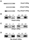
Expression and translocation of epitope-tagged Hmp31 in T. vaginalis. (A) Epitope-tagged full-length Hmp31 and mutant Hmp31 proteins. The C-terminal (HA)2 epitope is denoted by the two adjacent shaded boxes. The Hmp31 presequence is indicated by the hatched box and the Fd presequence by the black box. (B to D) Western blot analysis of fractions from the in vivo transformants shown in panel A with anti-HA antibody. WC, whole cell; S, soluble cellular fraction; P, crude pelleted organelles. Equivalent amounts of each fraction were loaded on the gels so that WC = S + P. HY, purified hydrogenosomes; HS, soluble organellar fraction; P, insoluble membrane pellet from sodium carbonate-extracted hydrogenosomes. Equivalent amounts of soluble and insoluble fractions were loaded so that HY = HS + HP.
FIG. 3
FIG. 3
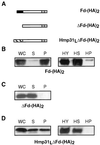
Expression and translocation of epitope-tagged Fd in T. vaginalis. (A) Epitope-tagged full-length Fd and mutant Fd proteins. The C-terminal (HA)2 epitope is denoted by the two adjacent shaded boxes. The Hmp31 presequence is indicated by the hatched box and the Fd presequence is indicated by the black box. (B to D) Western analysis of fractions from the above in vivo transformants with anti-HA antibody. WC, whole cell; S, soluble cellular fraction; P, crude pelleted organelles. Equivalent amounts of each fraction were loaded on the gels so that WC = S + P. HY, purified hydrogenosomes; HS, soluble organellar fraction; HP, insoluble membrane pellet from sodium carbonate-extracted hydrogenosomes. Equivalent amounts of soluble and insoluble fractions were loaded so that HY = HS + HP.
FIG. 4
FIG. 4
Endogenous Hmp31 is protected from trypsin digestion in intact hydrogenosomes. Intact hydrogenosomes in SM (250 mM sucrose, 10 mM MOPS [pH 7.2]) at a concentration of 0.5 μg of total protein per ml were subjected to increasing concentrations of trypsin, from 0.02 to 1.0 mg/ml, for 30 min on ice. Two aliquots were solubilized with 2% Triton X-100 prior to trypsin treatment at concentrations of 0.02 and 1.0 mg/ml, respectively. Soybean trypsin inhibitor was added to a final concentration of 2.0 mg/ml to all samples. Following a 5-min incubation on ice, intact hydrogenosomes were washed with SM supplemented with soybean trypsin inhibitor and reisolated by centrifugation. The solubilized samples were precipitated with trichloroacetic acid at a final concentration of 10% and recovered by centrifugation. The pellets and precipitates were resuspended in Laemmli buffer for SDS–12% PAGE separation, followed by Western blotting and analysis using a polyclonal antibody against Hmp31.
FIG. 5
FIG. 5
Phylogenetic analysis of Hmp31. A maximum-parsimony analysis using PAUP (48) was performed to display the relationship between the hydrogenosomal membrane protein Hmp31 and representative members of MCFs. The topology of the tree was identical to that obtained from the Fitch-Margoliash distance method (9). Bootstrap values for the maximum-parsimony analysis are given in percentages at each branch node. The bootstrap values from the distance (FM) tree are displayed where they differ from the maximum-parsimony (MP) tree. The organelles to which the proteins localize are indicated in brackets next to the protein name: H, hydrogenosome; M, mitochondrion; and P, peroxisome. Accession numbers of proteins from top to bottom are AF216971, AF004161, P18239, X15712, AF049130, P12236, S51132, P16260, 016261, AJ223983, Y11220, P55851, S44091, Q02978, Y08499, AF062383, P40614, B53737, P10566, P23500, P21245, AF002109, X87417, Q01356, P38152, and P53007.
FIG. 6
FIG. 6
Alignment of Hmp31 with S. cerevisiae AAC2. Identities are indicated by an asterisk (∗) and conserved substitutions by a dot (•). Sequences in boldface correspond to the MCF degenerate signature sequence P (Hy) (D,E) X X (K, R). Essential charge-pair network residues in AAC2 (33, 34) and their counterparts in the hydrogenosomal protein are indicated in color: positive charges are shown in red and negative charges are shown in green. Charges are conserved at all positions except for residue E179 in the hydrogenosomal protein aligning with R204, resulting in an opposite charge at this position. The horizontal arrowed lines show putative transmembrane domains TM1 to TM6.
FIG. 7
FIG. 7
Expression and translocation of epitope-tagged yeast AAC1 in T. vaginalis. S. cerevisiae AAC1 is imported into T. vaginalis hydrogenosomes in vivo. Western analysis of fractions from the ScAAC1-(HA)2 in vivo transformant with anti-HA antibody. WC, whole cell; S, soluble cellular fraction; P, crude pelleted organelles. Equivalent amounts of each fraction were loaded on the gels so that WC = S + P. HY, purified hydrogenosomes; HS, soluble organellar fraction; HP, insoluble membrane pellet from sodium carbonate extracted hydrogenosomes. Equivalent amounts of soluble and insoluble fractions were loaded so that HY = HS + HP.
FIG. 8
FIG. 8
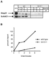
The T. vaginalis Hmp31 protein is imported into isolated yeast mitochondria in vitro. (A) Radiolabelled T. vaginalis Hmp31 and S. cerevisiae AAC1 were incubated for 1, 2, and 4 min at 25°C with yeast wild-type (WT) and tim10-1 mutant mitochondria (21) in the presence (+) or absence (−) of a membrane potential (ΔΨ). Following trypsin treatment, the organelles were reisolated and subjected to sodium carbonate extraction. The fractions were then separated by SDS-PAGE, followed by fluorography. STD, 10% of the radiolabelled precursor present in each assay. (B) Quantitation of Hmp31 import data shown in panel A. The densitometer reading at the 4-min time point was set at 100%.

Similar articles

Cited by

References

    1. Adam A, Endres M, Sirrenberg C, Lottspeich F, Neupert W, Brunner M. Tim9, a new component of the TIM22.54 translocase in mitochondria. EMBO J. 1999;18:313–319. - PMC - PubMed
    1. Andersson S G E, Kurland C G. Origins of mitochondria and hydrogenosomes. Curr Opin Microbiol. 1999;2:535–541. - PubMed
    1. Andersson S G E, Zomorodipour A, Andersson J P, Sicheritz-Ponten T, Alsmark U C M, Podowski R M, Naslund A K, Eriksson A-S, Winkler H H, Kurland C G. The genome sequence of Rickettsia prowazekii and the origin of mitochondria. Nature. 1998;396:133–140. - PubMed
    1. Bradley P J, Lahti C J, Plümper E, Johnson P J. Targeting and translocation of proteins into the hydrogenosome of the protist Trichomonas: similarities with mitochondrial protein import. EMBO J. 1997;16:3484–3493. - PMC - PubMed
    1. Bui E T N, Bradley P J, Johnson P J. A common evolutionary origin for mitochondria and hydrogenosomes. Proc Natl Acad Sci USA. 1996;93:9651–9656. - PMC - PubMed

Publication types

MeSH terms

Associated data

LinkOut - more resources