. 2000 Apr;20(7):2446-54.
doi: 10.1128/MCB.20.7.2446-2454.2000.

DNA interstrand cross-links induce futile repair synthesis in mammalian cell extracts

Affiliations

DNA interstrand cross-links induce futile repair synthesis in mammalian cell extracts

D Mu et al. Mol Cell Biol. 2000 Apr.

Abstract

DNA interstrand cross-links are induced by many carcinogens and anticancer drugs. It was previously shown that mammalian DNA excision repair nuclease makes dual incisions 5' to the cross-linked base of a psoralen cross-link, generating a gap of 22 to 28 nucleotides adjacent to the cross-link. We wished to find the fates of the gap and the cross-link in this complex structure under conditions conducive to repair synthesis, using cell extracts from wild-type and cross-linker-sensitive mutant cell lines. We found that the extracts from both types of strains filled in the gap but were severely defective in ligating the resulting nick and incapable of removing the cross-link. The net result was a futile damage-induced DNA synthesis which converted a gap into a nick without removing the damage. In addition, in this study, we showed that the structure-specific endonuclease, the XPF-ERCC1 heterodimer, acted as a 3'-to-5' exonuclease on cross-linked DNA in the presence of RPA. Collectively, these observations shed some light on the cellular processing of DNA cross-links and reveal that cross-links induce a futile DNA synthesis cycle that may constitute a signal for specific cellular responses to cross-linked DNA.


Figures

FIG. 1
FIG. 1
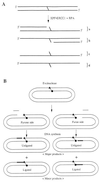
Schematic drawing of the covalently closed circular DNA substrate containing a psoralen interstrand cross-link. Restriction sites around the interstrand cross-link are indicated, and the sizes of the corresponding restriction fragments are shown.
FIG. 2
FIG. 2
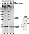
Interstrand psoralen cross-link induces futile DNA synthesis. The covalently closed circular DNA containing a site-specific psoralen interstrand cross-link (XL) was incubated with wild-type rodent extract (AA8) in the presence of [α-32P]dCTP under repair synthesis conditions. After incubation at 30°C for 60 min, the DNA was digested with HindIII (lane 2), PvuI (lane 3), or both (lane 4) and examined on a 7% denaturing polyacrylamide gel. W indicates the signal representing the ligated fraction of DNA that underwent futile repair synthesis. The unligated 130-nt fragment arose from the repair synthesis to fill the gap adjacent to the furan-side adducted thymine, and the 40-nt fragment was generated by filling in the gap adjacent to the pyrone-side adducted thymine. Lane 4 contains psoralen furan-side monoadducted DNA (MA) digested with HindIII and PvuI following the repair synthesis reaction. The 168-nt fragment carrying the 26-nt repair patch (1) is indicated by an arrow. The high-molecular-weight DNA near the origin represents nonspecific incorporation of radiolabel randomly throughout the plasmid. When normalized for fragment length, the background synthesis is about 20% of the damage-induced synthesis.
FIG. 3
FIG. 3
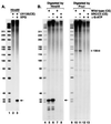
Cross-link-induced DNA synthesis and ligation without cross-link repair. After the “repair synthesis” reaction, the DNA was digested with HindIII and SacI and analyzed on a 7% denaturing polyacrylamide gel (lanes 2 to 4). The samples in lanes 6 to 8 were subjected to photoreversal (PR) treatment to convert interstrand cross-links to monoadducts prior to gel electrophoresis. To confirm that the fragment marked “ligated product” observed in lanes 2 to 4 contains a psoralen cross-link, the DNA was excised from the gel, purified, and subjected to photoreversal (lanes 10 and 11). Note that because of the pyrone-side preference of excision and synthesis, the 49-nt strand containing the pyrone-side adducted thymine is radiolabeled whereas the 41-nt complementary fragment with the furan-side adducted thymine (Fig. 1) is undetectable. DNA size markers (φX174 digested with HinfI) are shown in lanes 1, 5, and 9 (M).
FIG. 4
FIG. 4
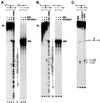
(A) Futile DNA synthesis is dependent on the nucleotide excision repair nuclease. XPG mutant cell extract (UV135) was incubated with the cross-linked substrate in the presence (lane 3) or absence (lane 2) of purified XPG protein (40 ng) under repair synthesis conditions. After incubation at 30°C for 90 min, the DNA was digested by HindIII and analyzed on a 7% denaturing polyacrylamide gel. Lane 1 contains DNA size markers. The 40-nt fragment arising from futile DNA synthesis is indicated by an arrow. (B) Futile DNA synthesis is independent of the XRCC3 function. The cross-link-containing plasmid was incubated with either wild-type (AA8) or XRCC3 mutant (irs1SF) cell extracts (CE) with or without the nonhydrolyzable ATP analog γ-S-ATP (2 mM), as indicated. Following incubation at 30°C for 60 min, the DNA was digested by either HindIII or PvuI and analyzed on 7% denaturing polyacrylamide gels. Lanes 1 and 6 contain size markers. The bands arising from futile DNA synthesis are indicated by arrows.
FIG. 5
FIG. 5
Linear cross-linked substrates for the XPF-ERCC1 nuclease. (A) Structure of the psoralen used in the present study. Both the furan side and pyrone side of the psoralen molecule can be adducted to thymine through a cyclobutane ring. (B) Structure of an interstrand psoralen cross-link. Only the two adducted thymines are shown. dR, deoxyribose. (C) Malondialdehyde. (D) TBG interstrand cross-link. This is a synthetic analog of a malondialdehyde-induced interstrand cross-link. The trimethylene group is linked to N2 of guanines on both strands. (E) Linear psoralen cross-link substrate. The oligomers used to assemble the duplex and the side of the cross-link are indicated. Both strands have a 1-base protruding 5′ end and are 149 nt long. (F) Linear TBG cross-link substrate. The oligomers used to assemble the duplex and the position of the TBG cross-link are shown. Both strands have a 1-base protruding 5′ terminus and are 147 nt long. (G) Nucleotide sequences of some of the oligomers used to make linear cross-link substrates. The thymine base of oligomer 3 linked to the furan side of a psoralen molecule is indicated by an asterisk. Oligomer 8 contains a TBG moiety cross-linking a 19-mer and an 11-mer, as shown. The sequences of oligomers 1, 4, 5, and 7 have been published (23).
FIG. 6
FIG. 6
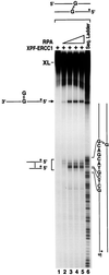
Specific degradation of a linear duplex containing a psoralen cross-link by XPF-ERCC1 and RPA. (A) Reactions performed with a substrate containing 5′ label on the furan-side adducted strand. The triangle and circle represent pyrone-side and furan-side adducted thymines of the cross-link (XL), respectively. Asterisks indicate 5′-terminally labeled strands. The reactions in lanes 2 and 3 were carried out with XPF-ERCC1 (30 ng) alone or in combination with RPA (60 ng) as indicated. Lane 4 shows the Maxam-Gilbert purine sequencing ladder of the same DNA. The nucleotide sequence around the cross-linked thymine is indicated to the right of lane 4. The psoralen-adducted thymine is circled. In the sequence ladder, a significant portion of the cross-linked substrate was converted to monoadduct by the hot alkali used in the sequencing reaction (4). Control experiments using a monoadducted psoralen substrate (MA) are shown in lanes 7 and 8. (B) Cleavage reactions of the same substrate radiolabeled at the 5′ terminus of the pyrone-side adducted strand. The experiments were carried out as described in panel A. Lanes 14 to 16 contain control reactions with monoadducted DNA. (C) The exonucleolytic activity on cross-linked DNA is intrinsic to XPF-ERCC1. The substrate, radiolabeled at the 5′ end of the pyrone-adducted strand, is shown schematically at the top. Where indicated, XPF-ERCC1 (50 ng) was mixed with either anti-ERCC1 (α-ERCC1) or anti-XPB (α-XPB) antibodies linked to protein A-agarose beads, and following removal of the beads by centrifugation, the supernatant was incubated with the substrate. Schematic drawings to the right of lane 4 show the long and short cleavage products generated by XPF-ERCC1.
FIG. 7
FIG. 7
(A) Experimental design to determine whether XPF-ERCC1 nicks DNA 5′ to a cross-link. A 3′-terminally labeled psoralen cross-linked substrate is treated with XPF-ERCC1 plus RPA and then irradiated with 254-nm light to reverse the cross-link. A specific nick 5′ to the cross-link would cause the release of a 71- to 75-nt oligomer upon photoreversal. (B) Lack of specific nicking 5′ to the cross-link by XPF-ERCC1. The reactions in lanes 2 to 5 are control experiments with 5′-terminally labeled substrate. Brackets A and B (lane 4) indicate the short and long products, respectively, as seen in lane 3 of Fig. 6. Bracket C (lane 9) defines the area where, with 3′-labeled substrate, the products indicative of endonucleolytic cutting 5′ to the cross-link would have migrated. The bands marked with asterisks are background fragments arising from partial ligation during the construction of the cross-linked substrates by ligating seven oligomers (Fig. 5E and F).
FIG. 8
FIG. 8
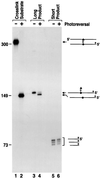
Analysis of XPF-ERCC1 reaction products from linear substrates with a psoralen cross-link. In lanes 1 and 2, the conversion of the control substrate to the monoadducted form with 254-nm light at 10 kJ/m2 for 3 min is shown. The long product (lane 3) and the short product (lane 5) generated from this substrate by XPF-ERCC1 plus RPA were gel purified from band A and band B in Fig. 7B, lane 4, and subjected to photoreversal under the same conditions. The photoreversed products are shown in lanes 4 and 6, respectively. DNA fragment sizes in nucleotides are indicated to the left of the figure. Drawings illustrating the corresponding species are shown to the right of lane 6.
FIG. 9
FIG. 9
Specific degradation of a linear substrate with a malondialdehyde-induced interstrand cross-link by XPF-ERCC1 plus RPA. The 147-mer duplex containing a TBG interstrand cross-link and a 5′ label in one strand (top) was used as the substrate. No nucleolytic degradation was observed when the substrate was incubated with XPF-ERCC1 (30 ng, lane 1). Addition of increasing amounts of RPA (10, 30, 60, and 90 ng from lane 2 to lane 5) to the reaction mixtures conferred cross-link-specific nuclease activity, which gave rise to the indicated specific reaction products. Drawings representing the long and short cleavage products are shown to the left of lane 1. Lane 6 shows the Maxam-Gilbert purine chemical sequencing ladder of the substrate. The sequence 5′ to the adducted guanine is shown to the right of lane 7. Because of the stability of the TBG cross-link, fragments hydrolyzed at the purines 3′ to the cross-linked guanine remained attached to the complementary strand, migrated near the full-length cross-link substrate, and thus were not discernible in the sequence ladder.
FIG. 10
FIG. 10
Summary of the main findings of this study. (A) Cross-link-specific exonucleolytic degradation of a linear duplex by XPF-ERCC1 in the presence of RPA. Four types of products are generated. Products a and b result from the cross-link-attenuated progression of a 3′-to-5′ exonuclease activity of XPF-ERCC1. Products c and d represent terminal digestion. (B) Futile DNA synthesis induced by the cross-link. Nucleotide excision repair nuclease removes oligonucleotides of 22 to 28 nt from the immediate 5′ vicinity of the cross-link (1). The pyrone-side adducted strand is preferred over the furan-side adducted strand with the particular substrate used in this study. The gap is filled in by DNA polymerases. Following filling in, ligation to the unremoved cross-link is inefficient, leaving behind mostly unligated repair patch as the major product and a small fraction of molecules in which the repair patch is ligated to regenerate the original substrate.

Similar articles

Cited by

References

    1. Bessho T, Mu D, Sancar A. Initiation of DNA interstrand cross-link repair in humans: the nucleotide excision repair system makes dual incisions 5′ to the cross-linked base and removes a 22- to 28-nucleotide-long damage-free strand. Mol Cell Biol. 1997;17:6822–6830. - PMC - PubMed
    1. Bessho T, Sancar A, Thompson L H, Thelen M P. Reconstitution of human excision nuclease with recombinant XPF-ERCC1 complex. J Biol Chem. 1997;272:3833–3837. - PubMed
    1. Cheng S A, Sancar A, Hearst J E. RecA-dependent incision of psoralen-DNA crosslinked DNA by (A)BC excinuclease. Nucleic Acids Res. 1991;19:657–663. - PMC - PubMed
    1. Cimino G D, Gamper H B, Isaacs S T, Hearst J E. Psoralens as photoactive probes of nucleic acid structure and function: organic chemistry, photochemistry, and biochemistry. Annu Rev Biochem. 1985;54:1151–1193. - PubMed
    1. Cole R S. Repair of DNA containing interstrand cross-links in Escherichia coli: sequential excision and recombination. Proc Natl Acad Sci USA. 1973;70:1064–1068. - PMC - PubMed

Publication types

LinkOut - more resources