Functional reconstitution of thymopoiesis after human immunodeficiency virus infection
- PMID: 10684316
- PMCID: PMC111790
- DOI: 10.1128/jvi.74.6.2943-2948.2000
Functional reconstitution of thymopoiesis after human immunodeficiency virus infection
Abstract
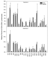
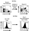
We have utilized combination antiretroviral therapy following human immunodeficiency virus type 1-induced human CD4(+) thymocyte depletion in the SCID-hu mouse to examine the immune competence of reconstituting thymocytes which appear following administration of combination therapy. These cells express a normal distribution of T-cell receptor variable gene families and are responsive to costimulatory signals. These results suggest that normal thymic function may be restored following antiretroviral treatment.
Figures


Similar articles
-
Reconstitution of human thymic implants is limited by human immunodeficiency virus breakthrough during antiretroviral therapy.J Virol. 1999 Aug;73(8):6361-9. doi: 10.1128/JVI.73.8.6361-6369.1999. J Virol. 1999. PMID: 10400728 Free PMC article.
-
Transient renewal of thymopoiesis in HIV-infected human thymic implants following antiviral therapy.Nat Med. 1997 Oct;3(10):1102-9. doi: 10.1038/nm1097-1102. Nat Med. 1997. PMID: 9334721
-
Development of a human thymic organ culture model for the study of HIV pathogenesis.AIDS Res Hum Retroviruses. 1995 Sep;11(9):1073-80. doi: 10.1089/aid.1995.11.1073. AIDS Res Hum Retroviruses. 1995. PMID: 8554904
-
Thymic function in HIV-infection.Dan Med J. 2013 Apr;60(4):B4622. Dan Med J. 2013. PMID: 23651726 Review.
-
[Antiretroviral therapy and immune reconstitution].J Soc Biol. 1999;193(1):5-10. J Soc Biol. 1999. PMID: 10851549 Review. French.
Cited by
-
Developmental regulation of P-glycoprotein activity within thymocytes results in increased anti-HIV protease inhibitor activity.J Leukoc Biol. 2011 Oct;90(4):653-60. doi: 10.1189/jlb.0111-009. Epub 2011 Apr 19. J Leukoc Biol. 2011. PMID: 21504949 Free PMC article.
-
Studies of retroviral infection in humanized mice.Virology. 2015 May;479-480:297-309. doi: 10.1016/j.virol.2015.01.017. Epub 2015 Feb 11. Virology. 2015. PMID: 25680625 Free PMC article. Review.
-
HIV disease and advanced age: an increasing therapeutic challenge.Drugs Aging. 2002;19(9):647-69. doi: 10.2165/00002512-200219090-00003. Drugs Aging. 2002. PMID: 12381235 Review.
-
Engineering antigen-specific T cells from genetically modified human hematopoietic stem cells in immunodeficient mice.PLoS One. 2009 Dec 7;4(12):e8208. doi: 10.1371/journal.pone.0008208. PLoS One. 2009. PMID: 19997617 Free PMC article.
References
-
- Aldrovandi G M, Feuer G, Gao L, Jamieson B, Kristeva M, Chen I S, Zack J A. The SCID-hu mouse as a model for HIV-1 infection. Nature. 1993;363:732–736. - PubMed
-
- Autran B, Carcelain G, Li T S, Blanc C, Mathez D, Tubiana R, Katlama C, Debre P, Leibowitch J. Positive effects of combined antiretroviral therapy on CD4+ T cell homeostasis and function in advanced HIV disease. Science. 1997;277:112–116. - PubMed
-
- Berkowitz R D, Beckerman K P, Schall T J, McCune J M. CXCR4 and CCR5 expression delineates targets for HIV-1 disruption of T cell differentiation. J Immunol. 1998;161:3702–3710. - PubMed
-
- Bertho J M, Demarquay C, Moulian N, Van Der Meeren A, Berrih-Aknin S, Gourmelon P. Phenotypic and immunohistological analyses of the human adult thymus: evidence for an active thymus during adult life. Cell Immunol. 1997;179:30–40. - PubMed
Publication types
MeSH terms
Substances
Grants and funding
LinkOut - more resources
Full Text Sources
Medical
Research Materials

