. 2000 Feb 15;97(4):1695-700.
doi: 10.1073/pnas.97.4.1695.

Human cytomegalovirus harbors its own unique IL-10 homolog (cmvIL-10)

Affiliations

Human cytomegalovirus harbors its own unique IL-10 homolog (cmvIL-10)

S V Kotenko et al. Proc Natl Acad Sci U S A. .

Abstract

We identified a viral IL-10 homolog encoded by an ORF (UL111a) within the human cytomegalovirus (CMV) genome, which we designated cmvIL-10. cmvIL-10 can bind to the human IL-10 receptor and can compete with human IL-10 for binding sites, despite the fact that these two proteins are only 27% identical. cmvIL-10 requires both subunits of the IL-10 receptor complex to induce signal transduction events and biological activities. The structure of the cmvIL-10 gene is unique by itself. The gene retained two of four introns of the IL-10 gene, but the length of the introns was reduced. We demonstrated that cmvIL-10 is expressed in CMV-infected cells. Thus, expression of cmvIL-10 extends the range of counter measures developed by CMV to circumvent detection and destruction by the host immune system.


Figures
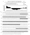
Figure 1
Figure 1
Fragment of the CMV genome encoding the IL-10 homolog. (A) Two coding frames starting at nucleotides 159678 and 159979, respectively, of the CMV genome (GenBank accession no. X17403) and identified by the tblastn homology search with human IL-10 as a query sequence (top lines). Letters in the center lines indicate identical amino acids; +, similar amino acids. Numbering is as follows. Upper lines, amino acids 1–52 and 75–112 of human IL-10 starting from Met1; lower lines, nucleotide numbering starting at 159678 and 159979, respectively, and corresponding to reading frames from the CMV genome. (B) Hydropathy plot of the whole first reading frame of the CMV genome with homology to the N-terminal portion of IL-10. ↓, end of the first exon. The predicted signal peptide is underlined. (C) Fragment of the CMV genome encoding the IL-10 homolog. Fragments of the two coding frames that were identified by the tblastn search program to have homology to human IL-10 are shown in boldface. Nucleotides of the two introns are shown in lowercase letters. Nucleotides of the three exons are shown in uppercase letters, and exon-encoded protein sequences are in shaded boxes. The predicted signal peptide is in an open box. Nucleotide numbering corresponds to the numbers of the CMV genome (GenBank accession no. X17403). Amino acid residues in the boxed regions are numbered starting from first Met residue in the first reading frame with only exon-encoded amino acid residues (boxes) counted.
Figure 2
Figure 2
cmvIL-10 expression. (A) Western blotting analysis of COS-1 cell-conditioned media. COS-1 cells were transiently transfected with the pEF-SPFL [lane 1 (mock)], the pEF-SPFL-cmv1 [lane 2 (FL-cmv1IL-10)], the pEF-SPFL-cmv1SP [lane 3 (FL-cmv1SPIL-10)], the pEF-SPFL-cmv2 [lane 4 (FL-cmv2IL-10)], or the pEF-SPFL-cmvIL-10 [lane 5 (FL-cmvIL-10)] expression vectors. Three days later, 1 ml of the conditioned media was subjected to immunoprecipitation and Western blotting experiments with anti-FLAG antibody. The molecular weight markers are shown on the left. (B) CMV-infected cells express cmvIL-10. PCR (lanes 3 and 4) or RT-PCR (lanes 6 and 7) with the same sets of primers was performed with DNA or RNA isolated from virus-infected (lanes 4 and 7) or uninfected (lanes 3 and 6) cells as described in Materials and Methods. Plasmids pEF-cmv3 (lane 2) and pEF-SPFL-cmvIL-10 (lane 5) were used for PCR as positive controls. A 1-kb ladder was run in lanes 1 and 10.
Figure 3
Figure 3
Alignment of amino acid sequences of human IL-10 and its viral homologs. The alignment of the amino acid sequences of cellular IL-10 encoded by the human genome (23) and viral IL-10s encoded by EBV [ebvIL-10 (10)], OV [ovIL-10 (12)], and CMV [cmvIL-10 (this study)] are shown. A consensus sequence is shown on the bottom. Identical amino acids corresponding to the consensus sequence are shown in black outline with white lettering. Similar amino acids are shown in gray outline with white lettering. Amino acid residues are numbered starting from first Met residue (signal peptide amino acids are included). The α-helices A through F, taken from the crystal structure of IL-10 and ebvIL-10 (30, 36), are underlined. Symbols: ① and ② designate Cys residues of IL-10 that form two intramolecular disulfide bridges (30). Asterisks denote amino acids predicted to be involved in interaction with IL-10R1 (32). The bold asterisks represent those residues involved in the interaction with IL-10R1 that are conserved among all the IL-10s. ■ points to conserved amino acids within regions involved in interaction with IL-10R1. □ points to conserved amino acids in the middle of IL-10 homologs that may be involved in interaction with IL-10R2. Arrows indicate positions of introns within IL-10 and cmvIL-10 genes. Numbers in parentheses represent the number of introns in IL-10 and cmvIL-10 (intron number within IL-10/intron number within cmvIL-10); the minuses at (2/−) and (4/−) denote no intron in cmvIL-10 at these positions. The triangle represents the position of Ala-98 of ebvIL-10. The program pileup of the Wisconsin Package, Version 9.1, Genetics Computer Group, Madison, WI, was used with the following parameters: the gap creation penalty 10, the gap extension penalty 2. The boxshade 3.21 program was used for shading of the alignment file.
Figure 4
Figure 4
Ligand binding and MHC class I antigen induction. (row I) Schematic of four cell lines used in these experiments: the parental Chinese hamster 16–9 cells and three 16–9-based cell lines expressing human IL-10R1/γR1 chimeric receptor or human IL-10R2 alone or both receptors together, which were created and described in detail (21). (Row II, A–D) The cells described in row I were incubated for 30 min at 4°C with conditioned medium from COS-1 cells transfected with one of the following plasmids: the control vector pEF-SPFL (open areas, thick lines); the pEF-SPFL-IL-10 (open areas, thin lines), or the pEF-SPFL-cmvIL-10 (shaded areas, thin lines). Ligand binding to the cell surface was determined by flow cytometry with anti-FLAG antibody (Sigma) as the primary antibody and FITC-conjugated goat anti-mouse IgG (Santa Cruz) as the secondary antibody. Here and in row III, the ordinate represents relative cell number, and the abscissa is relative fluorescence. (Row III, E–H). The ability of IL-10 and cmvIL-10 to induce MHC class I antigen expression was demonstrated by flow cytometry as described (21). The cells described in row I were left untreated (open areas, thick lines) or treated with conditioned media (100 μl) from COS-1 cells transfected with the pEF-SPFL-cmvIL-10 plasmid (shaded areas, thin lines) or with Hu-IL-10 (100 units/ml; open areas, thin lines).
Figure 5
Figure 5
Ligand-binding competition. Cells expressing both chains of the IL-10 receptor complex were incubated with FL-cmvIL-10 alone [30 μl of conditioned media from COS-1 cells expressing FL-cmvIL-10; (A) thick line] or with increased concentrations of IL-10 (expressed as ng/ml; A, thin lines) or with FL-IL-10 alone [30 μl of conditioned media from COS-1 cells expressing FL-IL-10; B, thick line] and with increased quantities of conditioned media from COS-1 cells expressing cmvIL-10 [expressed as μl; B, thin lines]. Cells were treated as described for row II in the legend to Fig. 4.
Figure 6
Figure 6
Stat activation induced by cmvIL-10 in hamster cells and PBMCs. EMSAs were used to measure activation of Stat1 and Stat3. Hamster cells expressing both receptor chains (Fig. 4) and PBMCs were used. Cells were left untreated or treated with recombinant IL-10 (100 units/ml) or with conditioned media (200 μl) from COS cells transfected with the pEF-SPFL-cmvIL-10 plasmid or from uninfected or CMV-infected cells. Cellular lysates were prepared and assayed for Stat activation in the EMSA as described in Materials and Methods. Positions of Stat DNA-binding complexes are indicated by arrows. Antibodies against Stat1 and Stat3 were added as indicated to reduce the mobility of complexes containing these proteins.
Figure 7
Figure 7
Schematic map of the CMV genome. The CMV genome is organized as two regions of unique sequences, unique long (UL) and unique short (US), flanked by two sets of inverted repeats (TRL/IRL) and (IRS/TRS) (light shaded boxes). mtrI, mtrII, and mtrIII represent three morphological transforming regions identified within the CMV genome (24, 48). XbaI, BanII, and XhoI are sites for digestion with restriction endonucleases. The 79 ORF (dark shaded boxes) is an ORF of 79 aa whose disruption abolishes mtrII transforming ability. cmvIL-10 is encoded by three exons (see text). The position of two introns within the mtrII region is shown. The junction of the segments of the cmvIL-10 protein encoded by each exon is shown by the arrows. SP represents the signal peptide of the cmvIL-10 (light shaded box). The 79-aa ORF is slightly larger than the cmvIL-10 segment encoded by the first exon; it is the product of an mRNA that includes the first exon and intron 1 (Fig. 1).

Similar articles

Cited by

References

    1. Moore K W, O'Garra A, de Waal M, Vieira P, Mosmann T R. Annu Rev Immunol. 1993;11:165–190. - PubMed
    1. Fossa S D, Aamdal S, Naume B, Gallati H. Acta Oncol. 1995;34:599–603. - PubMed
    1. Boulland M L, Meignin V, Leroy-Viard K, Copie-Bergman C, Briere J, Touitou R, Kanavaros P, Gaulard P. Am J Pathol. 1998;153:1229–1237. - PMC - PubMed
    1. Mori N, Prager D. Leuk Lymphoma. 1998;29:239–248. - PubMed
    1. Halak B K, Maguire H C, Lattime E C. Cancer Res. 1999;59:911–917. - PubMed

Publication types

MeSH terms

LinkOut - more resources