. 2000 Feb 15;19(4):598-611.
doi: 10.1093/emboj/19.4.598.

The Drosophila caspase DRONC is regulated by DIAP1

Affiliations

The Drosophila caspase DRONC is regulated by DIAP1

P Meier et al. EMBO J. .

Abstract

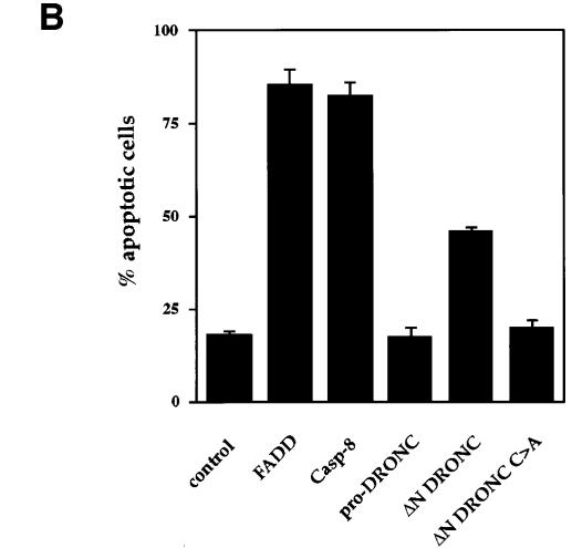
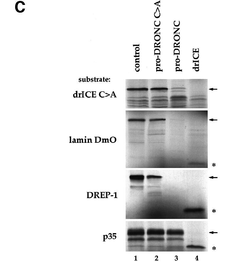
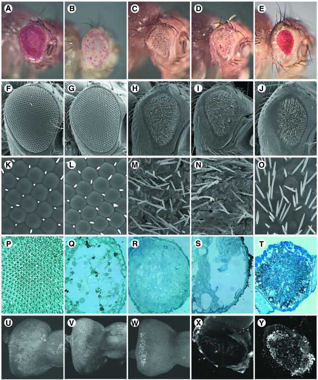
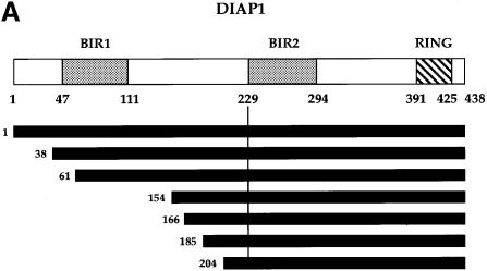
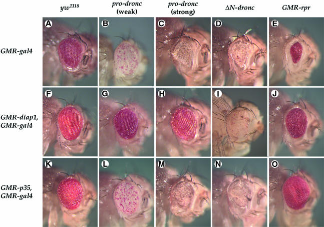
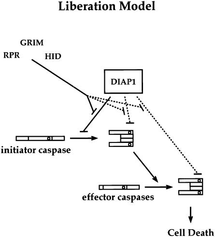
We have isolated the recently identified Drosophila caspase DRONC through its interaction with the effector caspase drICE. Ectopic expression of DRONC induces cell death in Schizosaccharomyces pombe, mammalian fibroblasts and the developing Drosophila eye. The caspase inhibitor p35 fails to rescue DRONC-induced cell death in vivo and is not cleaved by DRONC in vitro, making DRONC the first identified p35-resistant caspase. The DRONC pro-domain interacts with Drosphila inhibitor of apoptosis protein 1 (DIAP1), and co-expression of DIAP1 in the developing Drosophila eye completely reverts the eye ablation phenotype induced by pro-DRONC expression. In contrast, DIAP1 fails to rescue eye ablation induced by DRONC lacking the pro-domain, indicating that interaction of DIAP1 with the pro-domain of DRONC is required for suppression of DRONC-mediated cell death. Heterozygosity at the diap1 locus enhances the pro-DRONC eye phenotype, consistent with a role for endogenous DIAP1 in suppression of DRONC activation. Both heterozygosity at the dronc locus and expression of dominant-negative DRONC mutants suppress the eye phenotype caused by reaper (RPR) and head involution defective (HID), consistent with the idea that DRONC functions in the RPR and HID pathway.


Figures

None
Fig. 1. DRONC is a drICE-interacting caspase. (A) The dendrogram shows the phylogenetic relationships of the core region of caspase family members (i.e. the protein sequence without the pro-domain). ClustalX was used for the sequence analysis. (B) Yeast two-hybrid analysis showing that DRONC and drICE interact with each other through their core regions. The extent of the β-galactosidase staining, as detected in filter tests, is indicated: +++, intense blue staining of large colonies; ++, light blue staining of medium size colonies. (C) Co-immunoprecipitation from 293T cell extracts. FLAG-tagged full-length DRONC (pro-DRONC C→A) and Bcl-10 (control) were co-expressed together with either Myc-tagged pro-drICE C→A, ΔN drICE C→A or Bcl-10 (control). Cell lysates were incubated with M2 anti-FLAG monoclonal antibody resin, washed, and the co-immunoprecipitated Myc epitope-tagged proteins were detected by immunoblot analysis using anti-Myc monoclonal antibody (9E10). Expression of FLAG-tagged and Myc-tagged proteins was confirmed. Molecular mass markers in kDa are shown.
None
Fig. 2.Ectopic expression of DRONC induces cell death in yeast and in mammalian Rat-1 cells. (A) Expression of DRONC is toxic to S.pombe. For cytotoxicity assays, yeast from two independent colonies were grown to log phase, the OD595 of the culture determined and the yeast then plated in serial 10-fold dilutions on selective, inducing media. Western blot analysis with anti-FLAG M2 antibody was used to confirm expression and autoproteolytic cleavage of C-terminally tagged DRONC. (B) Transient transfection of dronc leads to induction of apoptosis in mammalian Rat-1 fibroblast cells. Various expression constructs were co-transfected with a CMV-lacZ reporter plasmid in a ratio of 10:1. At 24 h post-transfection, cells were fixed and examined for β-galactosidase activity. Shown are the percentage of β-galactosidase-positive cells with apoptotic morphology from three independent experiments (mean ± SD). (C) DRONC is a cysteine protease that cleaves drICE C→A, lamin DmO and DREP-1 but not p35 in vitro. In vitro translated substrates were incubated with (1) control (no protease added); (2) pro-DRONC C→A purified from yeast; (3) pro-DRONC purified from yeast; and (4) purified bacterially expressed drICE. The unprocessed substrate is indicated by an arrow and the cleavage product is denoted by an asterisk.
None
Fig. 3. Ectopic expression of DRONC in the developing eye causes ablation of all retinal structures resulting in a hollow eye. Phenotypes were analysed by light microscopy of whole mounts (A–E), tangential thin sections of adult eyes (P–T), scanning electron microscopy (F–O) and acridine orange staining of eye discs of third instar larvae (U–W) and 60 h after puparium formation (X and Y). (A, F, K, P, U and X) Control flies (+/GMR-gal4). (B, G, L, Q, V and Y) The weak pro-droncW transgenic line (GMR-gal4/UAS-pro-droncW) displays a spotted eye phenotype (B) with an essentially normal eye morphology on the outside (G and L) but a severely malformed cell arrangement in the interior (Q). (C, H, M and R) Pro-droncS transgenic flies that show reduced eye size (C and H) with no defined interior eye structure (R) (GMR-gal4/UAS-pro-droncS). (D, I, N, S and W) Ectopic expression of ΔN DRONC (GMR-gal4/UAS-ΔN dronc) causes excessive cell death in the eye disc of third instar larvae (W) resulting in a small eye phenotype (D and I). (E, J, O and T) GMR-rpr flies display eyes of a reduced size (E and J) but unlike dronc transgenic fly eyes they are red instead of white (E) (GMR-rpr/+). (C, H, M and R) and (D, I, N and S) represent pictures from animals that were crossed with GMR-gal4 (815, weak) and raised at 18°C. All other images were obtained from animals crossed with GMR-gal4 (816, strong) and raised at 25°C. In this and the following figures, anterior is to the right and posterior to the left.
None
Fig. 4. DIAP1 physically interacts with DRONC. (A) Seventeen DRONC-interacting clones encoded full-length and N-terminal truncations of DIAP1 of which seven representative DIAP1 clones are indicated. The positions of the first amino acid of the clones relative to full-length DIAP1 are denoted on the left. (B) Various DIAP1 deletion mutants were used in a yeast two-hybrid assay to map the interaction domain between DIAP1 and the pro-domain of DRONC. The BIR2 region of DIAP1 was sufficient for the interaction with the pro-domain of DRONC (C) Co-immunoprecipitation of DRONC and DIAP1 from cellular extracts. 293T cells were transiently transfected with plasmids expressing FLAG-tagged DRONC, ΔN DRONC, DRONC-CARD or Bcl-10 (control) and Myc-tagged BIR1/2, BIR1, BIR2 or Bcl-10 in the indicated combinations. Cell lysates were immunoprecipitated with anti-FLAG and immunoblotted with anti-Myc as in Figure 1C. Expression of FLAG-tagged and Myc-tagged proteins was confirmed. Molecular mass markers in kDa are shown.
None
Fig. 5.The eye ablation phenotype caused by ectopic expression of DRONC in the developing eye can be suppressed by co-expression of DIAP1, but not by p35. The effect of overexpressing the different dronc constructs or rpr alone (AE), or in combination with DIAP1 (FJ) or p35 (KO) is shown. (F–J) Ectopic expression of DIAP1 suppresses the eye phenotype caused by pro-DRONC (G and H) but not by ΔN DRONC (I). (K–O) Co-expression of p35 is unable to rescue the eye phenotype caused by pro-DRONC (L and M) or ΔN DRONC (N) overexpression but blocks RPR-induced cell death (O). Flies from pro-droncS and ΔN dronc lines were crossed to GMR-gal4 (815), GMR-diap1-GMR-gal4 (815) or GMR-p35-GMR-gal4 (815) and kept at 18°C. For all other crosses, GMR-gal4 (816), GMR-diap1-GMR-gal4 (816) or GMR-p35-GMR-gal4 (816) were used and kept at 25°C. (P) Expression of p35 fails to suppress DRONC-mediated toxicity in yeast. Vectors to express p35, a non-cleavable p35 mutant (where the caspase recognition motif DQMD has been changed to DQME), CrmA and the CrmA mutant T291R were introduced into S.pombe transformed with pro-dronc or caspase-3-lacZ, respectively. The viability of the resultant transfected yeast cells following induction of DRONC expression was assessed as described above.
None
Fig. 6. Heterozygosity at the diap1 locus bearing the deficiency [Df(3L)th102] enhances the eye phenotype caused by DRONC overexpression. (A) Flies with a 50% reduced diap1 gene dose are viable and show a perfectly normal compound eye [+/SM6; Df(3L)th102/UAS-pro-droncW]. (B) Overexpression of GAL4 induces a rough eye phenotype in heterozygous diap1 flies [+/GMR-gal4; Df(3L)th102/TM3]. (C) Ectopic expression of pro-droncW induces a spotted eye phenotype (+/GMR-gal4; UAS-pro-droncW/TM6c). Note: these flies show a less prominent spotted eye phenotype when compared with the flies shown in Figure 5B due to their different genetic background. (D) Flies that express pro-droncW and are heterozygous for diap1 [Df(3L)th102] display severely deformed eyes and die trapped in their pupae case [+/GMR-gal4; Df(3L) th102/UAS-pro-droncW]. All flies were embedded in holocarbon oil and photographed using a stereo-microscope.
None
Fig. 7. DRONC is a rate-limiting caspase in the RPR and HID death pathway. (A–D) Cell death induced by RPR and HID is sensitive to dronc gene dosage. Flies with a chromosomal deletion that removes the dronc locus [Df(3L)AC1] show a suppressed RPR and HID eye phenotype. (A) GMR-rpr,+; (B) GMR-rpr,Df(3L)AC1; (C) GMR-hid,+; (D) GMR-hid,Df(3L)AC1. (E and F) The expression of dominant-negative DRONC mutants suppresses the RPR eye phenotype. (E) GMR-rpr/GMR-gal4, UAS-pro-dronc C→A; (F) GMR-rpr/GMR-gal4, UAS-dronc-card.
None
Fig. 8. The observation that DIAP1 interacts with the pro-caspase DRONC as well as with apoptotic inducers such as RPR, GRIM and HID places DIAP1 in a potentially pivotal position to regulate apoptosis. In the proposed model, DIAP1 functions as ‘guardian of the caspase machinery’ by binding to and suppressing spontaneous pro-caspase activation in the absence of RPR, GRIM and active HID. As indicated by studies using heterologous systems, DIAP1 may also act by directly inhibiting the proteolytic activity of spontaneously activated caspases (dotted lines) (Kaiser et al., 1998; Hawkins et al., 1999) According to the ‘liberation model’, RPR, GRIM or HID exert some, or all, of their pro-apoptotic action by liberating initiator caspases, such as DRONC, from the activation-inhibitory effect of DIAP1. This displacement of DIAP1 from DRONC could then result in the activation of DRONC through DARK, the Drosophila homologue of Apaf-1/CED-4 (Rodriguez et al., 1999)

Similar articles

Cited by

References

    1. Abrams J.M., White, K., Fessler, L.I. and Steller, H. (1993) Programmed cell death during Drosophila embryogenesis. Development, 117, 29–43. - PubMed
    1. Alnemri E.S. (1997) Mammalian cell death proteases: a family of highly conserved aspartate specific cysteine proteases. J. Cell. Biochem., 64, 33–42. - PubMed
    1. Ashburner M. (1989) Drosophila: A Laboratory Manual. Cold Spring Harbor Laboratory Press, Cold Spring Harbor, NY.
    1. Basler K., Christen, B. and Hafen, E. (1991) Ligand-independent activation of the sevenless receptor tyrosine kinase changes the fate of cells in the developing Drosophila eye. Cell, 64, 1069–1081. - PubMed
    1. Bateman A., Birney, E., Durbin, R., Eddy, S.R., Finn, R.D. and Sonnhammer, E.L. (1999) Pfam 3.1: 1313 multiple alignments and profile HMMs match the majority of proteins. Nucleic Acids Res., 27, 260–262. - PMC - PubMed

Publication types

MeSH terms