Comparative Study
. 2000 Feb 1;97(3):1038-43.
doi: 10.1073/pnas.97.3.1038.

A family of chromatin remodeling factors related to Williams syndrome transcription factor

Affiliations
Comparative Study

A family of chromatin remodeling factors related to Williams syndrome transcription factor

D A Bochar et al. Proc Natl Acad Sci U S A. .

Abstract

Chromatin remodeling complexes have been implicated in the disruption or reformation of nucleosomal arrays resulting in modulation of transcription, DNA replication, and DNA repair. Here we report the isolation of WCRF, a new chromatin-remodeling complex from HeLa cells. WCRF is composed of two subunits, WCRF135, the human homolog of Drosophila ISWI, and WCRF180, a protein related to the Williams syndrome transcription factor. WCRF180 is a member of a family of proteins sharing a putative heterochromatin localization domain, a PHD finger, and a bromodomain, prevalent in factors involved in regulation of chromatin structure.


Figures
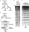
Figure 1
Figure 1
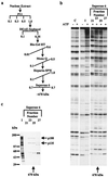
Identification of a new chromatin remodeling activity. (a) HeLa nuclear extract was fractionated by using phosphocellulose (P11) chromatography. The 0.5- and 0.75-M KCl elutions were concentrated on a DEAE cellulose (DE52) column and further fractionated on a Superose 6 gel filtration. (b) P11 D-fraction concentrated on DE52 displays a unique mononucleosome DNase I cleavage pattern. Fractions (3 μl) corresponding to the 0.5-M potassium chloride elution of P11 (C-fraction) concentrated on a DE52 or the 0.75-M potassium chloride elution of P11 (D-fraction) concentrated on a DE52 were analyzed for mononucleosome disruption assay and compared with the disruption activity seen by using purified yeast SWI/SNF complex (3 μl) (ref. 4). (c) Mononucleosome DNase I disruption assay for the fractions (3 μl) of the Superose 6 derived from the P11 D-fraction, as described in a. The arrow (Bottom) denotes the elution position of thyroglobulin (670 kDa). Superose 6 calibration is as follows: fraction 16, void; fraction 25, 670 kDa; fraction 29, 440 kDa; fraction 32, 158 kDa. (d) Human ISWI predominantly elutes from P11 at potassium chloride concentration of 1 M. Equal volumes (5 μl) of either nuclear extract (NE, 30 μg) or fractions of P11 (3 μg of 0.1 M/1.5 μg of 0.3/M/1.0 μg of 0.5 M/0.5 μg of 1.0 M) were separated in an SDS-polyacrylamide (8%) gel, and proteins were immunoblotted with anti-human ISWI antisera. (e) The 0.35-M potassium chloride eluate of DE52 fraction derived from P11 C-fraction contains a human ISWI complex larger than WCRF. Fractions of the Superose 6 chromatography shown in a (12 μl) were separated in an SDS-polyacrylamide (8%) gel, and proteins were immunoblotted with either anti-human SNF2L or anti-BRG1 antisera. The hSNF2L/h immunoreactivity in the D-fraction closely coelutes with the disrupting activity present in the same fractions (c), indicating that the majority of hSNF2L/h is associated with WCRF.
Figure 2
Figure 2
Isolation of WCRF. (a) Purification scheme. HeLa nuclear extract was fractionated by chromatography as described in Materials and Methods. The horizontal and diagonal lines indicate stepwise and gradient elution, respectively. Concentrations are given in molars. (b) Assay of mononucleosome DNase I cleavage of Superose 6 fractions (6 μl, 50 nM) was conducted as described in Materials and Methods. C denotes the addition of 0.1 M potassium chloride. I is the 0.55-M potassium chloride eluate from Heparin-5PW. The arrow (Bottom) denotes the elution position of thyroglobulin. (c) Resolution of WCRF as two subunits on a Superose 6. The 0.55-M potassium chloride eluate from Heparin-5PW (I) was loaded on a Superose 6 column, and proteins were eluted with 0.7 M potassium chloride. Fractions (5 μl, 50 nM) were separated in an SDS-polyacrylamide (4–12%) gel, and proteins were visualized by silver staining. Molecular masses of marker proteins (Left) and WCRF subunits (Right) are indicated. (Bottom) The arrow denotes the elution position of thyroglobulin.
Figure 3
Figure 3
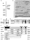
WCRF180 contains a bromodomain and a PHD finger. (a) SDS/PAGE of WCRF. WCRF (Superose 6, 15 μl) was separated in an SDS-polyacrylamide (4–12%) gel, and proteins were visualized by colloidal blue staining. Molecular masses of marker proteins are indicated (Left). (b) The primary amino acid sequence of WCRF180. The single-underlined peptides correspond to peptides obtained from comparison of MS/MS spectra against the EST database. The double-underlined peptides correspond to sequences that were confirmed by comparison of the MS/MS spectra against the full length clone. The conserved PHD finger and bromodomain are boxed. (c) Immunoprecipitation of WCRF. P11 1.0-M fraction was incubated with control antibodies (α-CSA), polyclonal WCRF180 antibodies (α-WCRF180), or polyclonal ISWI antibodies (α-ISWI). Immunoprecipitates were subjected to SDS/PAGE and immunoblotted with affinity-purified WCRF180 antibodies. (d) Northern blot analysis of WCRF180. RNA size markers are indicated. Human tissues are as indicated: H, heart; B, brain; Pl, placenta; Lg, lung; Lv, liver; M, skeletal muscle; K, kidney; Pc, pancreas; S, spleen; Ty, thymus; Pr, prostrate; Ts, testis; O, ovary; I, small intestine; C, colon; Lk, peripheral blood leukocytes. (e) Diagram of WCRF180 showing regions of homology with WSTF, KIAA0314, Acf1, YGL133w, YPL216w, and cbp146. The numbers (Right) correspond to the number of amino acids in each protein. The numbers in the boxes denote the percent of identity (Top) and similarity (Bottom) of each protein to WCRF, with the exception shown in shaded lines.
Figure 4
Figure 4
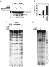
Functional analysis of WCRF. (a) DNA-dependent ATPase assays of WCRF. Fractions from Superose 6 (2 μl, 50 nM) corresponding to WCRF (fraction 23) or CSB (2 μl, 50 nM; fraction 27) were assayed for stimulation of ATPase activity, as described in Materials and Methods by using 100 ng of either double-stranded plasmid DNA, mononucleosomes or oligonucleosomes. (b) Quantification of fold stimulation of WCRF ATPase activity by DNA and oligonucleosomes. (c) Mononucleosome DNase I disruption assay comparing the mononucleosome disruption activity of yeast SWI/SNF (3 μl, 100 nM) to WCRF (heparin fraction, 3 μl, 70 nM). (d) WCRF remodels nucleosome arrays in an ATP-dependent fashion. Nucleosome array was reconstituted on a DNA fragment containing a dinucleosome length sequence with a synthetic GAL4-E4 promoter flanked by on either side by five repeats of a nucleosome-positioning sequence from sea urchin 5S rDNA. Purified WCRF (heparin fraction, 3 μl; 70 nM) was then incubated with the 12-nucleosome array in the presence or the absence of ATP, as indicated. Nucleosomal structure was then analyzed by DNase I digestion and migration on a 2% native agarose gel. Positions of the 5S and G4E4 nucleosomes are shown (Right) (opened and shaded ovals respectively).

Similar articles

Cited by

References

    1. Kornberg R D, Lorch Y. Cell. 1999;98:285–294. - PubMed
    1. Laurent B C, Yang X, Carlson M. Mol Cell Biol. 1992;12:1893–1902. - PMC - PubMed
    1. Cairns B R, Kim Y J, Sayre M H, Laurent B C, Kornberg R D. Proc Natl Acad Sci USA. 1994;91:1950–1954. - PMC - PubMed
    1. Côté J, Quinn J, Workman J L, Peterson C L. Science. 1994;265:53–60. - PubMed
    1. Eisen J A, Sweder K S, Hanawalt P C. Nucleic Acids Res. 1995;23:2715–2723. - PMC - PubMed

Publication types

MeSH terms

Substances

Associated data